
CHINESE JOURNAL OF PARASITOLOGY AND PARASITIC DISEASES ›› 2022, Vol. 40 ›› Issue (3): 299-304.doi: 10.12140/j.issn.1000-7423.2022.03.004
• ORIGINAL ARTICLES • Previous Articles Next Articles
ZHUO Yi-cheng1( ), YANG Hai-cheng1, LIU Cheng-hao1, ZHANG Bao-cai1, DUO Xiao-yong1, ZHANG Shi-jie2,*()
Received:2021-11-08
Revised:2022-01-25
Online:2022-06-30
Published:2022-07-06
Contact:
ZHANG Shi-jie
E-mail:zhuoyicheng1314@163.com;zhangshijie1@sina.com
Supported by:CLC Number:
ZHUO Yi-cheng, YANG Hai-cheng, LIU Cheng-hao, ZHANG Bao-cai, DUO Xiao-yong, ZHANG Shi-jie. Effect of osteopontin expression level on the growth and development of Echinococcus multilocularis protoscoleces[J]. CHINESE JOURNAL OF PARASITOLOGY AND PARASITIC DISEASES, 2022, 40(3): 299-304.
Add to citation manager EndNote|Ris|BibTeX
URL: https://www.jsczz.cn/EN/10.12140/j.issn.1000-7423.2022.03.004
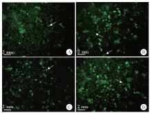
Fig. 1
Changes of EmOPN fluorescence of protoscoleces after infection with lentivirus in vitro for 72 hours(× 40) A: LV3-NC group; B: LV-EmOPN-734 group; C: LV5-NC group; D: LV-EmOPN-0423 group. The protoscoleces expressed green fluorescence in dots or circles in each group (shown by white arrows).
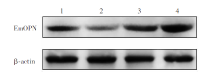
Fig. 2
Western blotting results of EmOPN after 72 hours of lentivirus infection with E. multilocularis protoscoleces 1: LV3-NC group; 2: LV-EmOPN-734 group; 3: LV5-NC group; 4: LV-EmOPN-0423 group.
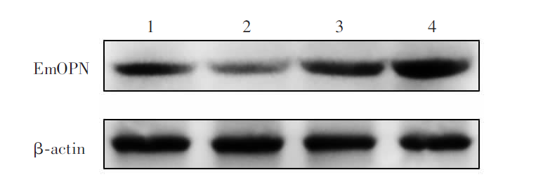
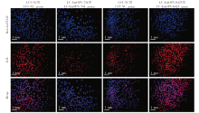
Fig. 3
Observation of EdU+ results of protoscoleces infected with lentivirus for 72 hours under a fluorescent microscope (× 40)

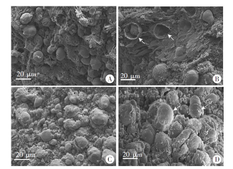
Fig. 4
Observation of vesicles formed after infection with lentivirus and cultured in vitro for 8 weeks by scanning electron microscopic (× 900) A: LV3-NC group; B: LV-EmOPN-734 group; C: LV5-NC group; D: LV-EmOPN-0423 group.
| [1] | Wen H,, Vuitton L,, Tuxun T, et al. Echinococcosis: advances in the 21st century[J]. Clin Microbiol Rev, 2019, 32(2): e00075-18. |
| [2] |
Fu MH,, Wang X,, Han S, et al. Advances in research on echinococcoses epidemiology in China[J]. Acta Trop, 2021, 219: 105921.
doi: 10.1016/j.actatropica.2021.105921 |
| [3] | Han S,, Kui Y,, Xue CZ, et al. The endemic status of echinococcosis in China from 2004 to 2020[J/OL]. Chin J Parasitol Parasit Dis, 2021. https://kns.cnki.net/kcms/detail/31.1248.R.20211015.1639.012.html. (in Chinese) |
| 韩帅,, 蒉嫣,, 薛垂召, 等. 2004—2020年全国棘球蚴病疫情分析[J/OL]. 中国寄生虫学与寄生虫病杂志, 2021. https://kns.cnki.net/kcms/detail/31.1248.R.20211015.1639.012.html. | |
| [4] | Craig PS,, Giraudoux P,, Wang ZH, et al. Echinococcosis transmission on the Tibetan Plateau[M]// Rollinson. Advances in Parasitology. Amsterdam: Elsevier, 2019: 165-246. |
| [5] |
Casulli A,, Barth TFE,, Tamarozzi F. Echinococcus multilocularis[J]. Trends Parasitol, 2019, 35(9): 738-739.
doi: 10.1016/j.pt.2019.05.005 |
| [6] | Conraths FJ,, Probst C,, Possenti A, et al. Potential risk factors associated with human alveolar echinococcosis: systematic review and meta-analysis[J]. PLoS Negl Trop Dis, 2017, 11(7): e0005801. |
| [7] | Brožová A,, Jankovská I,, Bejček V, et al. Echinococcus spp.: tapeworms that pose a danger to both animals and humans-a review[J]. Sci Agric Bohem, 2017, 48(4): 193-201. |
| [8] | Kern P,, da Silva AM,, Akhan O, et al. The echinococcoses: Diagnosis, clinical management and burden of disease[J]. Adv Parasitol, 2017, 96: 259-369. |
| [9] | Nikendei C,, Greinacher A,, Berkunova A, et al. Psychological burden and resilience factors in patients with alveolar echinococcosis: a cross-sectional study[J]. PLoS Negl Trop Dis, 2019, 13(1): e0007082. |
| [10] |
Akbulut S,, Sahin TT. Comment on surgical approaches for definitive treatment of hepatic alveolar echinococcosis: results of a survey in 178 patients[J]. Parasitology, 2020, 147(13): 1408-1410.
doi: 10.1017/S0031182020001390 |
| [11] |
Song ZL,, Chen W,, Athavale D, et al. Osteopontin takes center stage in chronic liver disease[J]. Hepatology, 2021, 73(4): 1594-1608.
doi: 10.1002/hep.31582 |
| [12] |
Lamort AS,, Giopanou I,, Psallidas I, et al. Osteopontin as a link between inflammation and cancer: the Thorax in the spotlight[J]. Cells, 2019, 8(8): 815.
doi: 10.3390/cells8080815 |
| [13] | Zhang L,, Zhang SJ,, Cao YW, et al. The correlation between osteopontin and metastasis of hepatic Echinococcus multilocularis infection[J]. Chin J Parasitol Parasit Dis, 2011, 29(1): 33-36. (in Chinese) |
| ( 张龙,, 张示杰,, 曹玉文, 等. 骨桥蛋白与肝泡型棘球蚴转移的相关性研究[J]. 中国寄生虫学与寄生虫病杂志, 2011, 29(1): 33-36.) | |
| [14] | Wu HX,, Wu XW,, Lian WB, et al. Effect of anti-osteopontin antibody on angiogenesis of hepatic alveolar hydatid tissue in gerbil[J]. J Pathog Biol, 2014, 9(10): 894-897. (in Chinese) |
| ( 吴何兴,, 吴向未,, 连文波, 等. 抗骨桥蛋白抗体对沙鼠肝多房棘球蚴组织周围血管生成的影响[J]. 中国病原生物学杂志, 2014, 9(10): 894-897.) | |
| [15] | Yang ZH,, Yang HC,, Zhang HW, et al. Cloning and biological analysis of osteopontin from Echinococcus multilocularis[J]. J Med Postgrad, 2020, 33(10): 1033-1038. (in Chinese) |
| ( 杨照辉,, 杨海成,, 张宏伟, 等. 泡状棘球蚴骨桥蛋白基因克隆及其生物学分析[J]. 医学研究生学报, 2020, 33(10): 1033-1038.) | |
| [16] |
Yang HC,, Zhang HW,, Yang J, et al. Autocrine osteopontin is involved in maintaining the growth and metastasis of Echinococcus multilocularis[J]. Acta Trop, 2022, 228: 106328.
doi: 10.1016/j.actatropica.2022.106328 |
| [17] |
Spiliotis M,, Brehm K. Axenic in vitro cultivation of Echinococcus multilocularis metacestode vesicles and the generation of primary cell cultures[J]. Methods Mol Biol, 2009, 470: 245-262.
doi: 10.1007/978-1-59745-204-5_17 pmid: 19089387 |
| [18] |
Zhang CS,, Lin RY,, Li ZD, et al. Immune exhaustion of T cells in alveolar echinococcosis patients and its reversal by blocking checkpoint receptor TIGIT in a murine model[J]. Hepatology, 2020, 71(4): 1297-1315.
doi: 10.1002/hep.30896 |
| [19] |
Chen DS,, Mellman I. Elements of cancer immunity and the cancer-immune set point[J]. Nature, 2017, 541(7637): 321-330.
doi: 10.1038/nature21349 |
| [20] |
Liu LL,, Zhang RY,, Deng JW, et al. Construction of TME and Identification of crosstalk between malignant cells and macrophages by SPP1 in hepatocellular carcinoma[J]. Cancer Immunol Immunother, 2022, 71(1): 121-136.
doi: 10.1007/s00262-021-02967-8 |
| [21] |
Icer MA,, Gezmen-Karadag M. The multiple functions and mechanisms of osteopontin[J]. Clin Biochem, 2018, 59: 17-24.
doi: 10.1016/j.clinbiochem.2018.07.003 |
| [22] |
Zhao HL,, Chen Q,, Alam A, et al. The role of osteopontin in the progression of solid organ tumour[J]. Cell Death Dis, 2018, 9(3): 356.
doi: 10.1038/s41419-018-0391-6 |
| [23] |
Phillips RJ,, Helbig KJ,, van der Hoek KH, et al. Osteopontin increases hepatocellular carcinoma cell growth in a CD44 dependant manner[J]. World J Gastroenterol, 2012, 18(26): 3389-3399.
doi: 10.3748/wjg.v18.i26.3389 |
| [24] |
Rangaswami H,, Bulbule A,, Kundu GC. Osteopontin: role in cell signaling and cancer progression[J]. Trends Cell Biol, 2006, 16(2): 79-87.
doi: 10.1016/j.tcb.2005.12.005 pmid: 16406521 |
| [25] |
Briones-Orta MA,, Avendaño-Vázquez SE,, Aparicio-Bautista DI, et al. Osteopontin splice variants and polymorphisms in cancer progression and prognosis[J]. Biochim Biophys Acta Rev Cancer, 2017, 1868(1): 93-108.
doi: 10.1016/j.bbcan.2017.02.005 |
| [26] | Zhang L,, Zhang SJ,, Cao YW, et al. The correlation between osteopontin and metastasis of hepatic Echinococcus multilocularis infection[J]. Chin J Parasitol Parasit Dis, 2011, 29(1): 33-36. (in Chinese) |
| ( 张龙,, 张示杰,, 曹玉文, 等. 骨桥蛋白与肝泡型棘球蚴转移的相关性研究[J]. 中国寄生虫学与寄生虫病杂志, 2011, 29(1): 33-36.) | |
| [27] | Yang HC,, Zhang HW,, Shi KJ, et al. Autocrine osteopontin promotes the growth and metastasis of Echinococcus multilocularis via the EGFR signaling pathway[J]. Chin J Parasitol Parasit Dis, 2021, 39(2): 226-232. (in Chinese) |
| ( 杨海成,, 张宏伟,, 史康杰, 等. 自分泌骨桥蛋白通过EGFR信号通路促进多房棘球蚴生长和转移的研究[J]. 中国寄生虫学与寄生虫病杂志, 2021, 39(2): 226-232.) | |
| [28] |
Cheng YG,, Wen G,, Sun Y, et al. Osteopontin promotes colorectal cancer cell invasion and the stem cell-like properties through the PI3K-AKT-GSK/3β-β/catenin pathway[J]. Med Sci Monit, 2019, 25: 3014-3025.
doi: 10.12659/MSM.913185 |
| [29] |
Fast EM,, Sporrij A,, Manning M, et al. External signals regulate continuous transcriptional states in hematopoietic stem cells[J]. eLife, 2021, 10: e66512.
doi: 10.7554/eLife.66512 |
| [30] | Wang M,, Han J,, Marcar L, et al. Radiation resistance in KRAS-mutated lung cancer is enabled by stem-like properties mediated by an osteopontin-EGFR pathway[J]. Cancer Res, 2017, 77(8): 2018-2028. |
| [31] |
Kiss T,, Jámbor K,, Koroknai V, et al. Silencing osteopontin expression inhibits proliferation, invasion and induce altered protein expression in melanoma cells[J]. Pathol Oncol Res, 2021, 27: 581395.
doi: 10.3389/pore.2021.581395 |
| [32] | Cheng Z,, Liu F,, Li X, et al. EGF-mediated EGFR/ERK signaling pathway promotes germinative cell proliferation in Echinococcus multilocularis that contributes to larval growth and development[J]. PLoS Negl Trop Dis, 2017, 11(2): e0005418. |
| [1] | CAO Deping, LI Jiajing, SONG Mengwei, MO Gang. Experimental observation on the changes of hepatic stellate cells stimulated in vitro with tissue protein of Echinococcus multilocularis [J]. CHINESE JOURNAL OF PARASITOLOGY AND PARASITIC DISEASES, 2023, 41(4): 440-445. |
| [2] | GUO Gang, REN Yuan, JIAO Hongjie, WU Juan, GUO Baoping, QI Wenjing, LI Jun, ZHANG Wenbao. Effect of intraperitoneal inoculation with Echinococcus microcysts on the infection and pathogenicity of E. multilocularis in mouse liver [J]. CHINESE JOURNAL OF PARASITOLOGY AND PARASITIC DISEASES, 2023, 41(2): 156-162. |
| [3] | DU Tao, HU Chunhui, GAN Xuehui, GAO Pan, ZHANG Fabin. Anti-Echinococcus multilocularis effect of total alkaloids of Sophora moorcroftiana in water solution and tablet forms in vitro and in vivo [J]. CHINESE JOURNAL OF PARASITOLOGY AND PARASITIC DISEASES, 2023, 41(1): 15-22. |
| [4] | WANG Xiao-ling, ZHANG Wei, YI Cun, CHEN Xiang-yu, YANG Wen-bin, XU Bin, HU Wei. The effect of SjGPR89 protein on the growth and development of Schistosoma japonicum [J]. CHINESE JOURNAL OF PARASITOLOGY AND PARASITIC DISEASES, 2022, 40(6): 701-707. |
| [5] | HOU Xin-ling, LI De-wei, SHI Yang, WANG Mao-lin, ZIBIGU Rousu, ABIDAN Ainiwaer, ZHENG Xu-ran, KANG Xue-jiao, WANG Hui, LI Jing, ZHANG Chuan-shan. Changes of ST2+ T cell subset function and their immune checkpoint molecule expression in the peritoneal cavity of mice infected with Echinococcus multilocularis [J]. CHINESE JOURNAL OF PARASITOLOGY AND PARASITIC DISEASES, 2022, 40(6): 708-716. |
| [6] | WANG Ji-peng. Research progress of stem cells in driving schistosome growth and development [J]. CHINESE JOURNAL OF PARASITOLOGY AND PARASITIC DISEASES, 2022, 40(4): 436-440. |
| [7] | WU Liang-liang, YANG Ling-fei, SONG Tao. Ultrasound and pathological manifestations of lesions in SD rats with hepatic Echinococcus multilocularis infection established by different methods [J]. CHINESE JOURNAL OF PARASITOLOGY AND PARASITIC DISEASES, 2022, 40(4): 549-552. |
| [8] | ZHONG Shun-hu, SUN Yue, GUO Xiao-la, ZHENG Ya-dong, CHEN Yi-xia. Identification and bioinformatics analysis of differentially expressed miRNAs in splenic lymphocytes in Echinococcus multilocularis-infected mice [J]. CHINESE JOURNAL OF PARASITOLOGY AND PARASITIC DISEASES, 2022, 40(3): 288-294. |
| [9] | ABUDUAINI Abulizi, PAIZULA Shalayiadang, TALAITI Tuergan, ZHANG Rui-qing, WANG Hui, ZHANG Chuan-shan, SHAO Ying-mei, TUERGANAILI Aji. Affect of Echinococcus multilocularis protein-mediated NK cell surface receptor NKG2A on the function of NK cells [J]. CHINESE JOURNAL OF PARASITOLOGY AND PARASITIC DISEASES, 2022, 40(1): 36-42. |
| [10] | HONG Yang, LIN Jiao-jiao. Research progress on proteomics in Schistosoma japonicum [J]. CHINESE JOURNAL OF PARASITOLOGY AND PARASITIC DISEASES, 2021, 39(6): 725-730. |
| [11] | HOU Jiao, WEN Hao, WANG Ming-kun, LI Wen-ding, LI liang, LI Jing, ZHANG Chuan-shan, WANG Hui. Changes of macrophage subsets and polarization in spleen of mice infected with Echinococcus multilocularis [J]. CHINESE JOURNAL OF PARASITOLOGY AND PARASITIC DISEASES, 2021, 39(6): 771-778. |
| [12] | SHI Qi-qi, LIU Cong-shan, HUO Le-le, WEI Yu-fen, JIANG Bin, YIN Meng, XUE Jian, TAO Yi, ZHANG Hao-bing. Affect of aminoalcohol compound HT24 on the expression of tubulin in Echinococcus multilocularis protoscoleces [J]. CHINESE JOURNAL OF PARASITOLOGY AND PARASITIC DISEASES, 2021, 39(4): 437-443. |
| [13] | LI Ling-hui, WANG Wei, HOU Xin-ling, SHI Yang, LI De-wei, LI Liang, WANG Hui, LI Jing, ZHANG Chuan-shan. Affects of Echinococcus multilocularis metacestode infection on the natural killer T cells and their subsets in mouse spleen [J]. CHINESE JOURNAL OF PARASITOLOGY AND PARASITIC DISEASES, 2021, 39(3): 311-317. |
| [14] | GUO Bao-ping, GUO Gang, ZHANG Li, XIANG Jing-jing, WANG Xiao-ping, REN Yuan, QI Wen-jing, ZHANG Hui, LI Jun, ZHANG Wen-bao, WANG Hai-yan. Investigation on infection of Echinococcus multilocularis metacestode in small rodents in Chabchar County, Xinjiang [J]. CHINESE JOURNAL OF PARASITOLOGY AND PARASITIC DISEASES, 2021, 39(3): 327-332. |
| [15] | YANG Hai-cheng, ZHANG Hong-wei, SHI Kang-jie, WEN Yu-peng, LIU Shi-wen, ZHANG Yong-guo, ZHU Zhi-qiang, ZHANG Shi-jie. Autocrine osteopontin promotes the growth and metastasis of Echinococcus multilocularis via the EGFR signaling pathway [J]. CHINESE JOURNAL OF PARASITOLOGY AND PARASITIC DISEASES, 2021, 39(2): 226-232. |
| Viewed | ||||||
|
Full text |
|
|||||
|
Abstract |
|
|||||