
CHINESE JOURNAL OF PARASITOLOGY AND PARASITIC DISEASES ›› 2023, Vol. 41 ›› Issue (1): 15-22.doi: 10.12140/j.issn.1000-7423.2023.01.003
• ORIGINAL ARTICLES • Previous Articles Next Articles
DU Tao1( ), HU Chunhui1, GAN Xuehui1, GAO Pan2, ZHANG Fabin1,*()
Received:2022-06-01
Revised:2022-08-23
Online:2023-02-28
Published:2023-02-26
Contact:
* E-mail: Supported by:CLC Number:
DU Tao, HU Chunhui, GAN Xuehui, GAO Pan, ZHANG Fabin. Anti-Echinococcus multilocularis effect of total alkaloids of Sophora moorcroftiana in water solution and tablet forms in vitro and in vivo[J]. CHINESE JOURNAL OF PARASITOLOGY AND PARASITIC DISEASES, 2023, 41(1): 15-22.

Fig. 1
Effect of TA-SM in water solution and tablet forms on the morphology of protoscolices of E. multilocularis co-culture in vitro for 7 days A, B: Culture medium and solvent control group, the protoscolices were oval, and each part was complete and clear; C, D: ABZ and ABZ-SO positive control group, the protoscolices body became longer and the suckers were visible; E, F, H, I: The low andthe medium concentration group of TA-SM in water form, the low and the medium concentration group of TA-SM in tablet form, the protoscolices were oval, and each part was complete and clear; G, J: The high concentration groups of TA-SM in water solution and in tablet form, the protoscolices became longer, and the suckers were visible. The white arrow shows the invaginated protoscolices, the red arrow shows the valgus protoscolices.
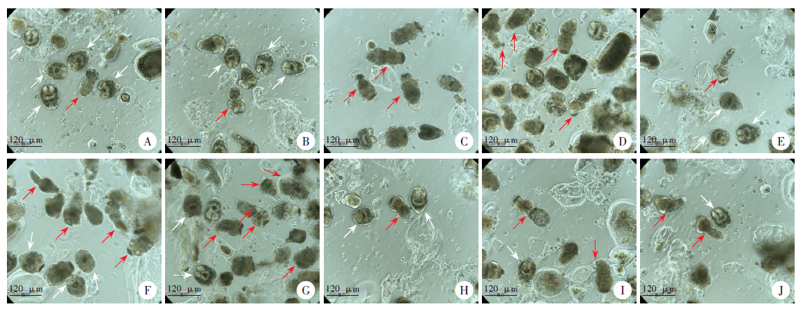

Fig. 2
Effects of TA-SM in water solution and tablet forms on the survival rate of protoscolices of E. multilocularis co-culture in vitro for 7 day

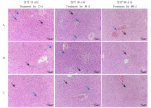
Fig. 3
Pathological changes of liver tissue in mice infected with E. multilocularis after treatment with TA-SM in water solution and tablet forms (HE staining, × 100) A: Saline group, after 15 and 30 days of treatment, the liver cells of mice were swollen, with a low level of inflammatory cells infiltrating around, and a small amount of vacuolar degeneration in the cytoplasm, after 60 days of treatment, the liver cells were more obviously swollen, with a large number of inflammatory cells infiltrating around, and the cytoplasmic vacuolar degeneration increased; B. C: The tablet group and the water solution group of TA-SM, there was no difference between the hepatocyte morphology and the normal saline group 15 and 30 days after treatment. After 60 days of treatment, the hepatocyte morphology tended to be normal, inflammatory cells decreased, and cytoplasmic vacuolar degeneration decreased. The black arrow shows damaged hepatocytes, and the blue arrow shows inflammatory cells.

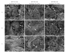
Fig. 4
Microstructure changes of liver tissue in mice infected with E. multilocularis after treatment with TA-SM in water solution and tablet forms A: Saline group, there were more typical vesicular structures, and with the extension of treatment time, the vesicular structures increased significantly; B. C: The tablet form group and the water solution group of TA-SM, the typical vesicular structure was significantly reduced, and with the extension of the treatment time, the vesicular structure did not change significantly. The white arrow indicates the typical structure of the vesicular cyst.

| [1] |
McManus DP, Zhang WB, Li J, et al. Echinococcosis[J]. Lancet, 2003, 362(9392): 1295-1304.
doi: 10.1016/S0140-6736(03)14573-4 pmid: 14575976 |
| [2] | Wen H, Vuitton L, Tuxun T, et al. Echinococcosis: advances in the 21st century[J]. Clin Microbiol Rev, 2019, 32(2): e00075-e00018. |
| [3] |
Wang ZH, Wang XM, Liu XQ. Echinococcosis in China, a review of the epidemiology of Echinococcus spp.[J]. EcoHealth, 2008, 5(2): 115-126.
doi: 10.1007/s10393-008-0174-0 |
| [4] |
Nunnari G, Pinzone MR, Gruttadauria S, et al. Hepatic echinococcosis: clinical and therapeutic aspects[J]. World J Gastroenterol, 2012, 18(13): 1448-1458.
doi: 10.3748/wjg.v18.i13.1448 |
| [5] | Tan XW, Yu XF, Jiang HJ, et al. Inhibitory effect of xanthohumol on the growth of Echinococcus multilocularis metacestode in the liver of mice[J]. Chin J Parasitol Parasit Dis, 2021, 39(3): 304-310. (in Chinese) |
| (谭小武, 俞晓凡, 姜慧娇, 等. 黄腐酚对小鼠肝多房棘球蚴生长的抑制作用[J]. 中国寄生虫学与寄生虫病杂志, 2021, 39(3): 304-310.) | |
| [6] | Tian CY, Chen B, Lu S, et al. Toxicity of doxorubicin on Echinococcus granulosus protoscoleces in vitro[J]. Chin J Parasitol Parasit Dis, 2019, 37(5): 571-575, 582. (in Chinese) |
| (田春艳, 陈蓓, 卢帅, 等. 阿霉素体外抗细粒棘球蚴原头节作用研究[J]. 中国寄生虫学与寄生虫病杂志, 2019, 37(5): 571-575, 582.) | |
| [7] | Qiao SY, Zhou X, Liu CH, et al. Effect of albendazole-loaded vesicles on the vitality of protoscoleces of Echinococcus granulosus[J]. Chin J Parasitol Parasit Dis, 2022, 40(3): 324-329, 336. (in Chinese) |
| (乔世源, 周雪, 刘程豪, 等. 阿苯达唑载药囊泡对细粒棘球蚴原头节活性的影响[J]. 中国寄生虫学与寄生虫病杂志, 2022, 40(3): 324-329, 336.) | |
| [8] | Lin SM. Sophora japonica in Tibet[J]. Pratacultural Sci, 2002, 19(3): 34. (in Chinese) |
| (林少敏. 西藏砂生槐[J]. 草业科学, 2002, 19(3): 34.) | |
| [9] | Luo YP, Zhang Y, Zhang HM, et al. Anti-parasitic effects of water-soluble alkaloid fractions from ethanolic extracts of Sophora moorcroftiana seeds in Caenorhabditis elegans[J]. Chin J Nat Med, 2018, 16(9): 665-673. |
| [10] |
Zhang G, Wang J, Luo Y,et al. In vivo evaluation of the efficacy of Sophora moorcroftiana alkaloids in combination with Bacillus Calmette-Guérin (BCG) treatment for cystic echinococcosis in mice[J]. J Helminthol, 2018, 92(6): 681-686.
doi: 10.1017/S0022149X1700089X pmid: 29061197 |
| [11] | Zhang FB, Hu CH, Cheng SL, et al. The investigation of the effect and mechanism of Sophora moorcroftiana alkaloids in combination with albendazole on echinococcosis in an experimental rats model[J]. Evid Based Complement Alternat Med, 2018, 2018: 3523126. |
| [12] |
Luo YP, Zhang GC, Liu X, et al. Therapeutic and immunoregulatory effects of water-soluble alkaloids E2-a from Sophora moorcroftiana seeds as a novel potential agent against echinococcosis in experimentally protoscolex-infected mice[J]. Vet Res, 2018, 49(1): 100.
doi: 10.1186/s13567-018-0596-9 |
| [13] |
Ma XM, Wan JM, et al. Therapeutic effects of Sophora moorcroftiana alkaloids in combination with albendazole in mice experimentally infected with protoscolices of Echinococcus granulosus[J]. Braz J Med Biol Res, 2007, 40(10): 1403-1408.
pmid: 17713646 |
| [14] |
Su G, Yang WK, Meng WB, et al. Anti-proliferation effects of ethanolic extracts from Sophora moorcroftiana seeds on human hepatocarcinoma HepG2 cell line[J]. Nat Prod Res, 2018, 32(12): 1472-1475.
doi: 10.1080/14786419.2017.1353503 |
| [15] |
Yuan RY, Dongzhi ZM, Guo W, et al. Hepatoprotective effect of Sophora moorcroftiana (Benth.) Benth. Ex baker seeds in vivo and in vitro[J]. Drug Chem Toxicol, 2022, 45(6): 2535-2544.
doi: 10.1080/01480545.2021.1962692 |
| [16] | Hu CH, Yang T, Li YP, et al. Optimization of the purification of total alkaloids from Sophora moorcroftiana seeds[J]. Chin Tradit Pat Med, 2016, 38(10): 2157-2162. (in Chinese) |
| (胡春晖, 杨涛, 李永平, 等. 砂生槐子总生物碱纯化工艺的优化[J]. 中成药, 2016, 38(10): 2157-2162.) | |
| [17] | Hu CH, Zhang FB. Optimization of extraction process of total alkaloids from Sophora japonica by box-behnken response surface methodology[J]. Chin Tradit Pat Med, 2016, 38(9): 2063-2066. (in Chinese) |
| (胡春晖, 张发斌. Box-Behnken响应面法优化砂生槐子中总生物碱的提取工艺[J]. 中成药, 2016, 38(9): 2063-2066.) | |
| [18] | Hu CH, Dang YJ, Sun MJ, et al. Preparation and quality evaluation of Tibetan medical Sophora moorcroftiana seed extract mini menadhesive tablets[J]. Guid J Tradit Chin Med Pharm, 2018, 24(9): 31-35. (in Chinese) |
| (胡春晖, 党云洁, 孙梦娟, 等. 藏药砂生槐子总生物碱生物黏附迷你肠溶片的制备与体内外评价[J]. 中医药导报, 2018, 24(9): 31-35.) | |
| [19] | Gao P, Pan RC, Zhang FB, et al. Therapeutic effects of bioadhesive tablets of total alkaloids from Sophora moorcroftiana and albendazole on secondary alveolar echinococcosis in mice[J]. Chin High Alt Med Biol, 2021, 42(1): 47-52. (in Chinese) |
| (高攀, 潘汝翀, 张发斌, 等. 砂生槐子总生物碱生物粘附片与阿苯达唑联合用药治疗继发性小鼠泡球蚴病的疗效[J]. 中国高原医学与生物学杂志, 2021, 42(1): 47-52.) | |
| [20] | Pan RC, Zhang FB, Cheng SL, et al. A study on treatment of secondary hepatic hydatid disease in mice with alkaloids extracted from Sophora moorcroftiana seeds[J]. J Pathog Biol, 2018, 13(3): 254-258. (in Chinese) |
| (潘汝翀, 张发斌, 程时磊, 等. 砂生槐生物碱治疗继发性小鼠肝包虫病的实验研究[J]. 中国病原生物学杂志, 2018, 13(3): 254-258.) | |
| [21] | Min H, Hu CH, Hu B, et al. Optimization of extraction technology of total alkaloids from Sophora moorcroftiana based on multi index comprehensive detection and its mechanism of anti-hepatic fibrosis[J]. Guid J Tradit Chin Med Pharm, 2018, 24(10): 14-18. (in Chinese) |
| (闵慧, 胡春晖, 胡斌, 等. 基于多指标综合检测优选藏药砂生槐子总生物碱提取工艺及其抗肝纤维化机制研究[J]. 中医药导报, 2018, 24(10): 14-18.) | |
| [22] | Gui XW. Effect of protoscolex of Echinococcus alveolaris on calcification of bone marrow mesenchymal stem cells[D]. Shihezi: Shihezi University, 2020: 4. (in Chinese) |
| (桂显伟. 泡状棘球蚴原头节对骨髓间充质干细胞钙化影响的研究[D]. 石河子: 石河子大学, 2020: 4.) | |
| [23] | Zhu JH, Cao DP, Qie YRZ, et al. Effect of Elsholtzia eriostachya in combination with albendazole in treatment of secondary Echinococcus multilocularis metacestode infection in rats[J]. Chin J Parasitol Parasit Dis, 2020, 38(6): 688-694. (in Chinese) |
| (朱吉海, 曹得萍, 切羊让中, 等. 藏药黄花香薷联合阿苯达唑治疗大鼠继发性多房棘球蚴感染的疗效研究[J]. 中国寄生虫学与寄生虫病杂志, 2020, 38(6): 688-694.) | |
| [24] |
Wu Y, Li Z, Xiu AY, et al. Carvedilol attenuates carbon tetrachloride-induced liver fibrosis and hepatic sinusoidal capillarization in mice[J]. Drug Des Devel Ther, 2019, 13: 2667-2676.
doi: 10.2147/DDDT |
| [25] |
Sharma U, Pal D, Prasad R. Alkaline phosphatase: an overview[J]. Ind J Clin Biochem, 2014, 29(3): 269-278.
doi: 10.1007/s12291-013-0408-y |
| [26] |
Choudhary GS, Al-Harbi S, Almasan A. Caspase-3 activation is a critical determinant of genotoxic stress-induced apoptosis[J]. Methods Mol Biol, 2015, 1219: 1-9.
doi: 10.1007/978-1-4939-1661-0_1 pmid: 25308257 |
| [27] |
Araya LE, Soni IV, Hardy JA, et al. Deorphanizing caspase-3 and caspase-9 substrates in and out of apoptosis with deep substrate profiling[J]. ACS Chem Biol, 2021, 16(11): 2280-2296.
doi: 10.1021/acschembio.1c00456 pmid: 34553588 |
| [28] |
Huang Q, Li F, Liu XJ, et al. Caspase 3-mediated stimulation of tumor cell repopulation during cancer radiotherapy[J]. Nat Med, 2011, 17(7): 860-866.
doi: 10.1038/nm.2385 pmid: 21725296 |
| [1] | CAO Deping, LI Jiajing, SONG Mengwei, MO Gang. Experimental observation on the changes of hepatic stellate cells stimulated in vitro with tissue protein of Echinococcus multilocularis [J]. CHINESE JOURNAL OF PARASITOLOGY AND PARASITIC DISEASES, 2023, 41(4): 440-445. |
| [2] | HOU Xin-ling, LI De-wei, SHI Yang, WANG Mao-lin, ZIBIGU Rousu, ABIDAN Ainiwaer, ZHENG Xu-ran, KANG Xue-jiao, WANG Hui, LI Jing, ZHANG Chuan-shan. Changes of ST2+ T cell subset function and their immune checkpoint molecule expression in the peritoneal cavity of mice infected with Echinococcus multilocularis [J]. CHINESE JOURNAL OF PARASITOLOGY AND PARASITIC DISEASES, 2022, 40(6): 708-716. |
| [3] | WU Liang-liang, YANG Ling-fei, SONG Tao. Ultrasound and pathological manifestations of lesions in SD rats with hepatic Echinococcus multilocularis infection established by different methods [J]. CHINESE JOURNAL OF PARASITOLOGY AND PARASITIC DISEASES, 2022, 40(4): 549-552. |
| [4] | ZHONG Shun-hu, SUN Yue, GUO Xiao-la, ZHENG Ya-dong, CHEN Yi-xia. Identification and bioinformatics analysis of differentially expressed miRNAs in splenic lymphocytes in Echinococcus multilocularis-infected mice [J]. CHINESE JOURNAL OF PARASITOLOGY AND PARASITIC DISEASES, 2022, 40(3): 288-294. |
| [5] | ZHUO Yi-cheng, YANG Hai-cheng, LIU Cheng-hao, ZHANG Bao-cai, DUO Xiao-yong, ZHANG Shi-jie. Effect of osteopontin expression level on the growth and development of Echinococcus multilocularis protoscoleces [J]. CHINESE JOURNAL OF PARASITOLOGY AND PARASITIC DISEASES, 2022, 40(3): 299-304. |
| [6] | ABUDUAINI Abulizi, PAIZULA Shalayiadang, TALAITI Tuergan, ZHANG Rui-qing, WANG Hui, ZHANG Chuan-shan, SHAO Ying-mei, TUERGANAILI Aji. Affect of Echinococcus multilocularis protein-mediated NK cell surface receptor NKG2A on the function of NK cells [J]. CHINESE JOURNAL OF PARASITOLOGY AND PARASITIC DISEASES, 2022, 40(1): 36-42. |
| [7] | HOU Jiao, WEN Hao, WANG Ming-kun, LI Wen-ding, LI liang, LI Jing, ZHANG Chuan-shan, WANG Hui. Changes of macrophage subsets and polarization in spleen of mice infected with Echinococcus multilocularis [J]. CHINESE JOURNAL OF PARASITOLOGY AND PARASITIC DISEASES, 2021, 39(6): 771-778. |
| [8] | SHI Qi-qi, LIU Cong-shan, HUO Le-le, WEI Yu-fen, JIANG Bin, YIN Meng, XUE Jian, TAO Yi, ZHANG Hao-bing. Affect of aminoalcohol compound HT24 on the expression of tubulin in Echinococcus multilocularis protoscoleces [J]. CHINESE JOURNAL OF PARASITOLOGY AND PARASITIC DISEASES, 2021, 39(4): 437-443. |
| [9] | LI Ling-hui, WANG Wei, HOU Xin-ling, SHI Yang, LI De-wei, LI Liang, WANG Hui, LI Jing, ZHANG Chuan-shan. Affects of Echinococcus multilocularis metacestode infection on the natural killer T cells and their subsets in mouse spleen [J]. CHINESE JOURNAL OF PARASITOLOGY AND PARASITIC DISEASES, 2021, 39(3): 311-317. |
| [10] | GUO Bao-ping, GUO Gang, ZHANG Li, XIANG Jing-jing, WANG Xiao-ping, REN Yuan, QI Wen-jing, ZHANG Hui, LI Jun, ZHANG Wen-bao, WANG Hai-yan. Investigation on infection of Echinococcus multilocularis metacestode in small rodents in Chabchar County, Xinjiang [J]. CHINESE JOURNAL OF PARASITOLOGY AND PARASITIC DISEASES, 2021, 39(3): 327-332. |
| [11] | XU Kai, WANG Hai-jiu, ZHANG Li, ZHANG Yao-gang, FAN Hai-ning, REN Li, REN Bin, WANG Zhi-xin. Research progress on the mechanisms underlying the impairment of host hepatocytes by Echinococcus multilocularis [J]. CHINESE JOURNAL OF PARASITOLOGY AND PARASITIC DISEASES, 2021, 39(2): 256-259. |
| [12] | ZHU Ji-hai, CAO De-ping, ZHAO Jun, LIU Jun, SHI Hu-xiang, LIU Yan. Investigation of differentially expressed genes in liver tissues of patients with alveolar echinococcosis [J]. CHINESE JOURNAL OF PARASITOLOGY AND PARASITIC DISEASES, 2021, 39(1): 48-54. |
| [13] | ZHU Ji-hai, CAO De-ping, QIE Yangrangzhong, LIU Jun, ZHAO Jun, LIU Yan. Effect of Elsholtzia eriostachya in combination with albendazole in treatment of secondary Echinococcus multilocularis metacestode infection in rats [J]. CHINESE JOURNAL OF PARASITOLOGY AND PARASITIC DISEASES, 2020, 38(6): 688-694. |
| [14] | SHANG Jing-ye, ZHANG Guang-jia, YU Wen-jie, HE Wei, LIAO Sha, LI Rui-rui, HUANG Yan, LIU Yang, ZHONG Bo. Advances in researches on the genetic diversity of Echinococcus multilocularis [J]. CHINESE JOURNAL OF PARASITOLOGY AND PARASITIC DISEASES, 2020, 38(5): 637-641. |
| [15] | ZHOU Hong-rang, MAO Guang-yao, WANG Xiao-ling, CHEN Mu-xin, YU Qing, WANG Ying, Ai Lin, XIAO Ning. Establishment and application of a multiplex recombinase-aided isothermal amplification technique for identifying Echinococcus granulosus and Echinococcus multilocularis [J]. CHINESE JOURNAL OF PARASITOLOGY AND PARASITIC DISEASES, 2020, 38(3): 310-316. |
| Viewed | ||||||||||||||||||||||||||||||||||||||||||||||||||
|
Full text 266
|
|
|||||||||||||||||||||||||||||||||||||||||||||||||
|
Abstract 398
|
|
|||||||||||||||||||||||||||||||||||||||||||||||||