

Bimonthly,Published Since February,1983
Publishing: National Institute of Parasitic Diseases,Chinese Center for Disease Control and Prevention(NIPD, China CDC)
Editor-in-Chief: LI Shizhu
Managing Editor: CHEN Qin
Publishing: National Institute of Parasitic Diseases,Chinese Center for Disease Control and Prevention(NIPD, China CDC)
Editor-in-Chief: LI Shizhu
Managing Editor: CHEN Qin
Wechat
News
- Acknowledgements to Reviewers 2025 | Thanks to Our Reviewers for Their Selfless Dedication to the Journal 2025-12-31
- The Handover Meeting of the Fifth Editorial Board of the Chinese Journal of Parasitology and Parasitic Diseases Successf... 2025-12-31
- Good News| 55 publications from Chinese Journal of Parasitology and Parasitic Diseases were selected as High-Im... 2025-11-27
- National Symposium on Innovative Development of Academic Journals in Tropical Medicine was successfully held in Hefei 2025-11-10
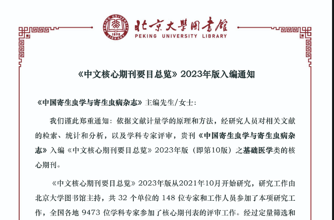
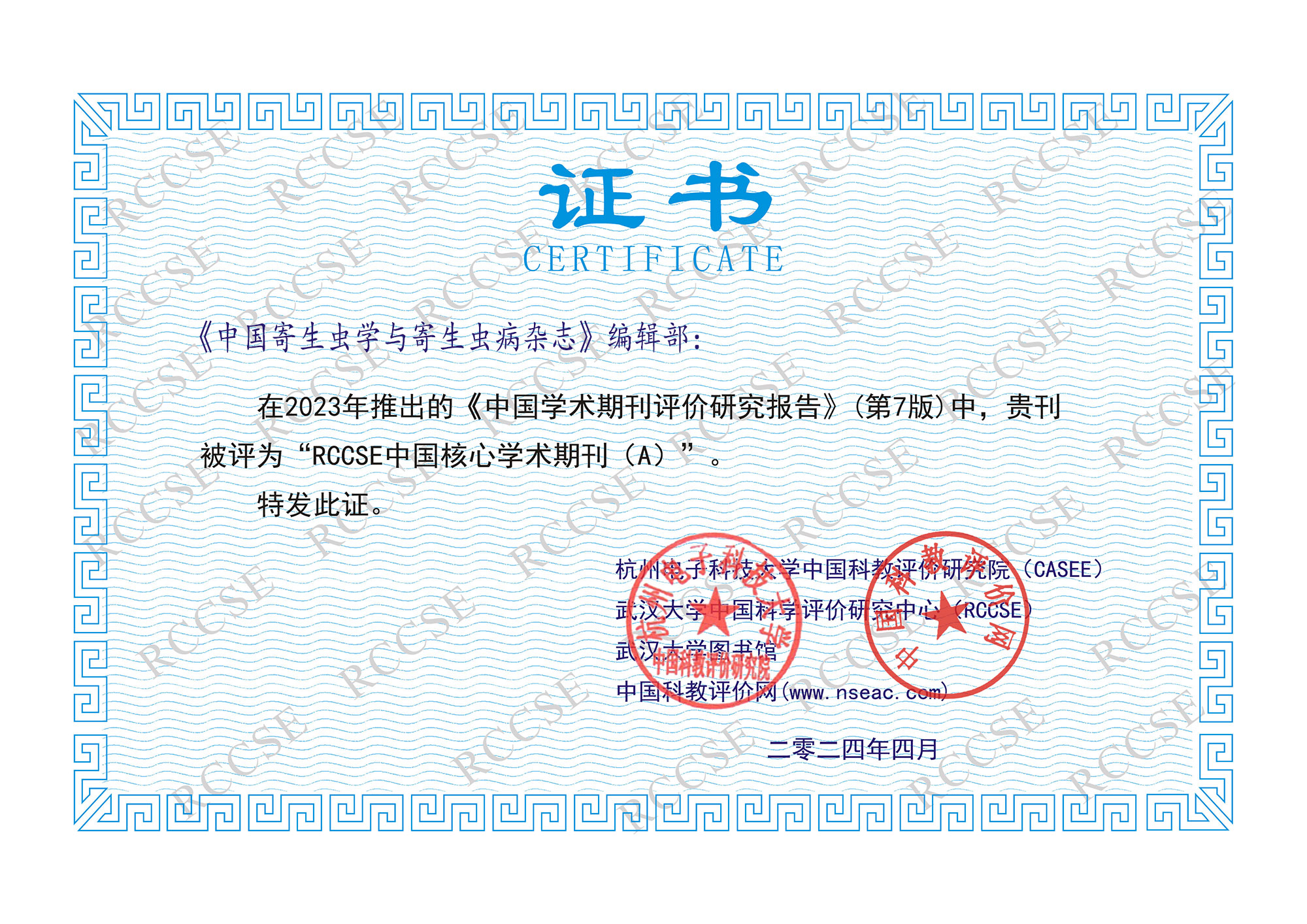
- Good News | Chinese Journal of Parasitology and Parasitic Diseases has been consecutively selected as a Chinese Core Jou... 2025-11-10
- Statement | Warning: Fraudulent Activities Impersonating Chinese Journal of Parasitology and Parasitic Diseases 2025-08-06
- Good News! Submission Inquiry for "Zhongguo Shuangji" Is Now Available! 2025-07-30
- Good News | Chinese Journal of Parasitology and Parasitic Diseases Selected in the "China S & T Journals Capacity Improv... 2025-07-30
- Acknowledgements to Reviewers 2024 Sincere thanks to all reviewers for their kind contributions 2025-02-20
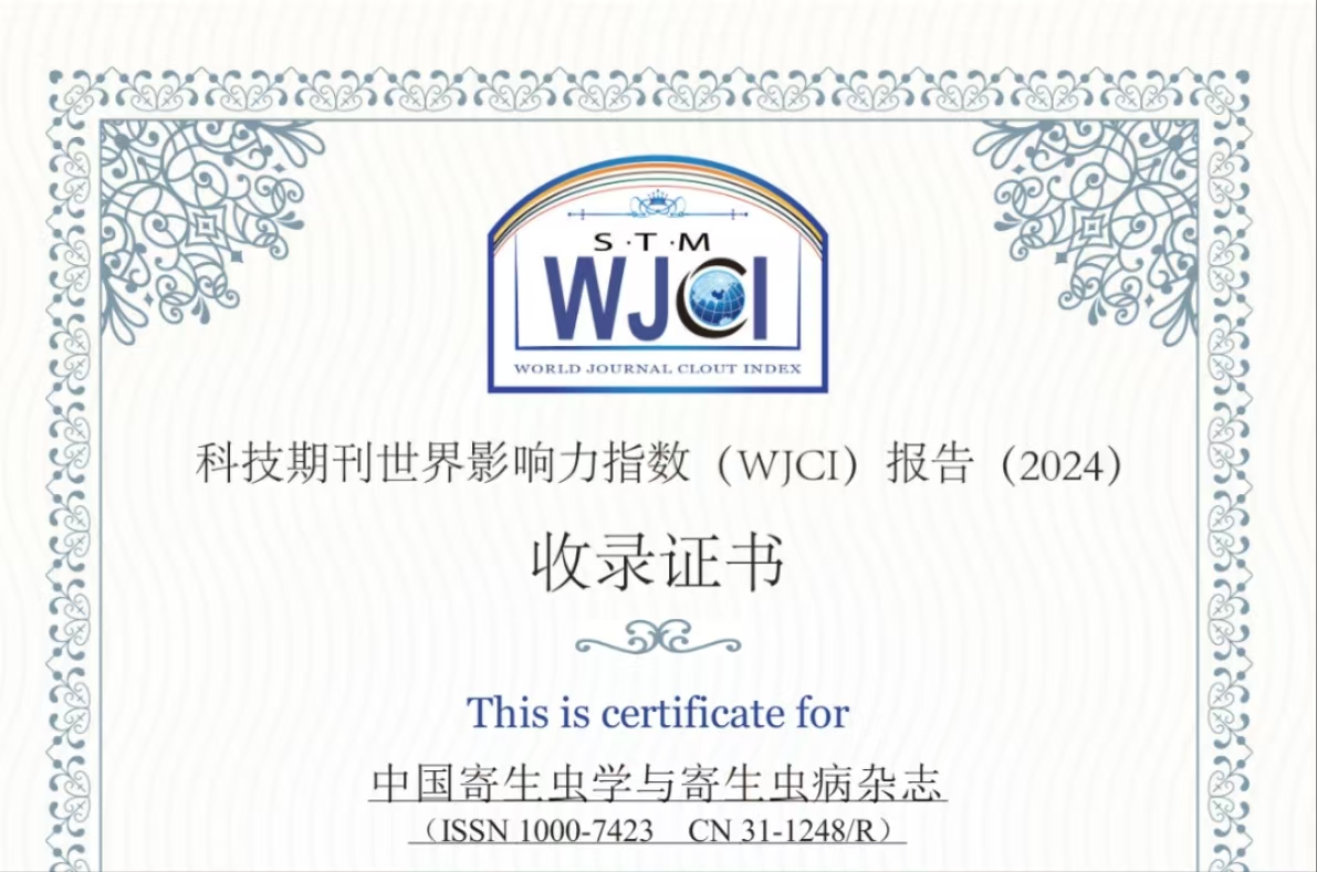
- Included in the World Journal Clout Index (WJCI)Report ((2023 STM) 2023-11-24
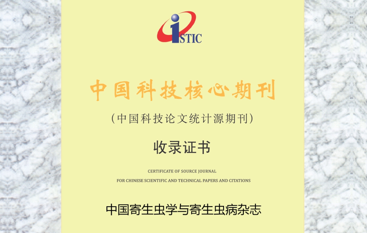
- In 2023, continue to be included in the Source Journal For Chinese Scientific and Technical Papers and Citations 2023-10-16
- In 2023, this journal was re-included in Embase 2023-07-18
- 2023-2024, Continue to Include in the Source Journals For Chinese Science Citation Database 2023-06-20
- In 2022,Zhang Zhengyan and Sheng Huifeng of editorial department were awarded “the seventh excellent periodical worker i... 2023-05-26
- In 2023, Celebratethe 40th anniversary of the Chinese Journal of Parasitology and Parasitic disease since its publicatio... 2023-02-28
- In 2022,the core impact factor set a new record 2022-12-29
- One paper has been awarded the seventh Outstanding Scientific & Technical Papers of CAST in 2022. 2022-12-13
- 2022 China Tropical Medicine Academic Journal Development Forum:A Summary 2022-12-13
- Included in the World Journal Clout Index (WJCI)Report ((2022 STM) 2022-12-12
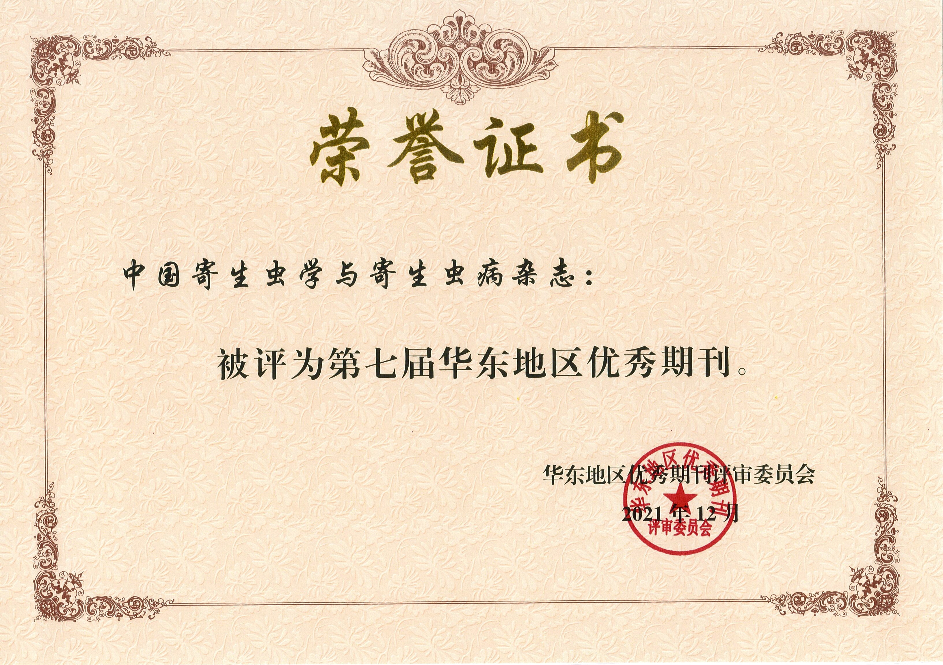
- In 2021,the Seventh Excellent Journal in Eastern China 2022-01-17
Important Information
-
World Malaria Report 2023
2023-12-10 -
2023-2024, Source Journals For Chinese Science Ci...
2023-06-15