
CHINESE JOURNAL OF PARASITOLOGY AND PARASITIC DISEASES ›› 2021, Vol. 39 ›› Issue (2): 226-232.doi: 10.12140/j.issn.1000-7423.2021.02.016
• ORIGINAL ARTICLES • Previous Articles Next Articles
YANG Hai-cheng1( ), ZHANG Hong-wei2, SHI Kang-jie1, WEN Yu-peng1, LIU Shi-wen3, ZHANG Yong-guo2, ZHU Zhi-qiang2, ZHANG Shi-jie2,*()
Received:2020-10-16
Revised:2021-01-05
Online:2021-04-30
Published:2021-04-30
Contact:
ZHANG Shi-jie
E-mail:1075660913@qq.com;zhangshijie1@sina.com
Supported by:CLC Number:
YANG Hai-cheng, ZHANG Hong-wei, SHI Kang-jie, WEN Yu-peng, LIU Shi-wen, ZHANG Yong-guo, ZHU Zhi-qiang, ZHANG Shi-jie. Autocrine osteopontin promotes the growth and metastasis of Echinococcus multilocularis via the EGFR signaling pathway[J]. CHINESE JOURNAL OF PARASITOLOGY AND PARASITIC DISEASES, 2021, 39(2): 226-232.
Add to citation manager EndNote|Ris|BibTeX
URL: https://www.jsczz.cn/EN/10.12140/j.issn.1000-7423.2021.02.016
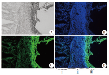
Fig. 1
The expression of lentivirus in E. multilocularis metacestode and liver tissues in mice(× 400) A: There was an obvious inflammatory zone between E. multilocularis metacestode tissue and liver tissue under a normal light microscope; B: DAPI staining showed aggregation of a great number of cells in the inflammatory zone; C: Of the inflammatory zone, external capsule and germinal layer exhibit strong green fluorescence, indicating the lentivirus invades into the tissue; D: Merge of A, B, C. Ⅰ: Liver cells; Ⅱ: Inflammatory zone; Ⅲ: E. multilocularis metacestode tissues.
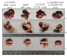
Fig. 2
Liver gross specimens at 1 month (A) and 4 months after infection of E. multilocularis metacestode (B), and the lung gross specimens at 4 months after infection in mice (C)
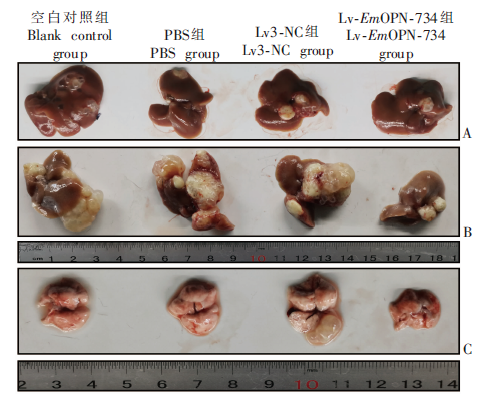
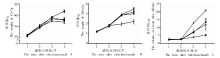
Fig. 3
Body weight (A), liver weight (B), and liver size (C) of mice at different times after infection
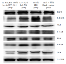
Fig. 4
Mice lung metastatic lesion E. multilocularis metacestode in the Lv3-NC group (HE staining, A: × 100; B: × 400) Note: Lung parenchyma of mice in the Lv3-NC group was eroded by E. multilocularis metacestode to certain extent.
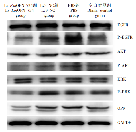
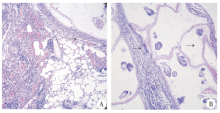
Fig. 5
Western blotting analysis of OPN and molecules involved in the EGFR signaling pathway in the mice E. multilocularis metacestode tissue
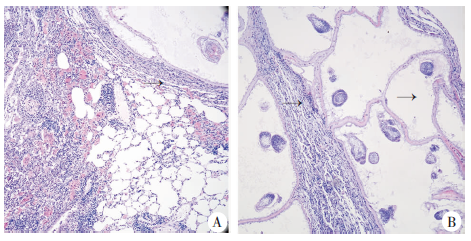
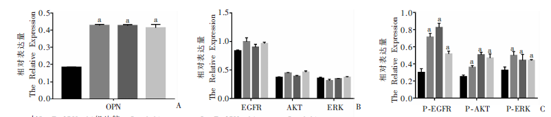
Fig. 6
Expression of molecules related with OPN and EGFR signaling pathways in the hepatic alveolar hydatid tissue of infected mice
| [1] |
Otero-Abad B, Torgerson PR. A systematic review of the epidemiology of echinococcosis in domestic and wild animals[J]. PLoS Negl Trop Dis, 2013,7(6):e2249.
doi: 10.1371/journal.pntd.0002249 |
| [2] |
Siles-Lucas M, Casulli A, Cirilli R, et al. Progress in the pharmacological treatment of human cystic and alveolar echinococcosis: compounds and therapeutic targets[J]. PLoS Negl Trop Dis, 2018,12(4):e0006422.
doi: 10.1371/journal.pntd.0006422 |
| [3] |
Vuitton DA, Azizi A, Richou C, et al. Current interventional strategy for the treatment of hepatic alveolar echinococcosis[J]. Expert Rev Anti Infect Ther, 2016,14(12):1179-1194.
doi: 10.1080/14787210.2016.1240030 |
| [4] |
Lanaya H, Natarajan A, Komposch K, et al. EGFR has a tumour-promoting role in liver macrophages during hepatocellular carcinoma formation[J]. Nat Cell Biol, 2014,16(10):972-977.
doi: 10.1038/ncb3031 |
| [5] | Miyamoto Y, Suyama K, Baba H. Recent advances in targeting the EGFR signaling pathway for the treatment of metastatic colorectal cancer[J]. Int J Mol Sci, 2017,18(4):E752. |
| [6] |
Tomas A, Futter CE, Eden ER. EGF receptor trafficking: consequences for signaling and cancer[J]. Trends Cell Biol, 2014,24(1):26-34.
doi: 10.1016/j.tcb.2013.11.002 |
| [7] |
Liu XD, Tian S, Liu M, et al. Wogonin inhibits the proliferation and invasion, and induces the apoptosis of HepG2 and Bel7402 HCC cells through NF-κB/Bcl-2, EGFR and EGFR downstream ERK/AKT signaling[J]. Int J Mol Med, 2016,38(4):1250-1256.
doi: 10.3892/ijmm.2016.2700 |
| [8] |
Spiliotis M, Lechner S, Tappe D, et al. Transient transfection of Echinococcus multilocularis primary cells and complete in vitro regeneration of metacestode vesicles[J]. Int J Parasitol, 2008,38(8/9):1025-1039.
doi: 10.1016/j.ijpara.2007.11.002 |
| [9] |
Spiliotis M, Brehm K. Axenic in vitro cultivation of Echinococcus multilocularis metacestode vesicles and the generation of primary cell cultures[J]. Methods Mol Biol, 2009,470:245-262.
doi: 10.1007/978-1-59745-204-5_17 pmid: 19089387 |
| [10] |
Anborgh PH, Mutrie JC, Tuck AB, et al. Role of the metastasis-promoting protein osteopontin in the tumour microenvironment[J]. J Cell Mol Med, 2010,14(8):2037-2044.
doi: 10.1111/j.1582-4934.2010.01115.x pmid: 20597997 |
| [11] |
Weber GF, Lett GS, Haubein NC. Osteopontin is a marker for cancer aggressiveness and patient survival[J]. Br J Cancer, 2010,103(6):861-869.
doi: 10.1038/sj.bjc.6605834 |
| [12] |
Lee SH, Park JW, Woo SH, et al. Suppression of osteopontin inhibits chemically induced hepatic carcinogenesis by induction of apoptosis in mice[J]. Oncotarget, 2016,7(52):87219-87231.
doi: 10.18632/oncotarget.v7i52 |
| [13] |
Matušan-Ilijaš K, Damante G, Fabbro D, et al. EGFR expression is linked to osteopontin and NF-κB signaling in clear cell renal cell carcinoma[J]. Clin Trans Oncol, 2013,15(1):65-71.
doi: 10.1007/s12094-012-0889-9 |
| [14] |
Lamour V, Henry A, Kroonen J, et al. Targeting osteopontin suppresses glioblastoma stem-like cell character and tumorigenicity in vivo[J]. Int J Cancer, 2015,137(5):1047-1057.
doi: 10.1002/ijc.v137.5 |
| [15] |
Peng XY, Li JH, Wu XW, et al. Detection of osteopontin in the pericyst of human hepatic Echinococcus granulosus[J]. Acta Trop, 2006,100(3):163-171.
doi: 10.1016/j.actatropica.2006.08.013 |
| [16] | Zhang L, Zhang SJ, Cao YW, et al. The correlation between osteopontin and metastasis of hepatic Echinococcus multilocularis infection[J]. Chin J Parasitol Parasit Dis, 2011,29(1):33-36. (in Chinese) |
| ( 张龙, 张示杰, 曹玉文, 等. 骨桥蛋白与肝泡型棘球蚴转移的相关性研究[J]. 中国寄生虫学与寄生虫病杂志, 2011,29(1):33-36.) | |
| [17] |
Simmerman E, Qin X, Yu JC, et al. Cannabinoids as a potential new and novel treatment for melanoma: a pilot study in a murine model[J]. J Surg Res, 2019,235:210-215.
doi: 10.1016/j.jss.2018.08.055 pmid: WOS:000456777000027 |
| [18] |
Hemer S, Konrad C, Spiliotis M, et al. Host insulin stimulates Echinococcus multilocularis insulin signalling pathways and larval development[J]. BMC Biol, 2014,12:5.
doi: 10.1186/1741-7007-12-5 |
| [19] |
Cheng Z, Liu F, Li X, et al. EGF-mediated EGFR/ERK signaling pathway promotes germinative cell proliferation in Echinococcus multilocularis that contributes to larval growth and development[J]. PLoS Negl Trop Dis, 2017,11(2):e0005418.
doi: 10.1371/journal.pntd.0005418 |
| [20] |
Förster S, Koziol U, Schäfer T, et al. The role of fibroblast growth factor signalling in Echinococcus multilocularis development and host-parasite interaction[J]. PLoS Negl Trop Dis, 2019,13(3):e0006959.
doi: 10.1371/journal.pntd.0006959 |
| [21] |
Roskoski R Jr. The ErbB/HER family of protein-tyrosine kinases and cancer[J]. Pharmacol Res, 2014,79:34-74.
doi: 10.1016/j.phrs.2013.11.002 |
| [22] |
Weihua WH, Tsan R, Huang WC, et al. Survival of cancer cells is maintained by EGFR independent of its kinase activity[J]. Cancer Cell, 2008,13(5):385-393.
doi: 10.1016/j.ccr.2008.03.015 pmid: 18455122 |
| [23] | Cheng Z, Xu ZJ, Tian HM, et al. In vitro and in vivo efficacies of inhibitors of the EGFR/MEK/ERK signaling in the treatment of alveolar echinococcosis[J]. Antimicrob Agents Chemother, 2020,64(8):e00341-20. |
| [24] |
Wang M, Han J, Marcar L, et al. Radiation resistance in KRAS-mutated lung cancer is enabled by stem-like properties mediated by an osteopontin-EGFR pathway[J]. Cancer Res, 2017,77(8):2018-2028.
doi: 10.1158/0008-5472.CAN-16-0808 |
| [25] | Li T, Wu XW, Zhang YG, et al. Effects of anti-osteopontin antibody on expression of MMP-2 and TGF-β1 in hepatic alveolar hydatid tissue of gerbil[J]. Chin J Parasitol Parasit Dis, 2013,31(6):450-453. (in Chinese) |
| ( 荔童, 吴向未, 张永国, 等. 抗骨桥蛋白抗体对沙鼠肝多房棘球蚴组织中的基质金属蛋白酶2和转化生长因子β1的影响[J]. 中国寄生虫学与寄生虫病杂志, 2013,31(6):450-453.) | |
| [26] |
Pietras A, Katz AM, Ekström EJ, et al. Osteopontin-CD44 signaling in the glioma perivascular niche enhances cancer stem cell phenotypes and promotes aggressive tumor growth[J]. Cell Stem Cell, 2014,14(3):357-369.
doi: 10.1016/j.stem.2014.01.005 |
| [27] |
Nardo AD, Grün NG, Zeyda M, et al. Impact of osteopontin on the development of non-alcoholic liver disease and related hepatocellular carcinoma[J]. Liver Int, 2020,40(7):1620-1633.
doi: 10.1111/liv.v40.7 |
| [28] | Wang HH, Guo DH, Li JJ, et al. Increased expression of osteopontin indicates poor prognosis in hepatocellular carcinoma[J]. Int J Clin Exp Pathol, 2018,11(12):5916-5922. |
| [29] |
Sun TT, Tang YR, Sun DW, et al. Osteopontin versus alpha-fetoprotein as a diagnostic marker for hepatocellular carcinoma: a meta-analysis[J]. Oncotargets Ther, 2018,11:8925-8935.
doi: 10.2147/OTT |
| [30] |
Cheng YG, Wen G, Sun Y, et al. Osteopontin promotes colorectal cancer cell invasion and the stem cell-like properties through the PI3K-AKT-GSK/3β-β/catenin pathway[J]. Med Sci Monit, 2019,25:3014-3025.
doi: 10.12659/MSM.913185 |
| [31] |
Tsai W, Tsai W, Lee H, et al. Association between osteopontin and EGFR expression with clinicopathological parameters in hepatocellular carcinoma[J]. Chin J Physiol, 2012,55(6):412-420.
doi: 10.4077/CJP.2012.BAA082 |
| [1] | ZHU Aiya, WANG Xu, WANG Jiangyou, WANG Ying, LI Yang, SONG Shan, GENG Yan, LAN Ziyao, DAI Jiarui. A child case of alveolar echinococcosis in Guizhou Province [J]. CHINESE JOURNAL OF PARASITOLOGY AND PARASITIC DISEASES, 2023, 41(4): 520-523. |
| [2] | RAOWAN Tuolehong, ABUDUSALAMU Abulikemu, YANG Lingfei, CHEN Lu, LI Zhao, JIA Fang, SONG Tao. Effect evaluation and factor analysis of ultrasonic manifestations in the diagnosis of hepatic alveolar echinococcosis [J]. CHINESE JOURNAL OF PARASITOLOGY AND PARASITIC DISEASES, 2023, 41(3): 312-318. |
| [3] | KUI Yan, XUE Chuizhao, WANG Xu, LIU Baixue, WANG Ying, WANG Liying, YANG Shijie, HAN Shuai, WU Weiping, XIAO Ning. Progress of echinococcosis control in China, 2021 [J]. CHINESE JOURNAL OF PARASITOLOGY AND PARASITIC DISEASES, 2023, 41(2): 142-148. |
| [4] | MA Hui, CHONG Shigui, CHEN Gen, ZHANG Linghui, QIN Junmei, ZHAO Yumin. Research progress on the cellular signal pathways associated in alveolar echinococcosis [J]. CHINESE JOURNAL OF PARASITOLOGY AND PARASITIC DISEASES, 2023, 41(2): 223-227. |
| [5] | AN Xiu-qing, WANG Miao-miao, ZHOU Hong-qian, MENG Kai, CAI Jian-ping, LIU Guang-hui, A Ji-de, YANG Jing-yu. Research progress on microvascular density in hepatic alveolar echinococcosis [J]. CHINESE JOURNAL OF PARASITOLOGY AND PARASITIC DISEASES, 2022, 40(6): 792-797. |
| [6] | ZHANG Ting-ting, DU Qiu-pei, GUO Xin-jian, ZHANG Ling-qiang, WANG Zhi-xin, CHANG Zheng-song, ZHAO Qian, WANG Hai-jiu, HOU Li-zhao. Research progress on vascular invasion of hepatic alveolar echinococcosis [J]. CHINESE JOURNAL OF PARASITOLOGY AND PARASITIC DISEASES, 2022, 40(4): 516-523. |
| [7] | WU Liang-liang, YANG Ling-fei, SONG Tao. Ultrasound and pathological manifestations of lesions in SD rats with hepatic Echinococcus multilocularis infection established by different methods [J]. CHINESE JOURNAL OF PARASITOLOGY AND PARASITIC DISEASES, 2022, 40(4): 549-552. |
| [8] | ZHUO Yi-cheng, YANG Hai-cheng, LIU Cheng-hao, ZHANG Bao-cai, DUO Xiao-yong, ZHANG Shi-jie. Effect of osteopontin expression level on the growth and development of Echinococcus multilocularis protoscoleces [J]. CHINESE JOURNAL OF PARASITOLOGY AND PARASITIC DISEASES, 2022, 40(3): 299-304. |
| [9] | ZHANG Ling-hui, CHEN Gen, CHONG Shi-gui, SHEN Hui, MA Hui, ZHAO Yu-min. Research progress on the immune regulation mechanism in alveolar echinococcosis [J]. CHINESE JOURNAL OF PARASITOLOGY AND PARASITIC DISEASES, 2022, 40(1): 109-113. |
| [10] | YAN Ji-can, YU Wen-hao, HOU Li-zhao, ZHANG Ling-qiang, XU Xiao-lei, WANG Hai-jiu, LU Qian, FAN Hai-ning. Diagnosis and treatment of celiac-subcutaneous echinococcosis granulosus in an elderly [J]. CHINESE JOURNAL OF PARASITOLOGY AND PARASITIC DISEASES, 2022, 40(1): 132-135. |
| [11] | SHAO Han, LI Si-yuan, LI Jun. The affect of metformin on autophagy and apoptosis of Echinococcus multilocularis cysts and protoscoleces [J]. CHINESE JOURNAL OF PARASITOLOGY AND PARASITIC DISEASES, 2022, 40(1): 43-49. |
| [12] | YANG Liu, HE Wei, WANG Qi, YU Wen-jie, ZHONG Bo, LIU Yang, XIAO Tong-guang, XIE Fei, YAO Ren-xin, HUANG Yan, LI Rui-rui, LIAO Sha, ZHANG Guang-jia, WANG Qian. The impact of reducing stray dog density on the prevalence of Echinococcus spp. in small mammals [J]. CHINESE JOURNAL OF PARASITOLOGY AND PARASITIC DISEASES, 2021, 39(2): 156-160. |
| [13] | XU Kai, WANG Hai-jiu, ZHANG Li, ZHANG Yao-gang, FAN Hai-ning, REN Li, REN Bin, WANG Zhi-xin. Research progress on the mechanisms underlying the impairment of host hepatocytes by Echinococcus multilocularis [J]. CHINESE JOURNAL OF PARASITOLOGY AND PARASITIC DISEASES, 2021, 39(2): 256-259. |
| [14] | JIANG Hui-jiao, GUI Xian-wei, GUO Li-jiao, YANG Xiong-feng, WANG Xiao-yi, CHEN Xue-ling, WU Xiang-wei. Expression and angiogenic effect of VEGFA/VEGFR2 in mice hepatic metacestode tissue of Echinococcus multilocularis [J]. CHINESE JOURNAL OF PARASITOLOGY AND PARASITIC DISEASES, 2020, 38(6): 673-681. |
| [15] | SHANG Jing-ye, ZHANG Guang-jia, YU Wen-jie, HE Wei, LIAO Sha, LI Rui-rui, HUANG Yan, LIU Yang, ZHONG Bo. Advances in researches on the genetic diversity of Echinococcus multilocularis [J]. CHINESE JOURNAL OF PARASITOLOGY AND PARASITIC DISEASES, 2020, 38(5): 637-641. |
| Viewed | ||||||
|
Full text |
|
|||||
|
Abstract |
|
|||||