
CHINESE JOURNAL OF PARASITOLOGY AND PARASITIC DISEASES ›› 2023, Vol. 41 ›› Issue (3): 312-318.doi: 10.12140/j.issn.1000-7423.2023.03.008
• ORIGINAL ARTICLES • Previous Articles Next Articles
RAOWAN Tuolehong1( ), ABUDUSALAMU Abulikemu2, YANG Lingfei1, CHEN Lu1, LI Zhao1, JIA Fang1, SONG Tao1,*()
Received:2022-10-21
Revised:2023-01-23
Online:2023-06-30
Published:2023-06-28
Contact:
*E-mail: Supported by:CLC Number:
RAOWAN Tuolehong, ABUDUSALAMU Abulikemu, YANG Lingfei, CHEN Lu, LI Zhao, JIA Fang, SONG Tao. Effect evaluation and factor analysis of ultrasonic manifestations in the diagnosis of hepatic alveolar echinococcosis[J]. CHINESE JOURNAL OF PARASITOLOGY AND PARASITIC DISEASES, 2023, 41(3): 312-318.
Add to citation manager EndNote|Ris|BibTeX
URL: https://www.jsczz.cn/EN/10.12140/j.issn.1000-7423.2023.03.008
Table 1
Ultrasonic observation indexes and assignment description of hepatic alveolar echinococcosis lesions
| 超声观察指标 Ultrasonic observation index | 变量名 Variable name | 赋值说明 Assignment instructions |
|---|---|---|
| 位置 Location | X1 | 肝右叶 = 1,肝左叶 = 2,同时累及肝左右叶 = 3 Right lobe of liver = 1, left lobe of liver = 2, both left and right lobes of liver = 3 |
| 最大径大小 Size of maximum diameter | X2 | < 5 cm = 1,5~10 cm = 2,≥ 10 cm = 3 < 5 cm = 1, 5~10 cm = 2, ≥ 10 cm = 3 |
| 边界和形态 Boundary and shape | X3 | 界尚清或界欠清、尚规整或欠规整 = 1,界不清、不规整 = 2 Boundary is still clear or less clear = 1, boundary is not clear = 2 |
| 实性部分回声 Solid partial echo | X4 | 中高回声 = 1,高回声或强回声 = 2,中低回声或低回声 = 3 Medium-high echo = 1, high or strong echo = 2, low and middle echo or low echo = 3 |
| 钙化 Calcification | X5 | 未见明显钙化 = 1,散在钙化 = 2,广泛钙化 = 3 No obvious calcification = 1, scattered calcification = 2, extensive calcification = 3 |
| 液化坏死 Liquefied necrosis | X6 | 未见明显液化坏死 = 1,部分液化坏死 = 2,广泛液化坏死 = 3 No obvious liquefied necrosis = 1, part of liquefied necrosis = 2, extensive liquefied necrosis = 3 |
| 血流信号 Blood flow signal | X7 | 未见明显血流 = 1,少许点状血流 = 2,点条状血流 = 3 No obvious blood flow = 1, a little dot blood flow = 2, point bar blood flow = 3 |
| 胆管和血管的侵犯 Bile duct and vascular invasion | X8 | 未见明显受侵 = 1,部分胆管或血管受侵 = 2,大部分胆管及血管均受侵 = 3 No invasion = 1, part of the invasion = 2, widely invaded = 3 |
| 超声诊断符合情况 Ultrasound diagnosis | Y1 | 符合 = 0,不符合 = 1 Conformity = 0, non-conformity = 1 |
Table 2
Results of univariate analysis of ultrasound findings of hepatic alveolar echinococcosis lesions
| 因素 Factor | 病灶数 No. lesion | 占比/% Propotion/% | 超声诊断符合情况 Ultrasound diagnosis | χ2值 χ2 value | P值 P value | |||
|---|---|---|---|---|---|---|---|---|
| 符合(n = 122) Conformity (n = 122) | 不符合(n = 48) Non-conformity (n = 48) | |||||||
| 病灶数 No. lesion | 占比/% Propotion/% | 病灶数 No. lesion | 占比/% Propotion/% | |||||
| 位置 Location | 01.952 | > 0.05 | ||||||
| 肝右叶 Right lobe of liver | 088 | 51.8 | 62 | 70.5 | 26 | 29.5 | ||
| 肝左叶 Left lobe of liver | 042 | 24.7 | 28 | 66.7 | 14 | 33.3 | ||
| 同时累及肝左右叶 Both left and right lobes | 040 | 23.5 | 32 | 80.0 | 8 | 20.0 | ||
| 最大径大小 Size of maximum diameter | 39.026 | < 0.01 | ||||||
| < 5 cm | 027 | 15.8 | 6 | 22.2 | 21 | 77.8 | ||
| 5~10 cm | 053 | 31.1 | 42 | 79.2 | 11 | 20.8 | ||
| ≥ 10 cm | 090 | 52.9 | 74 | 82.2 | 16 | 17.8 | ||
| 边界和形态 Boundary and shape | 18.601 | < 0.01 | ||||||
| 界尚清或界欠清、尚规整或欠规整 Boundary is still clear or less clear | 051 | 30.0 | 25 | 49.0 | 26 | 51.0 | ||
| 界不清、不规整 Boundary is not clear | 119 | 70.0 | 97 | 81.5 | 22 | 18.5 | ||
| 实性部分回声 Solid partial echo | 15.743 | < 0.01 | ||||||
| 中低或低回声 Low and middle echo or low echo | 013 | 07.6 | 4 | 4/13 | 9 | 9/13 | ||
| 中高回声 Medium-high echo | 130 | 76.5 | 102 | 78.5 | 28 | 21.5 | ||
| 高或强回声 High or strong echo | 027 | 15.9 | 16 | 59.3 | 11 | 40.7 | ||
| 钙化 Calcification | 47.205 | < 0.01 | ||||||
| 未见明显钙化 No obvious calcification | 017 | 10.0 | 1 | 1/17 | 16 | 16/17 | ||
| 散在钙化 Scattered calcification | 108 | 63.5 | 92 | 85.2 | 16 | 14.8 | ||
| 广泛钙化 Extensive calcification | 045 | 26.5 | 29 | 64.4 | 16 | 35.6 | ||
| 液化坏死 Liquefied necrosis | 34.151 | < 0.01 | ||||||
| 未见明显液化坏死 No obvious liquefied necrosis | 081 | 47.6 | 41 | 50.6 | 40 | 49.4 | ||
| 部分液化坏死 Part of liquefied necrosis | 046 | 27.1 | 42 | 91.3 | 4 | 08.7 | ||
| 广泛液化坏死 Extensive liquefied necrosis | 043 | 25.3 | 39 | 90.7 | 4 | 09.3 | ||
| 血流信号 Blood flow signal | 06.597 | < 0.05 | ||||||
| 未见明显血流 No obvious blood flow | 116 | 68.2 | 90 | 77.6 | 26 | 22.4 | ||
| 少许点状血流 A little dot blood flow | 037 | 21.8 | 23 | 62.2 | 14 | 37.8 | ||
| 点条状血流 Point bar blood flow | 017 | 10.0 | 9 | 9/17 | 8 | 8/17 | ||
| 胆管和血管的侵犯 Bile duct and vascular invasion | 21.766 | < 0.01 | ||||||
| 未见明显受侵 No invasion | 061 | 35.9 | 33 | 54.1 | 28 | 45.9 | ||
| 部分受侵 Part of the invasion | 071 | 41.8 | 52 | 73.2 | 19 | 26.8 | ||
| 广泛受侵 Widely invaded | 038 | 22.3 | 37 | 97.4 | 1 | 02.6 | ||
Table 3
Multivariate logistic regression analysis of ultrasound findings of hepatic alveolar echinococcosis lesions
| 变量 Variable | 回归系数 Regression coefficient | 标准误 Standard error | Wald χ2 | P | OR | 95% CI | |
|---|---|---|---|---|---|---|---|
| 下限 Lower limit | 上限 Upper limit | ||||||
| 常量 Constant | -0.609 | 1.754 | 0.121 | > 0.05 | 0.544 | — | — |
| 最大径大小 Size of maximum diameter | 6.736 | < 0.05 | |||||
| < 5 cm | -1.717 | 1.130 | 2.308 | > 0.05 | 0.180 | 0.020 | 1.645 |
| 5~10 cm | -0.712 | 0.692 | 1.057 | > 0.05 | 2.038 | 0.524 | 7.916 |
| 边界和形态 Boundary and shape | |||||||
| 界尚清或界欠清、尚规整或欠规整 Boundary is still clear or less clear | -0.378 | 0.763 | 0.245 | > 0.05 | 1.459 | 0.327 | 6.514 |
| 实性部分回声 Solid partial echo | 3.547 | > 0.05 | |||||
| 中高回声 Medium-high echo | -2.026 | 1.105 | 3.360 | > 0.05 | 7.587 | 0.869 | 66.225 |
| 高或强回声 High or strong echo | -1.492 | 1.342 | 1.236 | > 0.05 | 4.447 | 0.320 | 61.724 |
| 钙化 Calcification | 11.715 | < 0.01 | |||||
| 未见明显钙化 No obvious calcification | -3.298 | 1.414 | 5.441 | < 0.05 | 0.037 | 0.002 | 0.590 |
| 散在钙化 Scattered calcification | -0.960 | 0.631 | 2.312 | > 0.05 | 2.612 | 0.758 | 9.002 |
| 液化坏死 Liquefied necrosis | 6.026 | < 0.05 | |||||
| 未见明显液化坏死 No obvious liquefied necrosis | -1.267 | 0.965 | 1.724 | > 0.05 | 0.282 | 0.042 | 1.867 |
| 部分液化坏死 Part of liquefied necrosis | -0.654 | 1.019 | 0.412 | > 0.05 | 1.924 | 0.261 | 14.165 |
| 血流信号 Blood flow signal | 14.737 | < 0.01 | |||||
| 未见明显血流 No obvious blood flow | -3.032 | 0.877 | 11.960 | < 0.01 | 20.746 | 3.720 | 115.686 |
| 少许点状血流 A little dot blood flow | -1.234 | 0.896 | 1.896 | > 0.05 | 3.435 | 0.593 | 19.899 |
| 胆管和血管的侵犯 Bile duct and vascular invasion | 3.834 | > 0.05 | |||||
| 未见明显受侵 No invasion | -2.274 | 1.382 | 2.705 | > 0.05 | 0.103 | 0.007 | 1.546 |
| 部分受侵 Part of the invasion | -2.408 | 1.231 | 3.829 | = 0.05 | 0.090 | 0.008 | 1.004 |

Fig. 1
ROC curve of logistic regression model prediction accuracy analysis result
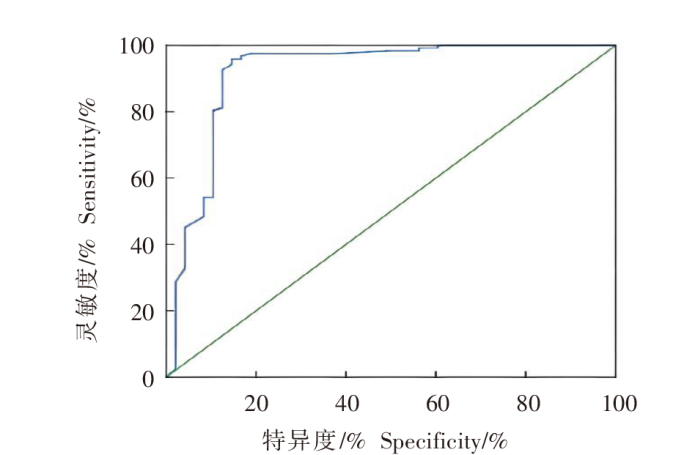
Fig. 2
A nomogram predicting coincidence factors for ultrasound diagnosis of hepatic alveolar echinococcosis lesions
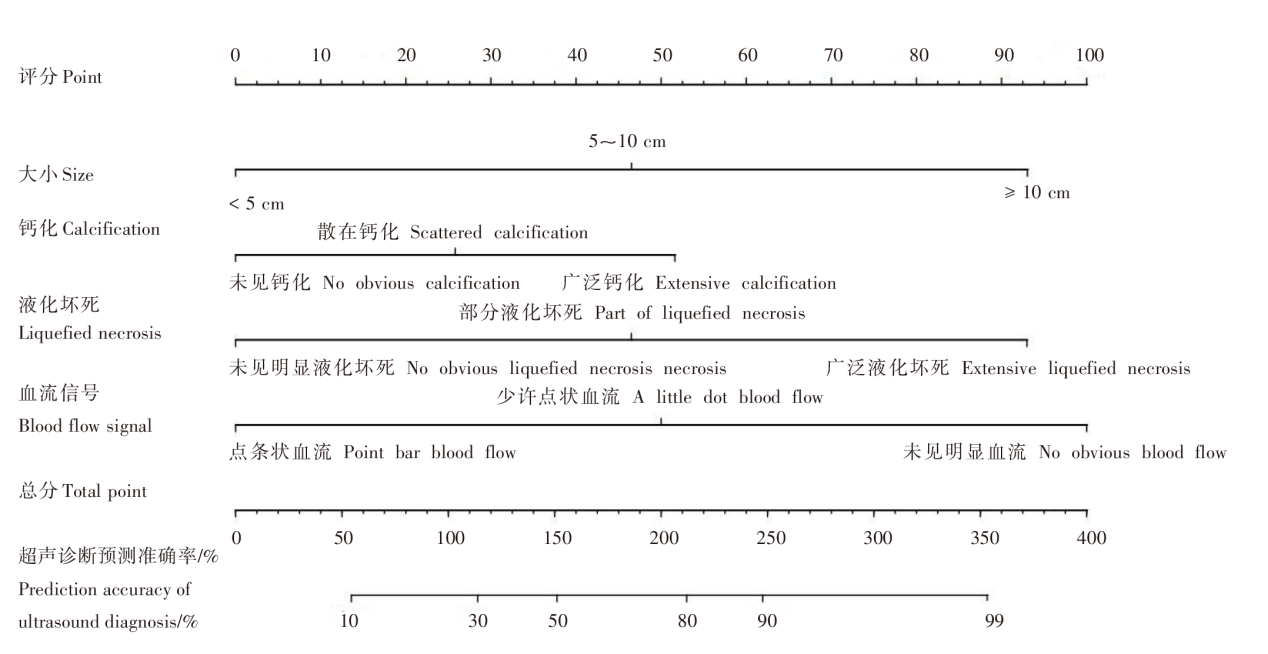
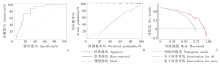
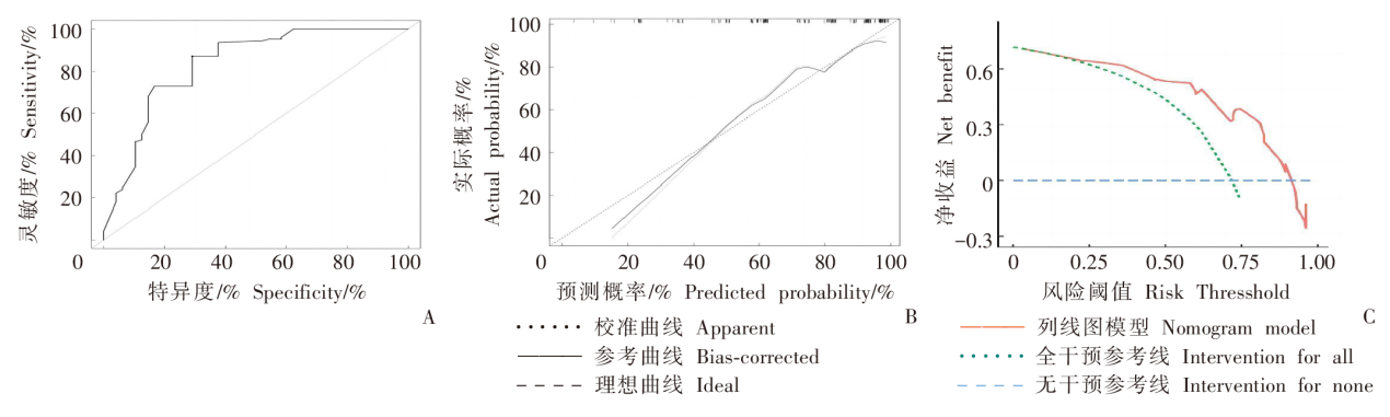
Fig. 3
Performance assessment with nomogram model A: ROC curve; B: calibration curve; C: decision analysis curve
| [1] |
Qian MB, Abela-Ridder B, Wu WP, et al. Combating echinococcosis in China: strengthening the research and development[J]. Infect Dis Poverty, 2017, 6: 161.
doi: 10.1186/s40249-017-0374-3 |
| [2] | Tuerhongjiang TX, Zhao YM, Tuerganaili AJ, et al. Consensus on Chinese terminology in the clinical field of echinococcosis[J]. Chin J Parasitol Parasit Dis, 2021, 39(1): 76-84. (in Chinese) |
| (吐尔洪江•吐逊, 邵英梅, 吐尔干艾力•阿吉, 等. 棘球蚴病临床领域相关中文专业术语专家共识[J]. 中国寄生虫学与寄生虫病杂志, 2021, 39(1): 76-84.) | |
| [3] |
Brunetti E, Kern P, Vuitton DA, et al. Expert consensus for the diagnosis and treatment of cystic and alveolar echinococcosis in humans[J]. Acta Trop, 2010, 114(1): 1-16.
doi: 10.1016/j.actatropica.2009.11.001 pmid: 19931502 |
| [4] | Liu R, He J, Wang H, et al. Comparative study of contrast-enhanced ultrasound and contrast-enhanced CT in measuring hepatic alveolar echinococcosis[J]. Clin J Med Offic, 2019, 47(7): 750-751. (in Chinese) |
| (刘荣, 赫娟, 王辉, 等. 超声造影及增强CT测量肝泡型包虫病病灶对比研究[J]. 临床军医杂志, 2019, 47(7): 750-751.) | |
| [5] | Zhao YM, Jing T, Ma SM, et al. A study of the prevalence of human echinococcosis in Maqu and Luqu Counties of Gannan Tibetan Autonomous Prefecture, China[J]. J Pathogen Biol, 2010, 5(1): 42-43, 56. (in Chinese) |
| (赵玉敏, 景涛, 马素美, 等. 甘南藏族自治州玛曲县和碌曲县人群包虫病流行情况调查[J]. 中国病原生物学杂志, 2010, 5(1): 42-43, 56.) | |
| [6] | Tao B, Zhang B, Du BB. Epidemiological investigation and control effect of echinococcosis in Inner Mongolia Autonomous Region[J]. Chin J Zoonoses, 2011, 27(7): 677-678. (in Chinese) |
| (涛波, 张斌, 杜宝彪. 内蒙古自治区包虫病流行病学调查与防治效果[J]. 中国人兽共患病学报, 2011, 27(7): 677-678.) | |
| [7] | Chen XL, Wu WG, Mi LG, et al. Application of immunological techniques in clinical immunological diagnosis of echinococcosis[J]. Prog Mod Biomed, 2011, 11(13): 2568-2571. (in Chinese) |
| (陈小林, 吴万贵, 米利古, 等. 免疫学技术在在棘球蚴病临床免疫诊断中的应用[J]. 现代生物医学进展, 2011, 11(13): 2568-2571.) | |
| [8] |
Feng XH, Wen H, Zhang ZX, et al. Dot immunogold filtration assay (DIGFA) with multiple native antigens for rapid serodiagnosis of human cystic and alveolar echinococcosis[J]. Acta Trop, 2010, 113(2): 114-120.
doi: 10.1016/j.actatropica.2009.10.003 pmid: 19836341 |
| [9] | Lyu YQ, Zhang ZL. Ultrasonographic diagnosis of hepatic alveolar echinococcosis[J]. Chin J Ultrasonog Med, 1998, 4(2): 78-80. (in Chinese) |
| (吕永泉, 张致凌. 肝泡状棘球蚴病的声像图诊断[J]. 中华超声医学杂志, 1998, 4(2): 78-80.) | |
| [10] | Song T, Lyu YQ, Yao LH, et al. Effects of contrast-enhanced grey scale ultrasonography on characterization of hepatic alveolar echinococcosis[J]. Chin J Ultrason, 2008, 17(2): 133-135. (in Chinese) |
| (宋涛, 吕永泉, 姚兰辉, 等. 灰阶超声造影在肝泡状棘球蚴病中的应用[J]. 中华超声影像学杂志, 2008, 17(2): 133-135.) | |
| [11] |
Song T, Zhao Q, Li HT, et al. Evaluation of color Dopplert ultrasonography on diagnosing hepatic alveolar echinococcusis[J]. Ultrasound Med Biol, 2012, 38(2): 183-189.
doi: 10.1016/j.ultrasmedbio.2011.11.010 pmid: 22230130 |
| [12] | Wang Y, Lyu YQ. Application of color Doppler energy in differentiating hepatic alveolar echinococcosis from hepatocellular carcinoma[J]. Chin J Ultrason, 2002, 11(1): 26-28. (in Chinese) |
| (王迎, 吕永泉. 彩色多普勒能量图在肝泡状棘球蚴病与肝癌鉴别诊断中的应用[J]. 中华超声影像学杂志, 2002, 11(1): 26-28.) | |
| [13] |
McManus DP, Gray DJ, Zhang WB, et al. Diagnosis, treatment, and management of echinococcosis[J]. BMJ, 2012, 344: e3866.
doi: 10.1136/bmj.e3866 |
| [14] | Zhang MZ, Zhou Y, Zhang LQ, et al. Pathological characteristics and clinical significance of marginal tissues of hepatic alveolar echinococcosis[J]. Chin J Curr Adv Gen Surg, 2020, 23(6): 502-504. (in Chinese) |
| (张蒙召, 周瀛, 张灵强, 等. 肝泡型包虫病灶边缘组织病理特点及临床意义的研究现状[J]. 中国现代普通外科进展, 2020, 23(6): 502-504.) |
| [1] | AN Xiu-qing, WANG Miao-miao, ZHOU Hong-qian, MENG Kai, CAI Jian-ping, LIU Guang-hui, A Ji-de, YANG Jing-yu. Research progress on microvascular density in hepatic alveolar echinococcosis [J]. CHINESE JOURNAL OF PARASITOLOGY AND PARASITIC DISEASES, 2022, 40(6): 792-797. |
| [2] | ZHANG Ting-ting, DU Qiu-pei, GUO Xin-jian, ZHANG Ling-qiang, WANG Zhi-xin, CHANG Zheng-song, ZHAO Qian, WANG Hai-jiu, HOU Li-zhao. Research progress on vascular invasion of hepatic alveolar echinococcosis [J]. CHINESE JOURNAL OF PARASITOLOGY AND PARASITIC DISEASES, 2022, 40(4): 516-523. |
| [3] | WU Liang-liang, YANG Ling-fei, SONG Tao. Ultrasound and pathological manifestations of lesions in SD rats with hepatic Echinococcus multilocularis infection established by different methods [J]. CHINESE JOURNAL OF PARASITOLOGY AND PARASITIC DISEASES, 2022, 40(4): 549-552. |
| [4] | KASIMU Aihaiti, ABUDUSALAMU Aini, TUERGANAILI Aji, SHAO Ying-mei, ZHANG Rui-qing, TALAITI Tuergan, JIANG Tie-min, RAN Bo, ABUDUAINI Abulizi, MIERADILI Aierken, WEN Hao. Analysis of hospital expenses for patients with end-stage hepatic alveolar echinococcosis receiving ex vivo liver resection and autotransplantation [J]. CHINESE JOURNAL OF PARASITOLOGY AND PARASITIC DISEASES, 2020, 38(1): 53-57. |
| [5] | Xiao-lei XU, Zhi-xin WANG, Zhan WANG, Hai-wen YE, Ming-quan PANG, Ying ZHOU, Hai-jiu WANG, Hai-ning FAN. Treatment of complicated hepatic alveolar echinococcosis: our experience of 98 cases [J]. CHINESE JOURNAL OF PARASITOLOGY AND PARASITIC DISEASES, 2018, 36(6): 552-559. |
| [6] | ABUDUSALAMU Aini1, TUERHONGJIANG Tuxun2, MA Hai-zhang3, ZHANG Heng2, ZHANG Hao1, ABUDUKAIYOUMU Maimaiti4, LI Yu-peng2, SHADIKE Apaer2, LIN Ren-yong5, SHAO Ying-mei1, WEN Hao5*. Changes of Toll-like Receptor mRNA and Related Cytokines in Patients with Hepatic Alveolar Echinococcosis [J]. , 2016, 34(6): 12-542-546. |
| [7] | ZHU Di-wen1,ZHANG Hai-xiao1,REN Wei-xin1 *,XIONG Jin2,XU Xiao-hui2,WEN Hao3. Pathological Changes of after Trans-Portal Vein Chemoembolization Echinococcus multilocularis in the Liver of Infected Rats [J]. , 2014, 32(1): 13-58-61. |
| [8] | FAN Yu-Xiang, LIN Wei-Xin, DI Li-Mu-La-Chi-·Ba-Wu-Dong, GU Dun-Feng, HU Xiao-Dong, ZHANG Hai-Xiao, JI Wei-Zheng, JIANG Chao, WEN Hao. Therapeutic Effect of Hepatic Artery Infusion with Albendazole Microspheres on Hepatic Alveolar Echinococcosis in Rats [J]. , 2011, 29(6): 4-415-418. |
| [9] | TANG Qun-Ke, ZHANG Ying, LI Yong-Shou, YUAN Chun-Ping, ZHANG Dong-Tian. Non-surgical Treatment for Nonresectable Advanced Hepatic Alveolar Echinococcosis [J]. , 2011, 29(1): 11-46-48. |
| [10] | WUWei-ping;LINDan-dan;HUFei;GUANYa-yi;WangYan-An;ZHUHong-qing;CAOChun-li;CHENHong-gen. Application of Multivariate Regression in Analyzing Factors of Schistosomiasis Japonica Transmission in Poyang Lake [J]. , 2003, 21(3): 11-166. |
| Viewed | ||||||
|
Full text |
|
|||||
|
Abstract |
|
|||||