
CHINESE JOURNAL OF PARASITOLOGY AND PARASITIC DISEASES ›› 2020, Vol. 38 ›› Issue (4): 412-416.doi: 10.12140/j.issn.1000-7423.2020.04.002
• ORIGINAL ARTICLES • Previous Articles Next Articles
YU Xiao-dong1, YALI Ya-sen1, WANG Jia-ling2, LI Meng2, YE Jian-rong1,*( )
)
Received:2020-02-25
Online:2020-08-30
Published:2020-09-09
Contact:
YE Jian-rong
E-mail:616227972@qq.com
Supported by:CLC Number:
YU Xiao-dong, YALI Ya-sen, WANG Jia-ling, LI Meng, YE Jian-rong. Establishment of BALB/c mouse model of Echinococcus granulosus-induced sensitization and changes of related immune cells[J]. CHINESE JOURNAL OF PARASITOLOGY AND PARASITIC DISEASES, 2020, 38(4): 412-416.
Add to citation manager EndNote|Ris|BibTeX
URL: https://www.jsczz.cn/EN/10.12140/j.issn.1000-7423.2020.04.002

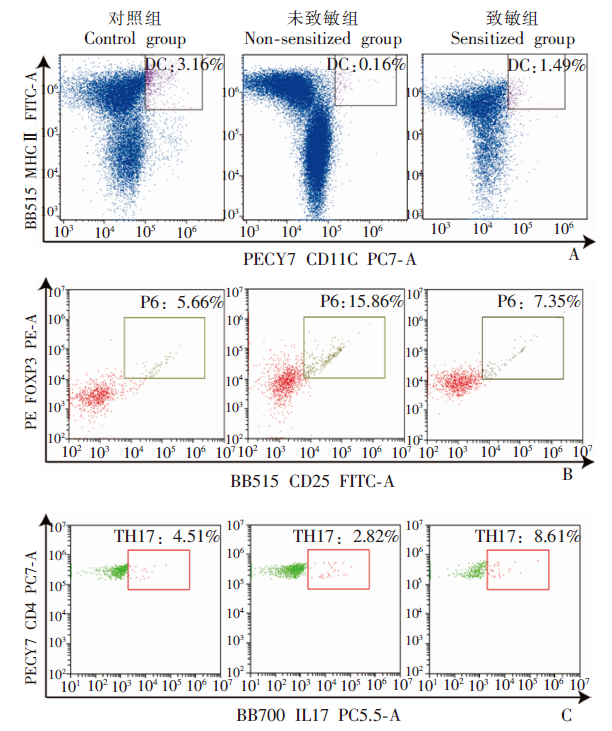
Fig. 1
The changes of DC, Treg and Th17 cell proportions in E. granulosus-induced sensitization BALB/c mice detected by flow cytometry
| [1] |
Budke CM, Deplazes P, Torgerson PR. Global socioeconomic impact of cystic echinococcosis[J]. Emerg Infect Dis, 2006,12(2):296-303.
doi: 10.3201/eid1202.050499 pmid: 16494758 |
| [2] | Zhang MY, Wu WP, Guan YY, et al. Analysis of disease burden of echinococcosis in China[J]. Chin J Parasitol Parasit Dis, 2018,36(1):15-19, 25. (in Chinese) |
| ( 张梦媛, 伍卫平, 官亚宜, 等. 我国棘球蚴病疾病负担分析[J]. 中国寄生虫学与寄生虫病杂志, 2018,36(1):15-19, 25.) | |
| [3] |
Zhang WB, Ross AG, McManus DP. Mechanisms of immunity in hydatid disease: implications for vaccine development[J]. J Immunol, 2008,181(10):6679-6685.
doi: 10.4049/jimmunol.181.10.6679 pmid: 18981082 |
| [4] | Xu S, Dang ZS, Zhang HB, et al. Advances in drug treatment of echinococcosis[J]. Chin J Parasitol Parasit Dis, 2018,36(3):297-302. (in Chinese) |
| ( 徐硕, 党志胜, 张皓冰, 等. 棘球蚴病药物治疗的研究进展[J]. 中国寄生虫学与寄生虫病杂志, 2018,36(3):297-302.) | |
| [5] | Wang TP, Cao ZG. Progress and problems in the prevention and control of echinococcosis in China[J]. Chin J Parasitol Parasit Dis, 2018,36(3):291-296. (in Chinese) |
| ( 汪天平, 操治国. 中国棘球蚴病防控进展及其存在的问题[J]. 中国寄生虫学与寄生虫病杂志, 2018,36(3):291-296.) | |
| [6] | Wang JC, You H. Advances in immunological studies of echinococcosis[J]. Chin J Vet Med, 2004,40(6):42-44. (in Chinese) |
| ( 王进成, 由弘. 棘球蚴病的免疫学研究进展[J]. 中国兽医杂志, 2004,40(6):42-44.) | |
| [7] | Xu LQ. The harm of major parasitic diseases in western China and the reflection on prevention and treatment[J]. Chin J Parasit Dis Control, 2002,15(1):1-3. (in Chinese) |
| ( 许隆祺. 我国西部地区重大寄生虫病的危害及对防治工作的反思[J]. 中国寄生虫病防治杂志, 2002,15(1):1-3.) | |
| [8] |
Li YM, Zheng H, Cao XH, et al. Demographic and clinical characteristics of patients with anaphylactic shock after surgery for cystic echinococcosis[J]. Am J Trop Med Hyg, 2011,85(3):452-455.
doi: 10.4269/ajtmh.2011.10-0448 |
| [9] | Zheng H, Xu ZX, Yang GX, et al. The study on the levels of IgG, IgG1 and IgE during anaphylactic shock in sheep infected with Echinococcus granulosus[J]. Chin J Parasitol Parasit Dis, 2003,21(1):42-45. (in Chinese) |
| ( 郑宏, 徐志新, 杨戈雄, 等. 感染细粒棘球蚴绵羊诱发过敏休克期间IgG、IgG1和IgE水平的探讨[J]. 中国寄生虫与寄生虫病杂志, 2003,21(1):42-45.) | |
| [10] |
Sun J, Arias K, Alvarez D, et al. Impact of CD40 ligand, B cells, and mast cells in peanut-induced anaphylactic responses[J]. J Immunol, 2007,179(10):6696-6703.
doi: 10.4049/jimmunol.179.10.6696 pmid: 17982059 |
| [11] | An R, Huang M, Zhang Q, et al. Preliminary study on the level of IL-10 in hypersensitive reaction of rats infected with Echinococcus granulosus[J]. Chin J Parasitol Parasit Dis, 2013,31(2):86-88. (in Chinese) |
| ( 安然, 黄谋, 张秦, 等. 细粒棘球蚴感染大鼠过敏反应中IL-10水平的初步研究[J]. 中国寄生虫学与寄生虫病杂志, 2013,31(2):86-88.) | |
| [12] |
Kim HJ, Kang SA, Yong TS, et al. Therapeutic effects of Echinococcus granulosus cystic fluid on allergic airway inflammation[J]. Exp Parasitol, 2019,198:63-70.
doi: 10.1016/j.exppara.2019.02.003 pmid: 30763570 |
| [13] |
De Wispelaere L, Vande VS, Schelstraete P, et al. Anaphylactic shock as a single presentation of Echinococcus cyst[J]. Acta Gastroenterol Belg, 2011,74(3):462-464.
pmid: 22103055 |
| [14] |
Vuitton DA. Echinococcosis and allergy[J]. Clin Rev Allergy Immunol, 2004,26(2):93-104.
doi: 10.1007/s12016-004-0004-2 pmid: 15146106 |
| [15] | Xu HW, Cao JP, Zheng KY, et al. Advances in the treatment of inflammatory bowel disease with parasitic infections[J]. Chin J Parasitol Parasit Dis, 2017,35(2):173-179. (in Chinese) |
| ( 徐慧雯, 曹建平, 郑葵阳, 等. 寄生虫感染治疗炎症性肠病的研究进展[J]. 中国寄生虫学与寄生虫病杂志, 2017,35(2):173-179.) | |
| [16] |
Mejri N, Müller J, Gottstein B. Intraperitoneal murine Echinococcus multilocularis infection induces differentiation of TGF-β-expressing DCs that remain immature[J]. Parasite Immunol, 2011,33(9):471-482.
doi: 10.1111/j.1365-3024.2011.01303.x pmid: 21609335 |
| [17] |
Simpson CR, Newton J, Hippisley-Cox J, et al. Incidence and prevalence of multiple allergic disorders recorded in a national primary care database[J]. J R Soc Med, 2008,101(11):558-563.
doi: 10.1258/jrsm.2008.080196 pmid: 19029357 |
| [18] |
Simpson CR, Newton J, Hippisley-Cox J, et al. Incidence and prevalence of multiple allergic disorders recorded in a national primary care database[J]. J R Soc Med, 2008,101(11):558-563.
doi: 10.1258/jrsm.2008.080196 pmid: 19029357 |
| [19] |
Everts B, Smits HH, Hokke CH, et al. Helminths and dendritic cells: sensing and regulating via pattern recognition receptors, Th2 and Treg responses[J]. Eur J Immunol, 2010,40(6):1525-1537.
doi: 10.1002/eji.200940109 pmid: 20405478 |
| [20] | Guo P, Zhang H, Li CF, , et al. Research progress of Toll-like receptor pathway regulating the function of Tregs[J/OL]. J Biol Eng. 2020: 1-12. [2020-04-20]. https://kns.cnki.net/KCMS/detail/11.1998.Q.20200407.1637.005.html?v=MjQwMTZa T3NOWXc5TXptUm42ajU3VDNmbHFXTTBDTEw3UjdxZForWm9 GeUhsVWJ6TkpWWT1OaVhjZTdHNEhOSE1xNDlD. (in Chinese) |
| ( 郭鹏, 张含, 李长菲, 等. Toll样受体通路调节Tregs功能的研究进展[J/OL]. 生物工程学报, 2020: 1-12 [2020-04-20]. https://kns.cnki.net/KCMS/detail/11.1998.Q.20200407.1637.005.html?v=MjQwMTZa T3NOWXc5TXptUm42ajU3VDNmbHFXTTBDTEw3UjdxZForWm9GeUhsVWJ6TkpWWT1OaVhjZTdHNEhOSE 1xNDlD.) | |
| [21] |
Bettelli E, Korn T, Oukka M, et al. Induction and effector functions of T(H)17 cells[J]. Nature, 2008,453(7198):1051-1057.
doi: 10.1038/nature07036 pmid: 18563156 |
| [1] | LU Junxia, XU Junying, ZHAO Bin, WANG Qianwen, LI Wenhua, GENG Yuqing, HOU Jun, WU Xiangwei, CHEN Xueling. Echinococcus granulosus infection induces macrophages to express CD73 and A2AR to suppress inflammatory response [J]. CHINESE JOURNAL OF PARASITOLOGY AND PARASITIC DISEASES, 2023, 41(5): 559-566. |
| [2] | WU Xiaoying, HU Yuan, CAO Jianping. Preparation of Echinococcus granulosus peptide embedded in chitosan quaternary ammonium salt nanoparticles [J]. CHINESE JOURNAL OF PARASITOLOGY AND PARASITIC DISEASES, 2023, 41(3): 300-305. |
| [3] | LI Benfu, WANG Zhengqing, XU Qian, ZI Jinrong, YAN Xinliu, PENG Jia, LI Jianxiong, CAI Xuan, WU Fangwei, YANG Yaming. Sequence analysis of mitochondrial co1 and nd1 genes in Echinococcus granulosus in Yunnan Province [J]. CHINESE JOURNAL OF PARASITOLOGY AND PARASITIC DISEASES, 2023, 41(3): 306-311. |
| [4] | GUO Gang, REN Yuan, JIAO Hongjie, WU Juan, GUO Baoping, QI Wenjing, LI Jun, ZHANG Wenbao. Effect of intraperitoneal inoculation with Echinococcus microcysts on the infection and pathogenicity of E. multilocularis in mouse liver [J]. CHINESE JOURNAL OF PARASITOLOGY AND PARASITIC DISEASES, 2023, 41(2): 156-162. |
| [5] | JIAO Hongjie, QI Wenjing, GUO Gang, BAO Jianling, WU Chuanchuan, SONG Chuanlong, LI Jun, ZHANG Wenbao, YAN Mei. Polarization effect of Echinococcus granulosus antigen B on the mouse macrophage RAW264.7 [J]. CHINESE JOURNAL OF PARASITOLOGY AND PARASITIC DISEASES, 2023, 41(1): 23-28. |
| [6] | WU De-fang, FU Yong, REN Bin, ZHANG Yao-gang, XU Xiao-lei, PANG Ming-quan, FAN Hai-ning. Genetic diversity and differentiation time of human isolates of Echinococcus granulosus and E. multilocularis from Qinghai [J]. CHINESE JOURNAL OF PARASITOLOGY AND PARASITIC DISEASES, 2022, 40(5): 610-615. |
| [7] | QIAO Shi-yuan, ZHOU Xue, LIU Cheng-hao, JIANG Hui-jiao, BU Yuan-yuan, CHEN Xue-ling, WU Xiang-wei. Effect of albendazole-loaded vesicles on the vitality of protoscoleces of Echinococcus granulosus [J]. CHINESE JOURNAL OF PARASITOLOGY AND PARASITIC DISEASES, 2022, 40(3): 324-329. |
| [8] | SUN Ye-ting, JIANG Nan, JIANG Yan-yan, LI Teng, JIANG Xiao-feng, CAO Jian-ping, SHEN Yu-juan. Study on the polarization of MDSC stimulated by Echinococcus granulosus protoscolex-derived exosomes in vitro [J]. CHINESE JOURNAL OF PARASITOLOGY AND PARASITIC DISEASES, 2022, 40(2): 175-180. |
| [9] | ZHOU Wen-zheng, SUN Jun-gang, ZHAO Xi-bin, CAO Li. Therapeutic effect of intensity modulated radiation therapy on secondary femur infection with Echinococcus granulosus in rats [J]. CHINESE JOURNAL OF PARASITOLOGY AND PARASITIC DISEASES, 2021, 39(4): 443-448. |
| [10] | TIAN Meng-xiao, ZANG Xiao-yan, GUO Gang, QI Wen-jing, GUO Bao-ping, REN Yuan, LI Jun, ZHANG Wen-bao. Expression and activity assay of serine protease in Echinococcus granulosus [J]. CHINESE JOURNAL OF PARASITOLOGY AND PARASITIC DISEASES, 2021, 39(2): 233-239. |
| [11] | FAN Jun-jie, HAN Xiu-min, Nur Fazleen Binti Idris, LI Kai, TAN Qing-qing, CAO Wen-qiao, LI Xiang, LIAO Peng, YE Bin. Bioinformatics characteristics and immunoreactivity of protein kinase A of Echinococcus granulosus [J]. CHINESE JOURNAL OF PARASITOLOGY AND PARASITIC DISEASES, 2020, 38(6): 682-687. |
| [12] | SHI Chun-li, YANG Hui, PAN Wen, ZHANG Xin, ZHU Xiao-ting, ZHAO Jia-qing. Proteomic analysis of human proteins in extracellular vesicles secreted by protoscoleces of Echinococcus granulosus [J]. CHINESE JOURNAL OF PARASITOLOGY AND PARASITIC DISEASES, 2020, 38(6): 695-701. |
| [13] | CAO Sheng-kui, ZHANG Xiao-fan, WEI Yu-huan, PAN Jia-ming, CAO Jian-ping, SHEN Yu-juan, CHEN Jia-xu. Expression and function of arginase in livers of mice infected with Echinococcus granulosus [J]. CHINESE JOURNAL OF PARASITOLOGY AND PARASITIC DISEASES, 2020, 38(3): 304-309. |
| [14] | ZHOU Hong-rang, MAO Guang-yao, WANG Xiao-ling, CHEN Mu-xin, YU Qing, WANG Ying, Ai Lin, XIAO Ning. Establishment and application of a multiplex recombinase-aided isothermal amplification technique for identifying Echinococcus granulosus and Echinococcus multilocularis [J]. CHINESE JOURNAL OF PARASITOLOGY AND PARASITIC DISEASES, 2020, 38(3): 310-316. |
| [15] | CHEN He-jie, JIANG Hui-jiao, LIANG Qian, Wu Jie, GUI Xian-wei, ZOU Hai-liang, XING Zhi-kun, WANG Er-qiang, CHEN Xue-ling, WU Xiang-wei. Effects of supernatant of different hepatoma cells on the vitality of Echinococcus granulosus protoscoleces in vitro [J]. CHINESE JOURNAL OF PARASITOLOGY AND PARASITIC DISEASES, 2020, 38(3): 317-323. |
| Viewed | ||||||
|
Full text |
|
|||||
|
Abstract |
|
|||||