
CHINESE JOURNAL OF PARASITOLOGY AND PARASITIC DISEASES ›› 2020, Vol. 38 ›› Issue (3): 304-309.doi: 10.12140/j.issn.1000-7423.2020.03.008
• ORIGINAL ARTICLES • Previous Articles Next Articles
CAO Sheng-kui, ZHANG Xiao-fan, WEI Yu-huan, PAN Jia-ming, CAO Jian-ping, SHEN Yu-juan, CHEN Jia-xu*( )
)
Received:2019-11-21
Online:2020-06-30
Published:2020-07-07
Contact:
Jia-xu CHEN
E-mail:chenjx@nipd.chinacdc.cn
Supported by:CLC Number:
CAO Sheng-kui, ZHANG Xiao-fan, WEI Yu-huan, PAN Jia-ming, CAO Jian-ping, SHEN Yu-juan, CHEN Jia-xu. Expression and function of arginase in livers of mice infected with Echinococcus granulosus[J]. CHINESE JOURNAL OF PARASITOLOGY AND PARASITIC DISEASES, 2020, 38(3): 304-309.
Add to citation manager EndNote|Ris|BibTeX
URL: https://www.jsczz.cn/EN/10.12140/j.issn.1000-7423.2020.03.008
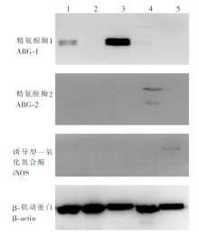
Fig. 1
The expression ARG-1, ARG-2 and iNOS proteins in liver leukocytes of mice infected with E. granulosus, as detected by Western blotting 1: Infection group; 2: Control group; 3: Peritoneal cells with IL-4 and IL-13 stimulation; 4: HepG2 cells; 5: Peritoneal cells with LPS stimulation


Fig. 2
Statistical analysis of protein expression abundance with the Image J software 1: Infection group; 2: Control group; 3: Peritoneal cells with IL-4 and IL-13 stimulation; 4: HepG2 cells; 5: Peritoneal cells with LPS stimulation. a: P < 0.01
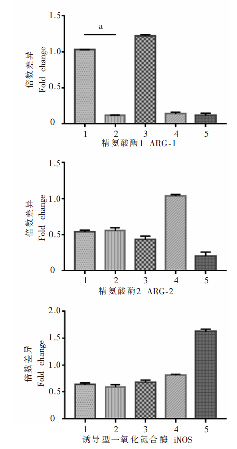
Table 1
Analysis of ARG-1 expression in multiple myeloid cells in livers of mice infected with E. granulosus
| 细胞类型Cell type | ARG-1表达的细胞比例/% Percentages of cells expressing ARG-1/% | ARG-1表达的相对平均荧光密度 Relative MFI of ARG-1 expression | ||
|---|---|---|---|---|
| 感染组Infection group | 对照组Control group | 感染组Infection group | 对照组Control group | |
| CD11b+CD11c+ | 1.85 ± 0.24 | 1.69 ± 0.16 | 1.41 ± 0.12 | 1.00 ± 0.14 |
| CD11b+F4/80+ | 5.39 ± 1.17 | 6.19 ± 0.40 | 1.21 ± 0.06 | 1.00 ± 0.05 |
| CD11b+Gr-1+ | 2.11 ± 0.55 | 1.27 ± 0.52 | 1.52 ± 0.16 | 1.00 ± 0.01 |
| CD11b+Gr-1+Ly-6C--Ly-6G+ | 1.63 ± 0.46 | 0.73 ± 0.18 | 1.30 ± 0.03 | 1.00 ± 0.07 |
| CD11b+Gr-1+Ly-6C+-Ly-6G- | 1.85 ± 0.40 | 0.89 ± 0.43 | 1.58 ± 0.12 | 1.00 ± 0.03 |
| CD11b+Ly-6G+ | 1.86 ± 0.45 | 1.15 ± 0.09 | 1.21 ± 0.04 | 1.00 ± 0.02 |
| [1] |
Budke CM, Carabin H, Ndimubanzi PC, et al. A systematic review of the literature on cystic echinococcosis frequency worldwide and its associated clinical manifestations[J]. Am J Trop Med Hyg, 2013,88(6):1011-1027.
pmid: 23546806 |
| [2] | Wu WP, Wang H, Wang Q, et al. A nationwide sampling survey on echinococcosis in China during 2012-2016[J]. Chin J Parasitol Parasit Dis, 2018,36(1):1-14. (in Chinese) |
| ( 伍卫平, 王虎, 王谦, 等. 2012-2016年中国棘球蚴病抽样调查分析[J]. 中国寄生虫学与寄生虫病杂志, 2018,36(1):1-14.) | |
| [3] | Vuitton DA. Echinococcosis and allergy[J]. Clinic Rev Allerg Immunol, 2004,26(2):93-104. |
| [4] |
Zheng YD. Strategies of Echinococcus species responses to immune attacks: implications for therapeutic tool development[J]. Int Immunopharmacol, 2013,17(3):495-501.
pmid: 23973651 |
| [5] |
Grubor NM, Jovanova-Nesic KD, Shoenfeld Y. Liver cystic echinococcosis and human host immune and autoimmune follow-up: a review[J]. World J Hepatol, 2017,9(30):1176-1189.
pmid: 29109850 |
| [6] | Caldwell RW, Rodriguez PC, Toque HA, et al. Arginase: amultifaceted enzyme important in health and disease[J]. Physiol Rev, 2018,98(2):641-665. |
| [7] | Alderton WK, Cooper CE, Knowles RG. Nitric oxide synthases: structure, function and inhibition[J]. Biochem J, 2001,357(3):593-615. |
| [8] |
Amri M, Touil-Boukoffa C. A protective effect of the laminated layer on Echinococcus granulosus survival dependent on upregulation of host arginase[J]. Acta Trop, 2015,149:186-194.
pmid: 26048557 |
| [9] | Zea AH, Rodriguez PC, Culotta KS, et al. L-arginine modulates CD3ζ expression and T cell function in activated human T lymphocytes[J]. Cell Immunol, 2004,232(1/2):21-31. |
| [10] | Xiao N, Qiu JM, Nakao M, et al. Short report: identification of Echinococcus species from a yak in the Qinghai-Tibet plateau region of China[J]. Am J Trop Med Hyg, 2003,69(4):445-446. |
| [11] | Gong WC, Huang FJ, Sun L, et al. Toll-like receptor-2 regulates macrophage polarization induced by excretory-secretory antigens from Schistosoma japonicum eggs and promotes liver pathology in murine schistosomiasis[J]. PLoS Negl Trop Dis, 2018,12(12):e0007000. |
| [12] |
Díaz A, Casaravilla C, Allen JE, et al. Understanding the laminated layer of larval Echinococcus Ⅱ: immunology[J]. Trends Parasitol, 2011,27(6):264-273.
doi: 10.1016/j.pt.2011.01.008 pmid: 21376669 |
| [13] | Paredes R, Godoy P, Rodríguez B, et al. Bovine (Bos taurus) humoral immune response against Echinococcus granulosus and hydatid cyst infertility[J]. J Cell Biochem, 2011,112(1):189-199. |
| [14] | El Kasmi KC, Qualls JE, Pesce JT, et al. Toll-like receptor-induced arginase 1 in macrophages thwarts effective immunity against intracellular pathogens[J]. Nat Immunol, 2008,9(12):1399-1406. |
| [15] |
Rodriguez PC, Quiceno DG, Zabaleta J, et al. Arginase Ⅰ production in the tumor microenvironment by mature myeloid cells inhibits T-cell receptor expression and antigen-specific T-cell responses[J]. Cancer Res, 2004,64(16):5839-5849.
pmid: 15313928 |
| [16] | Bowcutt R, Bell LV, Little M, et al. Arginase-1-expressing macrophages are dispensable for resistance to infection with the gastrointestinal helminth Trichuris muris[J]. Parasite Immunol, 2011,33(7):411-420. |
| [17] | Paduch K, Debus A, Rai B, et al. Resolution of cutaneous leishmaniasis and persistence of Leishmania major in the absence of arginase 1[J]. J Immunol, 2019,202(5):1453-1464. |
| [18] | Cao SK, Pan W, Liu H, et al. Expression and activity of arginase from monocytic-type myeloid-derived suppressor cells in rats infected with Echinococcus granulosus[J]. Chin J Parasitol Parasit Dis, 2016,34(1):27-31. (in Chinese) |
| ( 曹胜魁, 潘伟, 刘华, 等. 细粒棘球蚴感染小鼠单核髓源抑制性细胞精氨酸酶的表达和活性研究[J]. 中国寄生虫学与寄生虫病杂志, 2016,34(1):27-31.) | |
| [19] | Peng SS, Yu T, Wang L, et al. Influence of type 2 macrophages (M2) in echinococcosis[J]. Int J Clin Exp Pathol, 2016,9(3):4110-4116. |
| [20] | Munder M, Eichmann K, Morán JM, et al. Th1/Th2-regulated expression of arginase isoforms in murine macrophages and dendritic cells[J]. J Immunol, 1999,163(7):3771-3777. |
| [21] | Oberlies J, Watzl C, Giese T, et al. Regulation of NK cell function by human granulocyte arginase[J]. J Immunol, 2009,182(9):5259-5267. |
| [22] | Bronte V, Serafini P, Mazzoni A, et al. L-arginine metabolism in myeloid cells controls T-lymphocyte functions[J]. Trends Immunol, 2003,24(6):301-305. |
| [1] | LU Junxia, XU Junying, ZHAO Bin, WANG Qianwen, LI Wenhua, GENG Yuqing, HOU Jun, WU Xiangwei, CHEN Xueling. Echinococcus granulosus infection induces macrophages to express CD73 and A2AR to suppress inflammatory response [J]. CHINESE JOURNAL OF PARASITOLOGY AND PARASITIC DISEASES, 2023, 41(5): 559-566. |
| [2] | WU Xiaoying, HU Yuan, CAO Jianping. Preparation of Echinococcus granulosus peptide embedded in chitosan quaternary ammonium salt nanoparticles [J]. CHINESE JOURNAL OF PARASITOLOGY AND PARASITIC DISEASES, 2023, 41(3): 300-305. |
| [3] | LI Benfu, WANG Zhengqing, XU Qian, ZI Jinrong, YAN Xinliu, PENG Jia, LI Jianxiong, CAI Xuan, WU Fangwei, YANG Yaming. Sequence analysis of mitochondrial co1 and nd1 genes in Echinococcus granulosus in Yunnan Province [J]. CHINESE JOURNAL OF PARASITOLOGY AND PARASITIC DISEASES, 2023, 41(3): 306-311. |
| [4] | GUO Gang, REN Yuan, JIAO Hongjie, WU Juan, GUO Baoping, QI Wenjing, LI Jun, ZHANG Wenbao. Effect of intraperitoneal inoculation with Echinococcus microcysts on the infection and pathogenicity of E. multilocularis in mouse liver [J]. CHINESE JOURNAL OF PARASITOLOGY AND PARASITIC DISEASES, 2023, 41(2): 156-162. |
| [5] | JIAO Hongjie, QI Wenjing, GUO Gang, BAO Jianling, WU Chuanchuan, SONG Chuanlong, LI Jun, ZHANG Wenbao, YAN Mei. Polarization effect of Echinococcus granulosus antigen B on the mouse macrophage RAW264.7 [J]. CHINESE JOURNAL OF PARASITOLOGY AND PARASITIC DISEASES, 2023, 41(1): 23-28. |
| [6] | WU De-fang, FU Yong, REN Bin, ZHANG Yao-gang, XU Xiao-lei, PANG Ming-quan, FAN Hai-ning. Genetic diversity and differentiation time of human isolates of Echinococcus granulosus and E. multilocularis from Qinghai [J]. CHINESE JOURNAL OF PARASITOLOGY AND PARASITIC DISEASES, 2022, 40(5): 610-615. |
| [7] | QIAO Shi-yuan, ZHOU Xue, LIU Cheng-hao, JIANG Hui-jiao, BU Yuan-yuan, CHEN Xue-ling, WU Xiang-wei. Effect of albendazole-loaded vesicles on the vitality of protoscoleces of Echinococcus granulosus [J]. CHINESE JOURNAL OF PARASITOLOGY AND PARASITIC DISEASES, 2022, 40(3): 324-329. |
| [8] | GAO Yuan, ZHANG Xiao-cheng, HU Yuan, CAO Jian-ping. Study on the inhibitory effect of natural killer cells on liver fibrosis of schistosomiasis [J]. CHINESE JOURNAL OF PARASITOLOGY AND PARASITIC DISEASES, 2022, 40(2): 168-174. |
| [9] | SUN Ye-ting, JIANG Nan, JIANG Yan-yan, LI Teng, JIANG Xiao-feng, CAO Jian-ping, SHEN Yu-juan. Study on the polarization of MDSC stimulated by Echinococcus granulosus protoscolex-derived exosomes in vitro [J]. CHINESE JOURNAL OF PARASITOLOGY AND PARASITIC DISEASES, 2022, 40(2): 175-180. |
| [10] | ZHOU Wen-zheng, SUN Jun-gang, ZHAO Xi-bin, CAO Li. Therapeutic effect of intensity modulated radiation therapy on secondary femur infection with Echinococcus granulosus in rats [J]. CHINESE JOURNAL OF PARASITOLOGY AND PARASITIC DISEASES, 2021, 39(4): 443-448. |
| [11] | TIAN Meng-xiao, ZANG Xiao-yan, GUO Gang, QI Wen-jing, GUO Bao-ping, REN Yuan, LI Jun, ZHANG Wen-bao. Expression and activity assay of serine protease in Echinococcus granulosus [J]. CHINESE JOURNAL OF PARASITOLOGY AND PARASITIC DISEASES, 2021, 39(2): 233-239. |
| [12] | FAN Jun-jie, HAN Xiu-min, Nur Fazleen Binti Idris, LI Kai, TAN Qing-qing, CAO Wen-qiao, LI Xiang, LIAO Peng, YE Bin. Bioinformatics characteristics and immunoreactivity of protein kinase A of Echinococcus granulosus [J]. CHINESE JOURNAL OF PARASITOLOGY AND PARASITIC DISEASES, 2020, 38(6): 682-687. |
| [13] | SHI Chun-li, YANG Hui, PAN Wen, ZHANG Xin, ZHU Xiao-ting, ZHAO Jia-qing. Proteomic analysis of human proteins in extracellular vesicles secreted by protoscoleces of Echinococcus granulosus [J]. CHINESE JOURNAL OF PARASITOLOGY AND PARASITIC DISEASES, 2020, 38(6): 695-701. |
| [14] | YU Xiao-dong, YALI Ya-sen, WANG Jia-ling, LI Meng, YE Jian-rong. Establishment of BALB/c mouse model of Echinococcus granulosus-induced sensitization and changes of related immune cells [J]. CHINESE JOURNAL OF PARASITOLOGY AND PARASITIC DISEASES, 2020, 38(4): 412-416. |
| [15] | ZHOU Hong-rang, MAO Guang-yao, WANG Xiao-ling, CHEN Mu-xin, YU Qing, WANG Ying, Ai Lin, XIAO Ning. Establishment and application of a multiplex recombinase-aided isothermal amplification technique for identifying Echinococcus granulosus and Echinococcus multilocularis [J]. CHINESE JOURNAL OF PARASITOLOGY AND PARASITIC DISEASES, 2020, 38(3): 310-316. |
| Viewed | ||||||
|
Full text |
|
|||||
|
Abstract |
|
|||||