
CHINESE JOURNAL OF PARASITOLOGY AND PARASITIC DISEASES ›› 2019, Vol. 37 ›› Issue (3): 302-310.doi: 10.12140/j.issn.1000-7423.2019.03.011
• ORIGINAL ARTICLES • Previous Articles Next Articles
Qing ZHOU1( ), Xiong-feng YANG1, Huan-huan HAN1, Li-jiao GUO1, Hui-jiao JIANG1, Xiao-yi WANG1, Lin-lin LI2, Zhen-yu LIAO2, Xue-ling CHEN2, Xiang-wei WU1,*()
Received:2018-11-15
Online:2019-06-30
Published:2019-07-10
Contact:
Xiang-wei WU
E-mail:404915195@qq.com;wxwshz@126.com
Supported by:CLC Number:
Qing ZHOU, Xiong-feng YANG, Huan-huan HAN, Li-jiao GUO, Hui-jiao JIANG, Xiao-yi WANG, Lin-lin LI, Zhen-yu LIAO, Xue-ling CHEN, Xiang-wei WU. Correlation between angiogenesis and disease progression of hepatic Echinococcus multilocularis in C57BL/6 mice[J]. CHINESE JOURNAL OF PARASITOLOGY AND PARASITIC DISEASES, 2019, 37(3): 302-310.
Add to citation manager EndNote|Ris|BibTeX
URL: https://www.jsczz.cn/EN/10.12140/j.issn.1000-7423.2019.03.011
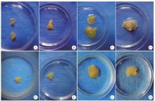
Fig. 1
Dynamic distribution of blood vessels around the infected Em in the livers of mice A-D: E. multilocularis infected mouse livers; E-H: Normal mouse livers. A: Em tissue was small, peripheral blood vessels were not clear at 30 d after infection; B: Em tissue was slightly larger, in irregular shape at 60 d after infection; C: Em tissue continued to grow, blood vessels were more obvious at 90 d after infection; D: Em tissue largely grew, clearly surrounded by peripheral blood vessels at 120 d after infection; E-H: No obvious abnormalities in the hepatic vasculature in control mice after 30, 60, 90, 120 d
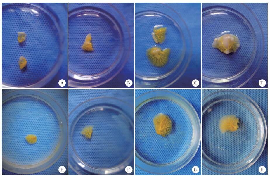
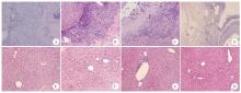
Fig. 2
Pathological changes of mouse livers after being infected with E. multilocularis(HE staining, × 100) A-D: E. multilocularis infected mouse livers; E-H: Normal mouse livers. A: Em tissure was surrounded by the filtration of inflammatory cells. The protoscoleces and calcification were visible inside Em at 30 d after infection; B: Em was seriously surrounded by the infiltration of inflammatory cells and some protoscoleces observed inside Em at 60 d after infection; C: Em tissue further grew in the liver, and necrosis was visible inside Em tissue at 90 d after infection; D: Serious necrosis and empty vesicles could be seen in Em tissue at 120 d after iinfection; E-H: Livers in control group for 30, 60, 90, 120 d, the liver tissue structure was basically normal
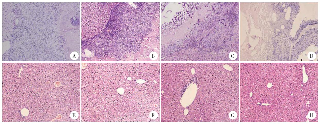
Fig. 3
Dynamic expression of VEGFA in E. multilocularis during its infection in mouse liver (EnVision, × 200) A-D: Em tissues in infected mouse liver; E-H: Liver tissues surrounding Em in infected mouse; I-L: Livers tissues in normal mouse. A: A small amount of VEGFA was stained in slight brownish yellow in the endothelial cells of Em at 30 d after infection; B: A medium amount of VEGFA stained in brownish yellow in the endothelial cells of Em at 60 d after infection; C: A large amount of VEGFA was stained in dark brown in the endothelial cells of Em at 90 d after infection; D: The VEGFA stained endothelial cells in the Em were significantly reduced, which were brownish brown at 120 d after infection; E-H: No obvious VEGFA staining was observed in the liver tissue surrounding tne infected Em tissues after being infected for 30, 60, 90 and 120 d, respectively; I-L: No obvious VEGFA staining was observed in the normal liver tissue of control mice after 30, 60, 90 and 120 d, respectively
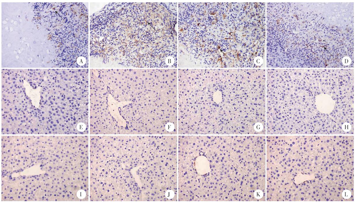
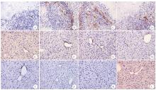
Fig. 4
Dynamic expression of CD34-MVD in E. multilocularis during its infection in mouse livers(EnVision, × 200) A-D: Em tissues in infected mouse liver; E-H: Liver tissues surrounding Em in infected mouse; I-L: Livers tissues in normal mouse. A: A small amount of CD34-MVD was stained in slight brownish yellow in the endothelial cells of Em at 30 d after infection; B: A medium amount of CD34-MVD stained in brownish yellow in the endothelial cells of Em at 60 d after infection; C: A large amount of CD34-MVD stained in dark brown in the endothelial cells of Em at 90 d after infection; D: The CD34-MVD stained endothelial cells in Em were reduced, which were brownish brown at 120 d after infection; E-H: No obvious CD34-MVD staining was observed in the liver tissue surrounding in infected Em tissues after being infected for 30, 60, 90 and 120 d, respectively; I-L: No obvious CD34-MVD staining was observed in the normal liver tissue of control mice after 30, 60, 90 and 120 d, respectively

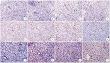
Fig. 5
Dynamic expression of CD31-MVD in E. multilocularis during its infection in mouse liver (EnVision, × 200) A-D: Em tissues in infected mouse liver; E-H: Liver tissues surrounding Em in infected mouse; I-L: Livers tissues in normal mouse. A: A small amount of CD31-MVD was stained in the endothelial cells of Em, with irregular shape at 30 d after infection; B: A medium amount of CD31-MVD stained in the endothelial cells of Em with fusiform or tubular shapes at 60 d after infection; C: A large amount of CD31-MVD stained in the endothelial cells of Em with regular shape at 90 d after infection; D: The CD31-MVD stained endothelial cells in Em were reduced with tubular shape at 120 d after infection; E-H: A large amount of CD31-MVD staining was observed in the liver nonepithelial cells surrounding infected Em tissues after being infected for 30, 60, 90 and 120 d, respectively; I-L: A large amount of CD31-MVD staining was observed in the normal liver nonepithelial cells after 30, 60, 90 and 120 d, respectively

| [1] | Wang J, Gottstein B.Immunoregulation in larval Echinococcus multilocularis infection[J]. Parasite Immunol, 2016, 38(3): 182-192. |
| [2] | Piarroux M, Piarroux R, Giorgi R, et al. Clinical features and evolution of alveolar echinococcosis in France from 1982 to 2007: results of a survey in 387 patients[J]. J Hepatol, 2011, 55(5): 1025-1033. |
| [3] | 张梦媛, 伍卫平, 官亚宜, 等. 我国棘球蚴病疾病负担分析[J]. 中国寄生虫学与寄生虫病杂志, 2018, 36(1): 15-19, 25. |
| [4] | Ferrara N, Gerber HP, LeCouter J. The biology of VEGF and its receptors[J]. Nat Med, 2003, 9(6): 669-676. |
| [5] | Namisaki T, Yoshiji H, Noguchi R, et al. The vascular endothelial growth factor(VEGF) receptor-2 is a major regulator of VEGF-mediated salvage effect in murine acute hepatic failure[J]. J Angiogenes Res, 2010, 2: 16. |
| [6] | Grothey A, Galanis E.Targeting angiogenesis: progress with anti-VEGF treatment with large molecules[J]. Nat Rev Clin Oncol, 2009, 6(9): 507-518. |
| [7] | Fina L, Molgaard HV, Robertson D, et al. Expression of the CD34 gene in vascular endothelial cells[J]. Blood, 1990, 75(12): 2417-2426. |
| [8] | Hristov M, Weber C.Endothelial progenitor cells in vascular repair and remodeling[J]. Pharmacol Res, 2008, 58(2): 148-151. |
| [9] | Deepak AV, Salimath BP.Antiangiogenic and proapoptotic activity of a novel glycoprotein from U. indica is mediated by NF-κB and caspase activated DNase in ascites tumor model[J]. Biochimie (Paris), 2006, 88(3/4): 297-307. |
| [10] | Weidner N, Folkman J, Pozza F, et al. Tumor angiogenesis: a new significant and independent prognostic indicator in early-stage breast carcinoma[J]. J Natl Cancer Inst, 1992, 84(24): 1875-1887. |
| [11] | 温浩, 徐明谦. 实用包虫病学[M]. 北京: 科学出版社, 2007: 16-35. |
| [12] | Fong GH.Mechanisms of adaptive angiogenesis to tissue hypoxia[J]. Angiogenesis, 2008, 11(2): 121-140. |
| [13] | Hanahan D.Rethinking the war on cancer[J]. Lancet, 2014, 383(9916): 558-563. |
| [14] | Mar SL, Adriano C, Roberto C, et al. Progress in the pharmacological treatment of human cystic and alveolar echinococcosis: compounds and therapeutic targets[J]. PLoS Neglect Trop Dis, 2018, 12(4): e0006422. |
| [15] | Yin J, Liu C, Yu A, et al. Pro-angiogenic activity of monocytic-type myeloid-derived suppressor cells from BALB/c mice infected with Echinococcus granulosus and the regulatory role of miRNAs[J]. Cell Physiol Biochem, 2018, 51: 1207-1220. |
| [16] | 姚冰, 王海涛, 刘文亚, 等. 肝泡球蚴病边缘区域CT灌注成像与组织病理对照研究[J]. 中国医学计算机成像杂志, 2010, 16(3): 215-220. |
| [17] | 李莉, 任伟新, 许晓冬. 活体碘油门静脉灌注大鼠肝Em感染模型的观察研究[J]. 新疆医科大学学报, 2006, 29(11): 1087-1089. |
| [18] | Rogan MT.T-cell activity associated with secondary infections and implanted cysts of Echinococcus granulosus in BALB/c mice[J]. Parasite Immunol, 2010, 20(11): 527-533. |
| [19] | 魏绪法, 邵英梅, 王俊华, 等. 泡球蚴感染中期小鼠肝脏病灶周围肉芽肿TGF-β1及其受体TβRⅠ和p-Smad2/3的表达及意义[J]. 中国病原生物学杂志, 2010, 5(12): 895-897. |
| [20] | Vuitton DA, Gottstein B.Echinococcus multilocularis and its intermediate host: a model of parasite-host interplay[J]. J Biomed Biotechnol, 2010, 2010(1): 923193. |
| [21] | Shibuya M.Vascular endothelial growth factor and its receptor system: physiological functions in angiogenesis and pathological roles in various diseases[J]. J Biochem, 2013, 153(1): 13-19. |
| [22] | 宋涛, 李海涛, 杨凌菲, 等. 大鼠肝泡球蚴病灶浸润增殖区微血管密度与超声造影的相关性研究[J]. 中国寄生虫学与寄生虫病杂志, 2014, 32(3): 200-204. |
| [23] | 魏晓丽, 丁剑冰, 许晏, 等. 小鼠感染Em后细胞因子水平的变化[J]. 中国寄生虫学与寄生虫病杂志, 2004, 22(6): 43-46. |
| [24] | 刘寒冬, 王宏兵, 樊海宁, 等. 多房棘球蚴病的免疫逃逸机制[J]. 中国寄生虫学与寄生虫病杂志, 2018, 36(6): 655-660. |
| [25] | 夏秀红, 范江涛, 李莉莉, 等. VEGF和CD31在子宫内膜样腺癌中的表达及其临床意义[J]. 实用妇产科杂志, 2018, 34(4): 282-286. |
| [26] | 崔晓楠, 侯力. 组织微阵列方法检测CD31、CD105、v-WF、PCNA在肝癌组织中的表达[J]. 中国肿瘤, 2007, 16(10): 801-804. |
| [1] | CAO Deping, LI Jiajing, SONG Mengwei, MO Gang. Experimental observation on the changes of hepatic stellate cells stimulated in vitro with tissue protein of Echinococcus multilocularis [J]. CHINESE JOURNAL OF PARASITOLOGY AND PARASITIC DISEASES, 2023, 41(4): 440-445. |
| [2] | DU Tao, HU Chunhui, GAN Xuehui, GAO Pan, ZHANG Fabin. Anti-Echinococcus multilocularis effect of total alkaloids of Sophora moorcroftiana in water solution and tablet forms in vitro and in vivo [J]. CHINESE JOURNAL OF PARASITOLOGY AND PARASITIC DISEASES, 2023, 41(1): 15-22. |
| [3] | HOU Xin-ling, LI De-wei, SHI Yang, WANG Mao-lin, ZIBIGU Rousu, ABIDAN Ainiwaer, ZHENG Xu-ran, KANG Xue-jiao, WANG Hui, LI Jing, ZHANG Chuan-shan. Changes of ST2+ T cell subset function and their immune checkpoint molecule expression in the peritoneal cavity of mice infected with Echinococcus multilocularis [J]. CHINESE JOURNAL OF PARASITOLOGY AND PARASITIC DISEASES, 2022, 40(6): 708-716. |
| [4] | WU Liang-liang, YANG Ling-fei, SONG Tao. Ultrasound and pathological manifestations of lesions in SD rats with hepatic Echinococcus multilocularis infection established by different methods [J]. CHINESE JOURNAL OF PARASITOLOGY AND PARASITIC DISEASES, 2022, 40(4): 549-552. |
| [5] | ZHONG Shun-hu, SUN Yue, GUO Xiao-la, ZHENG Ya-dong, CHEN Yi-xia. Identification and bioinformatics analysis of differentially expressed miRNAs in splenic lymphocytes in Echinococcus multilocularis-infected mice [J]. CHINESE JOURNAL OF PARASITOLOGY AND PARASITIC DISEASES, 2022, 40(3): 288-294. |
| [6] | ZHUO Yi-cheng, YANG Hai-cheng, LIU Cheng-hao, ZHANG Bao-cai, DUO Xiao-yong, ZHANG Shi-jie. Effect of osteopontin expression level on the growth and development of Echinococcus multilocularis protoscoleces [J]. CHINESE JOURNAL OF PARASITOLOGY AND PARASITIC DISEASES, 2022, 40(3): 299-304. |
| [7] | ABUDUAINI Abulizi, PAIZULA Shalayiadang, TALAITI Tuergan, ZHANG Rui-qing, WANG Hui, ZHANG Chuan-shan, SHAO Ying-mei, TUERGANAILI Aji. Affect of Echinococcus multilocularis protein-mediated NK cell surface receptor NKG2A on the function of NK cells [J]. CHINESE JOURNAL OF PARASITOLOGY AND PARASITIC DISEASES, 2022, 40(1): 36-42. |
| [8] | HOU Jiao, WEN Hao, WANG Ming-kun, LI Wen-ding, LI liang, LI Jing, ZHANG Chuan-shan, WANG Hui. Changes of macrophage subsets and polarization in spleen of mice infected with Echinococcus multilocularis [J]. CHINESE JOURNAL OF PARASITOLOGY AND PARASITIC DISEASES, 2021, 39(6): 771-778. |
| [9] | SHI Qi-qi, LIU Cong-shan, HUO Le-le, WEI Yu-fen, JIANG Bin, YIN Meng, XUE Jian, TAO Yi, ZHANG Hao-bing. Affect of aminoalcohol compound HT24 on the expression of tubulin in Echinococcus multilocularis protoscoleces [J]. CHINESE JOURNAL OF PARASITOLOGY AND PARASITIC DISEASES, 2021, 39(4): 437-443. |
| [10] | TAN Xiao-wu, YU Xiao-fan, JIANG Hui-jiao, XING Zhi-kun, CHEN Xue-ling, WU Xiang-wei. Inhibitory effect of xanthohumol on the growth of Echinococcus multilocularis metacestode in the liver of mice [J]. CHINESE JOURNAL OF PARASITOLOGY AND PARASITIC DISEASES, 2021, 39(3): 304-310. |
| [11] | LI Ling-hui, WANG Wei, HOU Xin-ling, SHI Yang, LI De-wei, LI Liang, WANG Hui, LI Jing, ZHANG Chuan-shan. Affects of Echinococcus multilocularis metacestode infection on the natural killer T cells and their subsets in mouse spleen [J]. CHINESE JOURNAL OF PARASITOLOGY AND PARASITIC DISEASES, 2021, 39(3): 311-317. |
| [12] | GUO Bao-ping, GUO Gang, ZHANG Li, XIANG Jing-jing, WANG Xiao-ping, REN Yuan, QI Wen-jing, ZHANG Hui, LI Jun, ZHANG Wen-bao, WANG Hai-yan. Investigation on infection of Echinococcus multilocularis metacestode in small rodents in Chabchar County, Xinjiang [J]. CHINESE JOURNAL OF PARASITOLOGY AND PARASITIC DISEASES, 2021, 39(3): 327-332. |
| [13] | XU Kai, WANG Hai-jiu, ZHANG Li, ZHANG Yao-gang, FAN Hai-ning, REN Li, REN Bin, WANG Zhi-xin. Research progress on the mechanisms underlying the impairment of host hepatocytes by Echinococcus multilocularis [J]. CHINESE JOURNAL OF PARASITOLOGY AND PARASITIC DISEASES, 2021, 39(2): 256-259. |
| [14] | ZHU Ji-hai, CAO De-ping, ZHAO Jun, LIU Jun, SHI Hu-xiang, LIU Yan. Investigation of differentially expressed genes in liver tissues of patients with alveolar echinococcosis [J]. CHINESE JOURNAL OF PARASITOLOGY AND PARASITIC DISEASES, 2021, 39(1): 48-54. |
| [15] | JIANG Hui-jiao, GUI Xian-wei, GUO Li-jiao, YANG Xiong-feng, WANG Xiao-yi, CHEN Xue-ling, WU Xiang-wei. Expression and angiogenic effect of VEGFA/VEGFR2 in mice hepatic metacestode tissue of Echinococcus multilocularis [J]. CHINESE JOURNAL OF PARASITOLOGY AND PARASITIC DISEASES, 2020, 38(6): 673-681. |
| Viewed | ||||||
|
Full text |
|
|||||
|
Abstract |
|
|||||