
CHINESE JOURNAL OF PARASITOLOGY AND PARASITIC DISEASES ›› 2022, Vol. 40 ›› Issue (2): 175-180.doi: 10.12140/j.issn.1000-7423.2022.02.007
• ORIGINAL ARTICLES • Previous Articles Next Articles
SUN Ye-ting( ), JIANG Nan, JIANG Yan-yan, LI Teng, JIANG Xiao-feng, CAO Jian-ping, SHEN Yu-juan*()
Received:2021-09-28
Revised:2021-12-30
Online:2022-04-30
Published:2022-04-20
Contact:
SHEN Yu-juan
E-mail:18863665089@163.com;shenyj12@nipd.chinacdc.cn
Supported by:CLC Number:
SUN Ye-ting, JIANG Nan, JIANG Yan-yan, LI Teng, JIANG Xiao-feng, CAO Jian-ping, SHEN Yu-juan. Study on the polarization of MDSC stimulated by Echinococcus granulosus protoscolex-derived exosomes in vitro[J]. CHINESE JOURNAL OF PARASITOLOGY AND PARASITIC DISEASES, 2022, 40(2): 175-180.
Add to citation manager EndNote|Ris|BibTeX
URL: https://www.jsczz.cn/EN/10.12140/j.issn.1000-7423.2022.02.007

Fig. 1
Flow cytometry analysis of the percentages of MDSC from healthy mice after 4 days of stimulation in vitro CD11b+Gr-1+ populations are MDSC.
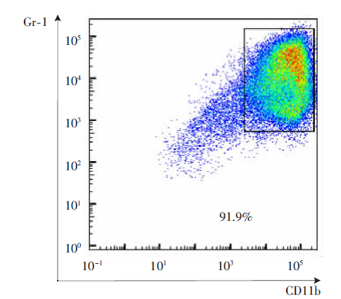
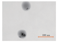
Fig. 2
The morphology of E. granulosus protoscolex-derived exosomes detected by transmission electron microscope(×50 000)


Fig. 3
Western blotting analysis of exosome-specific protein CD9 and enolase expression 1: PBS; 2, 3: Exosome.
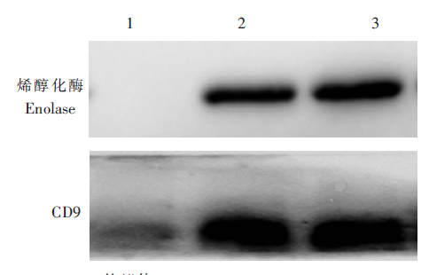
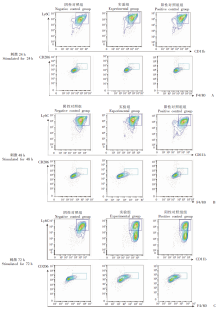
Fig. 4
Flow cytometry analysis of percentages of M-MDSC in MDSC and dynamic changes of molecular markers of M2 macrophages in 24 h (A), 48 h (B), and 72 h (C) post-stimulation by E. granulosus protoscolex-derived exosomes CD11b+Ly6C+ populations are M-MDSC, F4/80 and CD206 are specific markers of M2 macrophages.

| [1] |
Alvarez Rojas CA,, Romig T,, Lightowlers MW. Echinococcus granulosus sensu lato genotypes infecting humans: review of current knowledge[J]. Int J Parasitol, 2014, 44(1): 9-18.
doi: 10.1016/j.ijpara.2013.08.008 |
| [2] |
McManus DP. Current status of the genetics and molecular taxonomy of Echinococcus species[J]. Parasitology, 2013, 140(13): 1617-1623.
doi: 10.1017/S0031182013000802 pmid: 23750777 |
| [3] |
Grubor NM,, Jovanova-Nesic KD,, Shoenfeld Y. Liver cystic echinococcosis and human host immune and autoimmune follow-up: a review[J]. World J Hepatol, 2017, 9(30): 1176-1189.
doi: 10.4254/wjh.v9.i30.1176 |
| [4] |
Zhou XJ,, Wang W,, Cui F, et al. Myeloid-derived suppressor cells exert immunosuppressive function on the T helper 2 in mice infected with Echinococcus granulosus[J]. Exp Parasitol, 2020, 215: 107917.
doi: 10.1016/j.exppara.2020.107917 |
| [5] |
Lindau D,, Gielen P,, Kroesen M, et al. The immunosuppressive tumour network: myeloid-derived suppressor cells, regulatory T cells and natural killer T cells[J]. Immunology, 2013, 138(2): 105-115.
doi: 10.1111/imm.12036 |
| [6] |
Vetsika EK,, Koukos A,, Kotsakis A. Myeloid-derived suppressor cells: major figures that shape the immunosuppressive and angiogenic network in cancer[J]. Cells, 2019, 8(12): 1647.
doi: 10.3390/cells8121647 |
| [7] | Parker KH,, Beury DW,, Ostrand-Rosenberg S. Myeloid-derived suppressor cells: critical cells driving immune suppression in the tumor microenvironment[J]. Adv Cancer Res, 2015, 128:95-139. |
| [8] |
Mills CD,, Kincaid K,, Alt JM, et al. M-1/M-2 macrophages and the Th1/Th2 paradigm[J]. J Immunol, 2000, 164(12): 6166-6173.
doi: 10.4049/jimmunol.164.12.6166 pmid: 10843666 |
| [9] |
Mosser DM,, Edwards JP. Exploring the full spectrum of macrophage activation[J]. Nat Rev Immunol, 2008, 8(12):958-969.
doi: 10.1038/nri2448 pmid: 19029990 |
| [10] |
Funes SC,, Rios M,, Escobar-Vera J, et al. Implications of macrophage polarization in autoimmunity[J]. Immunology, 2018, 154(2): 186-195.
doi: 10.1111/imm.12910 |
| [11] |
Gordon SR,, Maute RL,, Dulken BW, et al. PD-1 expression by tumour-associated macrophages inhibits phagocytosis and tumour immunity[J]. Nature, 2017, 545(7655): 495-499.
doi: 10.1038/nature22396 |
| [12] |
Biswas S,, Mandal G,, Roy Chowdhury S, et al. Exosomes produced by mesenchymal stem cells drive differentiation of myeloid cells into immunosuppressive M2-polarized macrophages in breast cancer[J]. J Immunol, 2019, 203(12): 3447-3460.
doi: 10.4049/jimmunol.1900692 pmid: 31704881 |
| [13] | Xu QG,, Wang WH,, Zhu B, et al. Optimal conditions for the expansion of mouse myeloid-derived suppressor cells in vitro[J]. Curr Immunol, 2015, 35(2): 140-144. (in Chinese) |
| (许秋桂,, 王文红,, 朱波, 等. 小鼠髓样抑制细胞体外扩增体系的建立[J]. 现代免疫学, 2015, 35(2): 140-144.) | |
| [14] | Fan WX,, Diao YJ,, Ma YY, et al. Comparison of ultracentrifugation and membrane based-affinity column methods in exosome isolation from supernatants of prostate cancer cells[J]. J Mod Lab Med, 2019, 34(3): 6-9. (in Chinese) |
| (范维肖,, 刁艳君,, 马越云, 等. 超速离心法与QIAGEN膜亲和柱法提取前列腺癌细胞培养上清外泌体的方法学比较[J]. 现代检验医学杂志, 2019, 34(3): 6-9.) | |
| [15] | Sun YT,, Cao JP,, Shen YJ. Research progress on immunosuppressive function of myeloid-derived suppressor cells and its role in parasitic infection[J]. Chin J Parasitol Parasit Dis, 2021, 39 (4): 505-513. (in Chinese) |
| (孙叶挺,, 曹建平,, 沈玉娟. 髓源抑制性细胞免疫抑制功能及其在寄生虫感染领域的研究进展[J]. 中国寄生虫学与寄生虫病杂志, 2021, 39(4): 505-513.) | |
| [16] | E WJ,, Lu YL,, Zhang LQ, et al. Characteristics and clinical value of macrophage polarization in tissues and serum of patients with hepatic alveolar echinocococosis[J]. J Pract Med, 2020, 36(9): 1198-1202. (in Chinese) |
| (鄂维建,, 芦永良,, 张灵强, 等. 肝泡型包虫病肝组织和血清中巨噬细胞极化特点及临床意义[J]. 实用医学杂志, 2020, 36(9): 1198-1202.) | |
| [17] | Zhang XF,, Gong WC,, Cao SK, et al. Dynamic changes of myeloid-derived suppressor cells and regulatory T cells in livers of mice infected with Echinococcus granulosus[J]. Chin J Schisto Control, 2019, 31(6): 622-627. (in Chinese) |
| (张小凡,, 巩文词,, 曹胜魁, 等. 细粒棘球绦虫感染小鼠肝脏髓源抑制性细胞与调节性T细胞比例动态变化[J]. 中国血吸虫病防治杂志, 2019, 31(6): 622-627.) | |
| [18] |
Pritchard A,, Tousif S,, Wang Y, et al. Lung tumor cell-derived exosomes promote M2 macrophage polarization[J]. Cells, 2020, 9(5): 1303.
doi: 10.3390/cells9051303 |
| [19] |
Santos GB,, Monteiro KM,, da Silva ED, et al. Excretory/secretory products in the Echinococcus granulosus metacestode: is the intermediate host complacent with infection caused by the larval form of the parasite?[J]. Int J Parasitol, 2016, 46(13/14): 843-856.
doi: 10.1016/j.ijpara.2016.07.009 |
| [20] |
Siles-Lucas M,, Sánchez-Ovejero C,, González-Sánchez M, et al. Isolation and characterization of exosomes derived from fertile sheep hydatid cysts[J]. Vet Parasitol, 2017, 236: 22-33.
doi: S0304-4017(17)30032-8 pmid: 28288760 |
| [21] |
Van Ginderachter JA, Beschin A,, de Baetselier P, et al. Myeloid-derived suppressor cells in parasitic infections[J]. Eur J Immunol, 2010, 40(11): 2976-2985.
doi: 10.1002/eji.201040911 pmid: 21061431 |
| [22] |
Loke P,, MacDonald AS,, Allen JE. Antigen-presenting cells recruited by Brugia malayi induce Th2 differentiation of naïve CD4+ T cells[J]. Eur J Immunol, 2000, 30(4): 1127-1135.
pmid: 10760802 |
| [23] |
Kong YY,, Fuchsberger M,, Xiang SD, et al. Myeloid derived suppressor cells and their role in diseases[J]. Curr Med Chem, 2013, 20(11): 1437-1444.
pmid: 23409714 |
| [1] | LU Junxia, XU Junying, ZHAO Bin, WANG Qianwen, LI Wenhua, GENG Yuqing, HOU Jun, WU Xiangwei, CHEN Xueling. Echinococcus granulosus infection induces macrophages to express CD73 and A2AR to suppress inflammatory response [J]. CHINESE JOURNAL OF PARASITOLOGY AND PARASITIC DISEASES, 2023, 41(5): 559-566. |
| [2] | WU Xiaoying, HU Yuan, CAO Jianping. Preparation of Echinococcus granulosus peptide embedded in chitosan quaternary ammonium salt nanoparticles [J]. CHINESE JOURNAL OF PARASITOLOGY AND PARASITIC DISEASES, 2023, 41(3): 300-305. |
| [3] | LI Benfu, WANG Zhengqing, XU Qian, ZI Jinrong, YAN Xinliu, PENG Jia, LI Jianxiong, CAI Xuan, WU Fangwei, YANG Yaming. Sequence analysis of mitochondrial co1 and nd1 genes in Echinococcus granulosus in Yunnan Province [J]. CHINESE JOURNAL OF PARASITOLOGY AND PARASITIC DISEASES, 2023, 41(3): 306-311. |
| [4] | GUO Gang, REN Yuan, JIAO Hongjie, WU Juan, GUO Baoping, QI Wenjing, LI Jun, ZHANG Wenbao. Effect of intraperitoneal inoculation with Echinococcus microcysts on the infection and pathogenicity of E. multilocularis in mouse liver [J]. CHINESE JOURNAL OF PARASITOLOGY AND PARASITIC DISEASES, 2023, 41(2): 156-162. |
| [5] | JIAO Hongjie, QI Wenjing, GUO Gang, BAO Jianling, WU Chuanchuan, SONG Chuanlong, LI Jun, ZHANG Wenbao, YAN Mei. Polarization effect of Echinococcus granulosus antigen B on the mouse macrophage RAW264.7 [J]. CHINESE JOURNAL OF PARASITOLOGY AND PARASITIC DISEASES, 2023, 41(1): 23-28. |
| [6] | WU De-fang, FU Yong, REN Bin, ZHANG Yao-gang, XU Xiao-lei, PANG Ming-quan, FAN Hai-ning. Genetic diversity and differentiation time of human isolates of Echinococcus granulosus and E. multilocularis from Qinghai [J]. CHINESE JOURNAL OF PARASITOLOGY AND PARASITIC DISEASES, 2022, 40(5): 610-615. |
| [7] | QIAO Shi-yuan, ZHOU Xue, LIU Cheng-hao, JIANG Hui-jiao, BU Yuan-yuan, CHEN Xue-ling, WU Xiang-wei. Effect of albendazole-loaded vesicles on the vitality of protoscoleces of Echinococcus granulosus [J]. CHINESE JOURNAL OF PARASITOLOGY AND PARASITIC DISEASES, 2022, 40(3): 324-329. |
| [8] | ZHOU Wen-zheng, SUN Jun-gang, ZHAO Xi-bin, CAO Li. Therapeutic effect of intensity modulated radiation therapy on secondary femur infection with Echinococcus granulosus in rats [J]. CHINESE JOURNAL OF PARASITOLOGY AND PARASITIC DISEASES, 2021, 39(4): 443-448. |
| [9] | SUN Ye-ting, CAO Jian-ping, SHEN Yu-juan. Research progress on immunosuppressive function of myeloid-derived suppressor cells and its role in parasitic infection [J]. CHINESE JOURNAL OF PARASITOLOGY AND PARASITIC DISEASES, 2021, 39(4): 505-513. |
| [10] | TIAN Meng-xiao, ZANG Xiao-yan, GUO Gang, QI Wen-jing, GUO Bao-ping, REN Yuan, LI Jun, ZHANG Wen-bao. Expression and activity assay of serine protease in Echinococcus granulosus [J]. CHINESE JOURNAL OF PARASITOLOGY AND PARASITIC DISEASES, 2021, 39(2): 233-239. |
| [11] | FAN Jun-jie, HAN Xiu-min, Nur Fazleen Binti Idris, LI Kai, TAN Qing-qing, CAO Wen-qiao, LI Xiang, LIAO Peng, YE Bin. Bioinformatics characteristics and immunoreactivity of protein kinase A of Echinococcus granulosus [J]. CHINESE JOURNAL OF PARASITOLOGY AND PARASITIC DISEASES, 2020, 38(6): 682-687. |
| [12] | SHI Chun-li, YANG Hui, PAN Wen, ZHANG Xin, ZHU Xiao-ting, ZHAO Jia-qing. Proteomic analysis of human proteins in extracellular vesicles secreted by protoscoleces of Echinococcus granulosus [J]. CHINESE JOURNAL OF PARASITOLOGY AND PARASITIC DISEASES, 2020, 38(6): 695-701. |
| [13] | YU Xiao-dong, YALI Ya-sen, WANG Jia-ling, LI Meng, YE Jian-rong. Establishment of BALB/c mouse model of Echinococcus granulosus-induced sensitization and changes of related immune cells [J]. CHINESE JOURNAL OF PARASITOLOGY AND PARASITIC DISEASES, 2020, 38(4): 412-416. |
| [14] | CAO Sheng-kui, ZHANG Xiao-fan, WEI Yu-huan, PAN Jia-ming, CAO Jian-ping, SHEN Yu-juan, CHEN Jia-xu. Expression and function of arginase in livers of mice infected with Echinococcus granulosus [J]. CHINESE JOURNAL OF PARASITOLOGY AND PARASITIC DISEASES, 2020, 38(3): 304-309. |
| [15] | ZHOU Hong-rang, MAO Guang-yao, WANG Xiao-ling, CHEN Mu-xin, YU Qing, WANG Ying, Ai Lin, XIAO Ning. Establishment and application of a multiplex recombinase-aided isothermal amplification technique for identifying Echinococcus granulosus and Echinococcus multilocularis [J]. CHINESE JOURNAL OF PARASITOLOGY AND PARASITIC DISEASES, 2020, 38(3): 310-316. |
| Viewed | ||||||
|
Full text |
|
|||||
|
Abstract |
|
|||||