| [1] | MacConnachie A. Schistosomiasis[J]. J R Coll Physicians Edinb, 2012, 42(1): 47-50. | | [2] | Burke ML, Jones MK, Gobert GN, et al. Immunopathogenesis of human schistosomiasis[J]. Parasite Immunol, 2009, 31(4): 163-176. | | [3] | Wang YJ, Zhang J, Yin JH, et al. The formation of egg granulomas in the spleens of mice with late Schistosoma japonicum infection alters splenic morphology[J]. Parasit Vectors, 2015, 8: 375. | | [4] | Weinstock JV, Boros DL. Organ-dependent differences in composition and function observed in hepatic and intestinal granulomas isolated from mice with schistosomiasis mansoni[J]. J Immunol, 1983, 130(1): 418-422. | | [5] | Patton EA, La Flamme AC, Pedras-Vasoncelos JA, et al. Central role for interleukin-4 in regulating nitric oxide-mediated inhibition of T-cell proliferation and gamma interferon production in schistosomiasis[J]. Infect Immun, 2002, 70(1): 177-184. | | [6] | Cheever AW, Duvall RH, Hallack TA Jr, et al. Variation of hepatic fibrosis and granuloma size among mouse strains infected with Schistosoma mansoni[J]. Am J Trop Med Hyg, 1987, 37(1): 85-97. | | [7] | Cheever AW. Comparison of pathologic changes in mammalian hosts infected with Schistosoma mansoni, S. japonicum and S. haematobium[J]. Mem Inst Oswaldo Cruz, 1987, 82(Suppl 4): 39-45. | | [8] | Rumbley CA, Phillips SM. The schistosome granuloma: An immunoregulatory organelle[J]. Microbes Infect, 1999, 1(7): 499-504. | | [9] | King CL, Xianli J, Stavitsky AB. Murine schistosomiasis mansoni: Coordinate cytokine regulation and differences in cellular immune responses of granuloma cells and splenocytes to endogenous and exogenous schistosome egg antigens[J]. Parasite Immunol, 2001, 23(11): 607-615. | | [10] | Wang YJ, Hu Y, Zhang J, et al. Eggs of Schistosoma japonicum deposited in the spleen induce apoptosis of splenic T cells in C57BL/6 mice[J]. Parasitol Res, 2025, 124(3): 31. | | [11] | 王燕娟, 曹建平. 致小鼠脾脏T细胞凋亡日本血吸虫虫卵抗原蛋白的筛选[J]. 中国病原生物学杂志, 2023, 18(1): 37-41. | | | Wang YJ, Cao JP. Study on the screening of molecule(s) performing apoptotic effect on T lymphocytes from Schistosoma japonicum egg antigen[J]. J Pathog Biol, 2023, 18(1): 37-41. (in Chinese) | | [12] | Nolte MA, Hoen EN, van Stijn A, et al. Isolation of the intact white pulp. Quantitative and qualitative analysis of the cellular composition of the splenic compartments[J]. Eur J Immunol, 2000, 30(2): 626-634. | | [13] | Girkontaite I, Sakk V, Wagner M, et al. The sphingosine-1-phosphate (S1P) lysophospholipid receptor S1P3 regulates MAdCAM-1+ endothelial cells in splenic marginal sinus organization[J]. J Exp Med, 2004, 200(11): 1491-1501. | | [14] | MacKay F, Browning JL. Turning off follicular dendritic cells[J]. Nature, 1998, 395(6697): 26-27. | | [15] | Crotty S. Follicular helper CD4 T cells (TFH)[J]. Annu Rev Immunol, 2011, 29: 621-663. | | [16] | Fazilleau N, Mark L, McHeyzer-Williams LJ, et al. Follicular helper T cells: Lineage and location[J]. Immunity, 2009, 30(3): 324-335. | | [17] | Shi JW, Hou SY, Fang Q, et al. PD-1 controls follicular T helper cell positioning and function[J]. Immunity, 2018, 49(2): 264-274.e4. | | [18] | 孙钰浚, 李钊琪, 吕芳丽. 日本血吸虫虫卵肉芽肿免疫病理机制研究进展[J]. 中国寄生虫学与寄生虫病杂志, 2019, 37(6): 713-717, 722. | | | Sun YJ, Li ZQ, Lv FL. Research progress on the immunopathological mechanism of Schistosoma japonicum egg-induced granuloma[J]. Chin J Parasitol Parasit Dis, 2019, 37(6): 713-717, 722. (in Chinese) | | [19] | 刘蓉, 闻礼永. 晚期血吸虫病基础和临床研究新进展[J]. 中国寄生虫学与寄生虫病杂志, 2021, 39(4): 429-436. | | | Liu R, Wen LY. New progress in basic and clinical research of advanced schistosomiasis[J]. Chin J Parasitol Parasit Dis, 2021, 39(4): 429-436. (in Chinese) | | [20] | Jiang SY, Huang XQ, Ni LY, et al. Positive consequences of splenectomy for patients with schistosomiasis-induced variceal bleeding[J]. Surg Endosc, 2021, 35(5): 2339-2346. | | [21] | Angus DC, Wax RS. Epidemiology of sepsis: An update[J]. Crit Care Med, 2001, 29(7 Suppl): S109.116. |
|

 )(
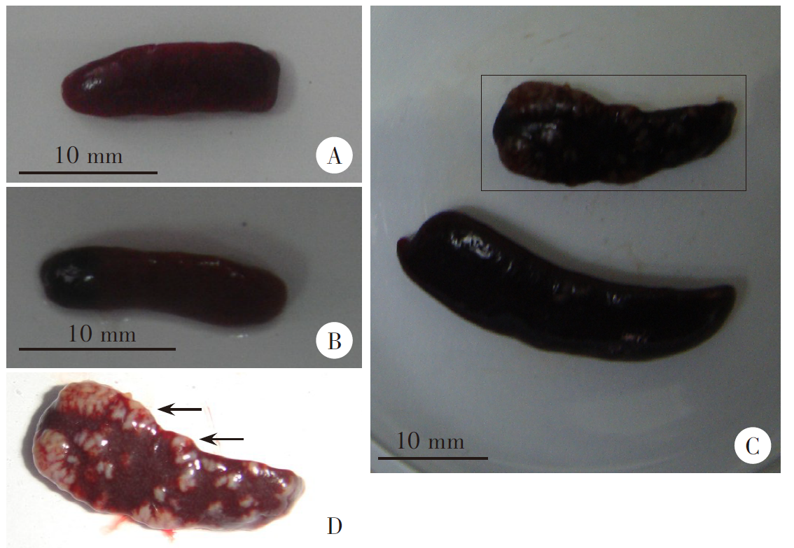
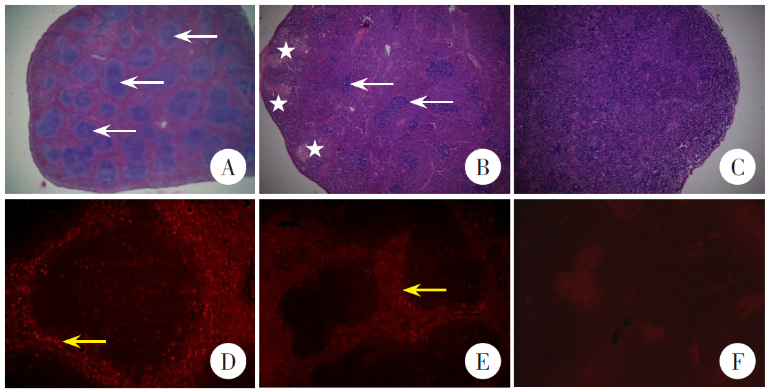
)( ), ZHANG Yanjun1, ZHOU Xiaojun1, CAO Jianping2,*(