
CHINESE JOURNAL OF PARASITOLOGY AND PARASITIC DISEASES ›› 2021, Vol. 39 ›› Issue (5): 621-626.doi: 10.12140/j.issn.1000-7423.2021.05.010
• ORIGINAL ARTICLES • Previous Articles Next Articles
HUANG Ai-long( ), ZHANG Bei, SHEN Han-yu, CHEN Guo, LI Jing, ZHU Dan-dan, DUAN Yi-nong*()
Received:2021-07-06
Revised:2021-07-26
Online:2021-10-30
Published:2021-11-10
Contact:
DUAN Yi-nong
E-mail:997243348@qq.com;yinongduan@aliyun.com
Supported by:CLC Number:
HUANG Ai-long, ZHANG Bei, SHEN Han-yu, CHEN Guo, LI Jing, ZHU Dan-dan, DUAN Yi-nong. Expression and function of triggering receptor expressed on myeloid cells 1 in the liver of mice infected with Schistosoma japonicum[J]. CHINESE JOURNAL OF PARASITOLOGY AND PARASITIC DISEASES, 2021, 39(5): 621-626.
Add to citation manager EndNote|Ris|BibTeX
URL: https://www.jsczz.cn/EN/10.12140/j.issn.1000-7423.2021.05.010
Table 1
Primers for real-time quantitative PCR
| 基因名称 Gene name | 引物序列(5′→3′) Primer sequence (5′→3′) |
|---|---|
| TREM-1 | F: CTGCTGTGCGTGTTCTTTGTCTC |
| R: AGGGTTCCTTCCCGTCTGGTA | |
| IL-1β | F: AATGACCTGTTCTTTGAAGTTGA |
| R: TGATGTGCTGCTGCGAGATTTGAAG | |
| GAPDH | F: TGGAAAGCTGTGGCGTGAT |
| R: TGCTTCACCACCTTCTTGAT |
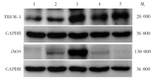
Fig. 1
Expression of TREM-1 and iNOS protein in mice liver infected with Schistosoma japonicum 1: Control group; 2-5: 3, 6, 9 and 12 weeks after infection. TREM-1: Triggering receptor expressed on myeloid cell 1; GAPDH:Glyceraldehyde-3-phosphate dehydrogenase; iNOS: Inducible NO synthase.
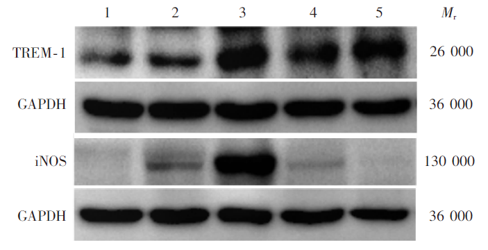
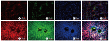
Fig. 2
Results of immunofluorescence staining in mice liver infected with S. japonicum A-D:Staining results of liver tissue sections from control group were successively presented as: F4/80 staining, TREM-1 staining, Hoechst nuclear staining and the combination of the three staining. The expression levels of TREM-1 and F4/80 were low in livers from control group; E-H:Staining results of liver tissue sections from mice at 6 weeks after infection were successively presented as: F4/80 staining, TREM-1 staining, Hoechst nuclear staining and the combination of the three staining. The expression levels of TREM-1 (green) and F4/80 (red) were both increased around hepatic granulomas. The number of the cells with the co-location (yellow) of TREM-1 and F4/80 was increased around the mouse hepatic granulomas.
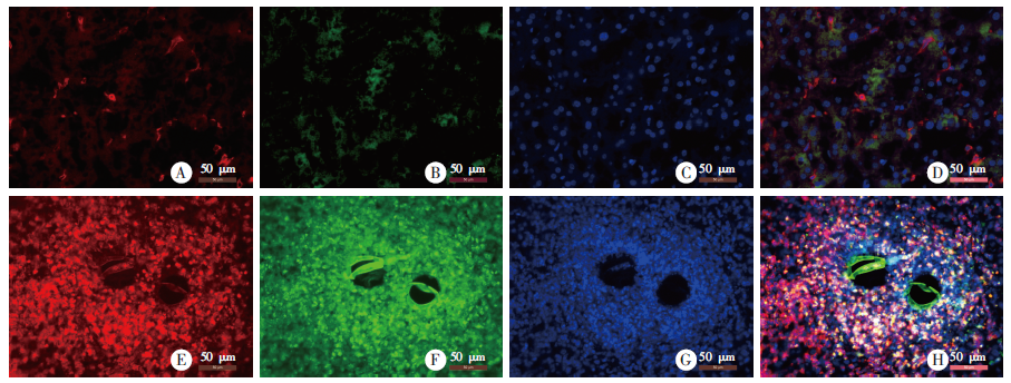
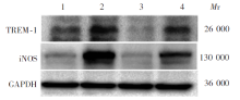
Fig. 3
Effects on expression of iNOS in RAW264.7 macrophage cells transfected with TREM-1 siRNA 1: NC siRNA group; 2: NC siRNA + SWA group; 3: TREM-1 siRNA group; 4: TREM-1 siRNA + SWA group. TREM-1: Triggering receptor expressed on myeloid cell 1; iNOS: Inducible NO synthase; GAPDH: Glyceraldehyde-3-phosphate dehydrogenase.

| [1] |
Zhang WJ, Fang ZM, Liu WQ. NLRP3 inflammasome activation from Kupffer cells is involved in liver fibrosis of Schistosoma japonicum-infected mice via NF-κB[J]. Parasit Vectors, 2019, 12(1):29.
doi: 10.1186/s13071-018-3223-8 |
| [2] |
Anthony BJ, Ramm GA, McManus DP. Role of resident liver cells in the pathogenesis of schistosomiasis[J]. Trends Parasitol, 2012, 28(12):572-579.
doi: 10.1016/j.pt.2012.09.005 pmid: 23099112 |
| [3] |
Ye Z, Huang S, Zhang Y, et al. Galectins, eosinophiles, and macrophages may contribute to Schistosoma japonicum egg-induced immunopathology in a mouse model[J]. Front Immunol, 2020, 11:146.
doi: 10.3389/fimmu.2020.00146 |
| [4] |
Nguyen-Lefebvre AT, Ajith A, Portik-Dobos V, et al. The innate immune receptor TREM-1 promotes liver injury and fibrosis[J]. J Clin Investig, 2018, 128(11):4870-4883.
doi: 10.1172/JCI98156 |
| [5] |
Zhang X, Yang Y, Zhao Y. Macrophage phenotype and its relationship with renal function in human diabetic nephropathy[J]. PLoS One, 2019, 14(9):e0221991.
doi: 10.1371/journal.pone.0221991 |
| [6] |
Wu J, Li J, Salcedo R, et al. The proinflammatory myeloid cell receptor TREM-1 controls Kupffer cell activation and development of hepatocellular carcinoma[J]. Cancer Res, 2012, 72(16):3977-3986.
doi: 10.1158/0008-5472.CAN-12-0938 |
| [7] | Zhao CS, Qin M, Tan MJ, et al. Effect of praziquantel on impaired renal function in mice with acute infection of Schistosoma japonicum[J]. Chin J Parasitol Parasit Dis, 2021, 39(2):200-209. (in Chinese) |
| ( 赵成思, 秦敏, 谭明娟, 等. 吡喹酮对日本血吸虫急性感染小鼠肾脏功能损伤的影响[J]. 中国寄生虫学与寄生虫病杂志, 2021, 39(2):200-209.) | |
| [8] | Bai X, Yu JL, Wang F, et al. Alternatively activated macrophages in helminth infections[J]. Chin J Parasitol Parasit Dis, 2011, 29(3):219-223. (in Chinese) |
| ( 白雪, 于建立, 王峰, 等. 替代性活化的巨噬细胞在蠕虫感染中的作用[J]. 中国寄生虫学与寄生虫病杂志, 2011, 29(3):219-223.) | |
| [9] | Chen C, Shen WT, Liu Y. Advances in research on the effect of the macrophage microenvironment on liver fibrosis due to schistosomiasis[J]. J Pathog Biol, 2020, 15(2):241-245. (in Chinese) |
| ( 陈聪, 沈文涛, 刘彦. 微环境中巨噬细胞影响血吸虫肝纤维化的研究进展[J]. 中国病原生物学杂志, 2020, 15(2):241-245.) | |
| [10] | Zhou HH, Lyu NY, Tong SJ, et al. Resveratrol regulates M1/M2 polarization of mice infected with Schistosoma japonicum through mitochondria[J]. Chin Pharmacol Bull, 2021, 37(1):98-106. (in Chinese) |
| ( 周慧慧, 吕年银, 佟书娟, 等. 白藜芦醇通过线粒体调控血吸虫感染小鼠M1/M2极化[J]. 中国药理学通报, 2021, 37(1):98-106.) | |
| [11] |
Burke ML, Jones MK, Gobert GN, et al. Immunopathogenesis of human schistosomiasis[J]. Parasite Immunol, 2009, 31(4):163-176.
doi: 10.1111/j.1365-3024.2009.01098.x pmid: 19292768 |
| [12] |
Jenkins SJ, Ruckerl D, Cook PC, et al. Local macrophage proliferation, rather than recruitment from the blood, is a signature of TH2 inflammation[J]. Science, 2011, 332(6035):1284-1288.
doi: 10.1126/science.1204351 |
| [13] |
Asahi H, Stadecker MJ. Analysis of egg antigens inducing hepatic lesions in schistosome infection[J]. Parasitol Int, 2003, 52(4):361-367.
doi: 10.1016/S1383-5769(03)00052-7 |
| [14] |
Zhu J, Xu Z, Chen X, et al. Parasitic antigens alter macrophage polarization during Schistosoma japonicum infection in mice[J]. Parasit Vectors, 2014, 7:122.
doi: 10.1186/1756-3305-7-122 |
| [15] |
Ribeiro de Jesus A, Araújo I, Bacellar O, et al. Human immune responses to Schistosoma mansoni vaccine candidate antigens[J]. Infect Immun, 2000, 68(5):2797-2803.
doi: 10.1128/IAI.68.5.2797-2803.2000 pmid: 10768975 |
| [16] |
Mosser DM, Edwards JP. Exploring the full spectrum of macrophage activation[J]. Nat Rev Immunol, 2008, 8(12):958-969.
doi: 10.1038/nri2448 pmid: 19029990 |
| [17] |
Martinez FO, Sica A, Mantovani A, et al. Macrophage activation and polarization[J]. Front Biosci, 2008, 13:453-461.
doi: 10.2741/2692 |
| [18] |
Martinez FO, Helming L, Gordon S. Alternative activation of macrophages: an immunologic functional perspective[J]. Annu Rev Immunol, 2009, 27:451-483.
doi: 10.1146/immunol.2009.27.issue-1 |
| [19] |
Yang FC, Chiu PY, Chen Y, et al. TREM-1-dependent M1 macrophage polarization restores intestinal epithelium damaged by DSS-induced colitis by activating IL-22-producing innate lymphoid cells[J]. J Biomed Sci, 2019, 26(1):46.
doi: 10.1186/s12929-019-0539-4 |
| [20] | Lao JF, Gong ST, Wu MH, et al. Role of TREM-1 on intestinal anti-tuberculosis immunity[J]. J Trop Med, 2021, 21(5): 566-570, 627, 673. (in Chinese) |
| ( 劳娟凤, 龚四堂, 吴敏昊, 等. 髓系细胞触发受体1增强肠道抗结核感染免疫的机制研究[J]. 热带医学杂志, 2021, 21(5): 566-570, 627, 673.) | |
| [21] |
Klesney-Tait J, Turnbull IR, Colonna M. The TREM receptor family and signal integration[J]. Nat Immunol, 2006, 7(12):1266-1273.
pmid: 17110943 |
| [22] |
El Mezayen R, El Gazzar M, Seeds MC, et al. Endogenous signals released from necrotic cells augment inflammatory responses to bacterial endotoxin[J]. Immunol Lett, 2007, 111(1):36-44.
pmid: 17568691 |
| [23] |
Cheng PC, Lin CN, Chen YJ, et al. Triggering receptor expressed on myeloid cells (TREM)-1 participates in Schistosoma mansoni inflammatory responses[J]. Parasite Immunol, 2011, 33(5):276-286.
doi: 10.1111/j.1365-3024.2011.01284.x pmid: 21332515 |
| [24] | Liu L, Yang Y, Zhou JY, et al. Porphyromonas gingivalis lipopolysaccharide regulates macrophage polarization via triggering receptors expressed on myeloid cells-1[J]. Chin J Stomatol, 2021, 56(2):175-181. (in Chinese) |
| ( 刘琳, 杨芸, 周婕妤, 等. 牙龈卟啉单胞菌脂多糖通过髓样细胞触发受体-1调控巨噬细胞极化状态的研究[J]. 中华口腔医学杂志, 2021, 56(2):175-181.) |
| [1] | TAN Xiao, ZHU Qi, LIU Zhongqi, LI Jia, PENG Dingjin. Immunogenicity of Schistosoma japonicum Sj26gst mRNA vaccine candidate [J]. CHINESE JOURNAL OF PARASITOLOGY AND PARASITIC DISEASES, 2023, 41(5): 546-551. |
| [2] | LIU Huaman, Bikash Giri, FANG Chuantao, ZHENG Yameng, WU Huixin, ZENG Minhao, LI Shan, CHENG Guofeng. Identification of gender associated m6A modified circRNA in Schistosoma japonicum [J]. CHINESE JOURNAL OF PARASITOLOGY AND PARASITIC DISEASES, 2023, 41(5): 552-558. |
| [3] | LU Junxia, XU Junying, ZHAO Bin, WANG Qianwen, LI Wenhua, GENG Yuqing, HOU Jun, WU Xiangwei, CHEN Xueling. Echinococcus granulosus infection induces macrophages to express CD73 and A2AR to suppress inflammatory response [J]. CHINESE JOURNAL OF PARASITOLOGY AND PARASITIC DISEASES, 2023, 41(5): 559-566. |
| [4] | LAN Weiming, XU Hui, XU Yin, QIU Tingting, XIE Shuying, DENG Fenglin, HU Shaoliang, LIU Huan, GUO Jiagang, ZENG Xiaojun. Study on early warning of high risk environment of Schistosoma japonicum infection by quantitative real-time PCR [J]. CHINESE JOURNAL OF PARASITOLOGY AND PARASITIC DISEASES, 2023, 41(4): 502-505. |
| [5] | JIAO Hongjie, QI Wenjing, GUO Gang, BAO Jianling, WU Chuanchuan, SONG Chuanlong, LI Jun, ZHANG Wenbao, YAN Mei. Polarization effect of Echinococcus granulosus antigen B on the mouse macrophage RAW264.7 [J]. CHINESE JOURNAL OF PARASITOLOGY AND PARASITIC DISEASES, 2023, 41(1): 23-28. |
| [6] | WANG Xiao-ling, ZHANG Wei, YI Cun, CHEN Xiang-yu, YANG Wen-bin, XU Bin, HU Wei. The effect of SjGPR89 protein on the growth and development of Schistosoma japonicum [J]. CHINESE JOURNAL OF PARASITOLOGY AND PARASITIC DISEASES, 2022, 40(6): 701-707. |
| [7] | CHEN Guo, ZHU Dan-dan, DUAN Yi-nong. Research progress of immune regulation protein B7 family on immune regulation during Schistosoma japonicum infection [J]. CHINESE JOURNAL OF PARASITOLOGY AND PARASITIC DISEASES, 2022, 40(6): 774-779. |
| [8] | YAN Xiao-lan, WEN Li-yong, XIONG Yan-hong, ZHENG Bin, ZHANG Jian-feng, WANG Tian-ping, YU Li-ling, XU Guo-zhang, LIN Dan-dan, ZHOU Xiao-nong. Interpretation of Criteria for Detection of Antibody against Schistosoma japonicum—Enzyme-linked Immunosorbent Assay [J]. CHINESE JOURNAL OF PARASITOLOGY AND PARASITIC DISEASES, 2022, 40(6): 798-800. |
| [9] | LI Jia-ming, WANG Yi-xuan, YANG Ning-ai, MA Hui-hui, LAN Min, LIU Chun-lan, ZHAO Zhi-jun. Effects of ROP16 protein of Toxoplasma gondii on polarization and apoptosis of MH-S cells and their related mechanisms [J]. CHINESE JOURNAL OF PARASITOLOGY AND PARASITIC DISEASES, 2022, 40(5): 579-586. |
| [10] | TANG Xian-shi, JI Wen-xiang, XIONG Chun-rong, ZHOU Yong-hua, XU Yong-liang, TONG De-sheng. Study on anxiety-like behavior of mice with late-stage infection of Schistosoma japonicum [J]. CHINESE JOURNAL OF PARASITOLOGY AND PARASITIC DISEASES, 2022, 40(5): 622-628. |
| [11] | LIANG Le, ZHANG Jing, SHEN Yu-juan, HU Yuan, CAO Jian-ping. Cyclic guanosine monophosphate-adenosine monophosphate promotes liver egg granuloma formation and fibrosis in mice infected with Schistosoma japonicum [J]. CHINESE JOURNAL OF PARASITOLOGY AND PARASITIC DISEASES, 2022, 40(4): 441-445. |
| [12] | WANG Jie, WEN Hong-yang, CHEN Ying, AN Ran, LUO Qing-li, SHEN Ji-long, DU Jian. Construction and identification of macrophage migration inhibitory factor gene knockout strain of Toxoplasma gondii [J]. CHINESE JOURNAL OF PARASITOLOGY AND PARASITIC DISEASES, 2022, 40(3): 349-354. |
| [13] | GAO Yuan, ZHANG Xiao-cheng, HU Yuan, CAO Jian-ping. Study on the inhibitory effect of natural killer cells on liver fibrosis of schistosomiasis [J]. CHINESE JOURNAL OF PARASITOLOGY AND PARASITIC DISEASES, 2022, 40(2): 168-174. |
| [14] | SUN Ye-ting, JIANG Nan, JIANG Yan-yan, LI Teng, JIANG Xiao-feng, CAO Jian-ping, SHEN Yu-juan. Study on the polarization of MDSC stimulated by Echinococcus granulosus protoscolex-derived exosomes in vitro [J]. CHINESE JOURNAL OF PARASITOLOGY AND PARASITIC DISEASES, 2022, 40(2): 175-180. |
| [15] | ZHANG Ling-hui, CHEN Gen, CHONG Shi-gui, SHEN Hui, MA Hui, ZHAO Yu-min. Research progress on the immune regulation mechanism in alveolar echinococcosis [J]. CHINESE JOURNAL OF PARASITOLOGY AND PARASITIC DISEASES, 2022, 40(1): 109-113. |
| Viewed | ||||||
|
Full text |
|
|||||
|
Abstract |
|
|||||