
CHINESE JOURNAL OF PARASITOLOGY AND PARASITIC DISEASES ›› 2021, Vol. 39 ›› Issue (2): 210-217.doi: 10.12140/j.issn.1000-7423.2021.02.014
• ORIGINAL ARTICLES • Previous Articles Next Articles
QIN Min( ), SHAO Tian-ye, ZHAO Cheng-si, MIAO Ting-ting, ZHANG Rong, QIU Jing-fan, WANG Yong*()
Received:2021-02-07
Revised:2021-03-12
Online:2021-04-30
Published:2021-04-30
Contact:
WANG Yong
E-mail:qm877209100@163.com;yongwsh@njmu.edu.cn
Supported by:CLC Number:
QIN Min, SHAO Tian-ye, ZHAO Cheng-si, MIAO Ting-ting, ZHANG Rong, QIU Jing-fan, WANG Yong. Effects of pyrimethamine on folate level and immune function of macrophages[J]. CHINESE JOURNAL OF PARASITOLOGY AND PARASITIC DISEASES, 2021, 39(2): 210-217.
Add to citation manager EndNote|Ris|BibTeX
URL: https://www.jsczz.cn/EN/10.12140/j.issn.1000-7423.2021.02.014
Table 1
Primer sequences for qRT-PCR
| 基因名称 Gene name | 引物序列(5′→3′) Primer sequence (5′→3′) |
|---|---|
| Mus-GAPDH | F: AGGTCGGTGTGAACGGATTTG |
| R: TGTAGACCATGTAGTTGAGGTCA | |
| Mus-DHFR | F: CGCTCAGGAACGAGTTCAAGT |
| R: TGCCAATTCCGGTTGTTCAATA | |
| Mus-TNF-α | F: CTGTAGCCCACGTCGTAGC |
| R: TTGAGATCCATGCCGTTG | |
| Mus-iNOS | F: GGAGCGAGTTGTGGATTGTC |
| R: GTGAGGGCTTGGCTGAGTGAG | |
| Mus-Arg-1 | F: CAGAAGAATGGAAGAGTCAG |
| R: CAGATATGCAGGGAGTCACC | |
| Mus-IL-1β | F: GCAACTGTTCCTGAACTCAACT |
| R: ATCTTTTGGGGTCCGTCAACT | |
| Mus-IL-10 | F: GACCAGCTGGACAACATACTGCTAA |
| R: GATAAGGCTTGGCAACCCAAGTAA | |
| Mus-TGF-β | F: GCAACAATTCCTGGCGTTACC |
| R: CGAAAGCCCTGTATTCCGTCT | |
| RH-DHFR | F: GGCATCGGCATCAACAAC |
| R: TCAGGCGACTGGCTTCTT |
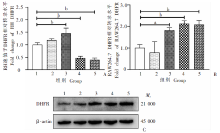
Fig. 1
Effects of pyrimethamine on DHFR A: mRNA levels of tachyzoite DHFR; B: mRNA levels of macrophage DHFR; C: Protein abundance of macrophage DHFR.1-5: The group of 0, 0.10, 1.00, 10.00, 100.00 μmol/L, respectively. a:P < 0.05;b:P < 0.01。
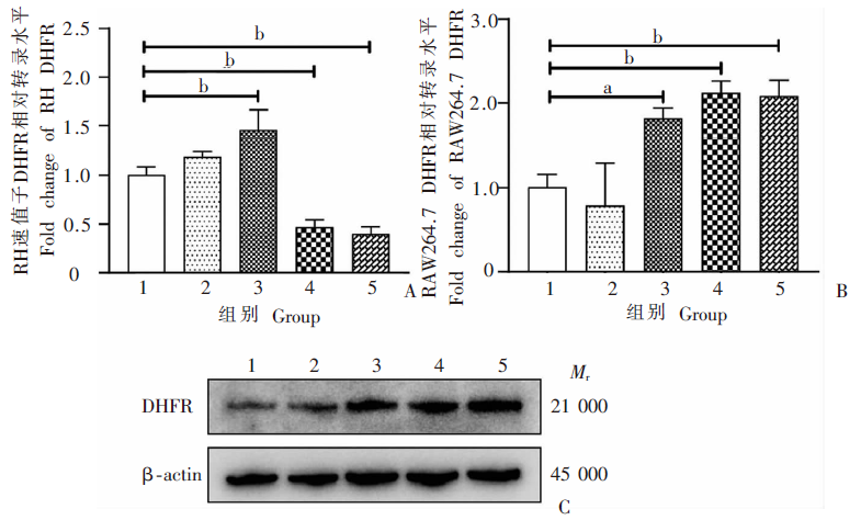
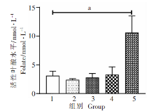
Fig. 2
Effects of pyrimethamine on levels of intracellular folate in macrophages 1-5: The group of 0, 0.10, 1.00, 10.00, 100.00 μmol/L, respectively.a:P < 0.01。
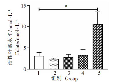
Fig. 3
Effects of pyrimidine on inflammatory gene expression in macrophages A-F: mRNA levels of TNF-α, iNOS, Arg-1, IL-1β, IL-10 and TGF-β, respectively.1-5: The group of 0, 0.10, 1.00, 10.00, 100.00 μmol/L, respectively. a:P < 0.05;b:P < 0.01。
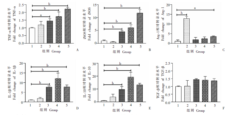
Fig. 4
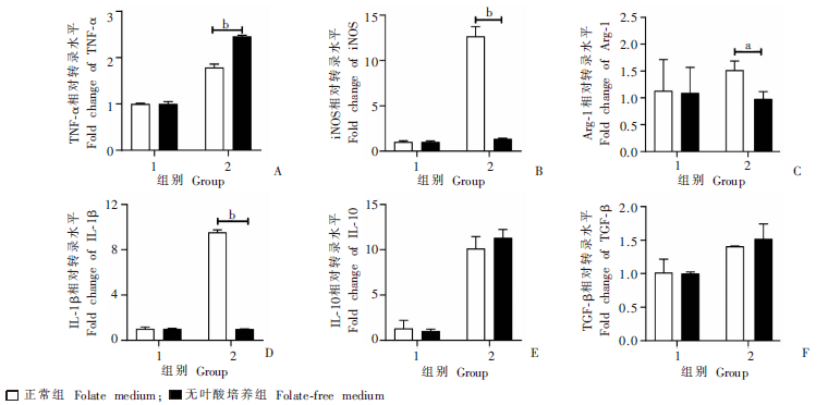
Effects of folate on inflammatory gene expression in macrophages A-F: mRNA levels of TNF-α, iNOS, Arg-1, IL-1β, IL-10 and TGF-β, repectively.
| [1] |
Zhou P, Chen ZG, Li HL, et al. Toxoplasma gondii infection in humans in China[J]. Parasit Vectors, 2011,4:165-173.
doi: 10.1186/1756-3305-4-165 |
| [2] | Wang ZZ, Xu R, Hong Y, et al. Fusion expression and identification of B cell epitopes of Toxoplasma gondii surface antigens (SAG) 1 and 2[J]. Chin J Parasitol Parasit Dis, 2017,35(6):575-579. (in Chinese) |
| ( 王钊哲, 许瑞, 洪炀, 等. 刚地弓形虫表面抗原1、2 B细胞表位基因的融合表达和鉴定[J]. 中国寄生虫学与寄生虫病杂志, 2017,35(6):575-579.) | |
| [3] |
Lewis JM, Clifford S, Nsutebu E. Toxoplasmosis in immunosuppressed patients[J]. Rheumatology, 2015,54(11):1939-1940.
doi: 10.1093/rheumatology/kev115 |
| [4] | Xie ZX, Jiang J, Wang WH, et al. Construction of the RSepitope-HPV16L1 fusion protein using the dominant epitope of ROP2-SAG1 antigen from Toxoplasma gondii and late structural protein 1 of HPV type 16 and its expression in eukaryotic cells[J]. Chin J Parasitol Parasit Dis, 2018,36(3):266-271. (in Chinese) |
| ( 谢自新, 姜洁, 汪文寰, 等. 弓形虫复合抗原ROP2-SAG1优势表位与人乳头瘤病毒16型晚期结构蛋白L1融合体的构建及其在真核细胞中的表达[J]. 中国寄生虫学与寄生虫病杂志, 2018,36(3):266-271.) | |
| [5] | Ma YL, Lyu FL. The role of type Ⅰ interferon in protozoan infections[J]. Chin J Parasitol Parasit Dis, 2018,36(4):399-404. (in Chinese) |
| ( 马远林, 吕芳丽. Ⅰ型干扰素在原虫感染中的作用[J]. 中国寄生虫学与寄生虫病杂志, 2018,36(4):399-404.) | |
| [6] |
Montazeri M, Sharif M, Sarvi S, et al. A systematic review of in vitro and in vivo activities of anti-Toxoplasma drugs and compounds (2006—2016)[J]. Front Microbiol, 2017,8:25-55.
doi: 10.3389/fmicb.2017.00025 pmid: 28163699 |
| [7] |
Meneceur P, Bouldouyre MA, Aubert D, et al. In vitro susceptibility of various genotypic strains of Toxoplasma gondii to pyrimethamine, sulfadiazine, and atovaquone[J]. Antimicrob Agents Chemother, 2008,52(4):1269-1277.
doi: 10.1128/AAC.01203-07 |
| [8] |
Martins-Duarte ÉS, de Souza W, Vommaro RC. Toxoplasma gondii: the effect of fluconazole combined with sulfadiazine and pyrimethamine against acute toxoplasmosis in murine model[J]. Exp Parasitol, 2013,133(3):294-299.
doi: 10.1016/j.exppara.2012.12.011 |
| [9] |
Borkowski PK, Brydak-Godowska J, Basiak W, et al. Adverse reactions in antifolate-treated toxoplasmic retinochoroiditis[J]. Adv Exp Med Biol, 2018,1108:37-48.
doi: 10.1007/5584_2018_262 pmid: 30191431 |
| [10] |
Ben-Harari RR, Goodwin E, Casoy J. Adverse event profile of pyrimethamine-based therapy in toxoplasmosis: a systematic review[J]. Drugs R D, 2017,17(4):523-544.
doi: 10.1007/s40268-017-0206-8 pmid: 28879584 |
| [11] |
Khan Assir MZ, Ahmad HI, Akram J, et al. An outbreak of pyrimethamine toxicity in patients with ischaemic heart disease in Pakistan[J]. Basic Clin Pharmacol Toxicol, 2014,115(3):291-296.
doi: 10.1111/bcpt.2014.115.issue-3 |
| [12] |
Maggini S, Wintergerst ES, Beveridge S, et al. Selected vitamins and trace elements support immune function by strengthening epithelial barriers and cellular and humoral immune responses[J]. Br J Nutr, 2007,98(Suppl 1):S29-S35.
doi: 10.1017/S0007114507832971 |
| [13] |
Troen AM, Mitchell B, Sorensen B, et al. Unmetabolized folic acid in plasma is associated with reduced natural killer cell cytotoxicity among postmenopausal women[J]. J Nutr, 2006,136(1):189-194.
pmid: 16365081 |
| [14] | Protiva P, Mason JB, Liu ZH, et al. Altered folate availability modifies the molecular environment of the human colorectum: implications for colorectal carcinogenesis[J]. Cancer Prev Res(Phila Pa), 2011,4(4):530-543. |
| [15] |
Meadows DN, Bahous RH, Best AF, et al. High dietary folate in mice alters immune response and reduces survival after malarial infection[J]. PLoS One, 2015,10(11):e0143738.
doi: 10.1371/journal.pone.0143738 |
| [16] |
Klee GG. Cobalamin and folate evaluation: measurement of methylmalonic acid and homocysteine vs vitamin B(12) and folate[J]. Clin Chem, 2000,46(8 pt 2):1277-1283.
pmid: 10926922 |
| [17] |
Kolb AF, Petrie L, Mayer CD, et al. Folate deficiency promotes differentiation of vascular smooth muscle cells without affecting the methylation status of regulated genes[J]. Biochem J, 2019,476(19):2769-2795.
doi: 10.1042/BCJ20190275 |
| [18] |
Ling YM, Shaw MH, Ayala C, et al. Vacuolar and plasma membrane stripping and autophagic elimination of Toxoplasma gondii in primed effector macrophages[J]. J Exp Med, 2006,203(9):2063-2071.
doi: 10.1084/jem.20061318 |
| [19] | Du KG, Zhuo XH, Lu SH. Research advances on the innate immunity mechanisms against Toxoplasma gondii[J]. Chin J Parasitol Parasit Dis, 2020,38(6):764-770. (in Chinese) |
| ( 杜凯歌, 卓洵辉, 陆绍红. 抗弓形虫固有免疫机制研究进展[J]. 中国寄生虫学与寄生虫病杂志, 2020,38(6):764-770.) | |
| [20] | Ying W, Cheruku PS, Bazer FW, et al. Investigation of macrophage polarization using bone marrow derived macrophages[J]. J Vis Exp, 2013(76):50323. |
| [21] |
Zhang AM, Shen Q, Li M, et al. Comparative studies of macrophage-biased responses in mice to infection with Toxoplasma gondii ToxoDB #9 strains of different virulence isolated from China[J]. Parasit Vectors, 2013,6(1):308-319.
doi: 10.1186/1756-3305-6-308 |
| [22] | Xu ZP, Zuo GP, Jin JL. Macrophage heterogeneity and its research progress in inflammation regulation[J]. Chin J Cell Mol Immunol, 2015,31(12):1711-1714. (in Chinese) |
| ( 徐志鹏, 左国平, 靳建亮. 巨噬细胞异质性及其在炎症调控中的研究进展[J]. 细胞与分子免疫学杂志, 2015,31(12):1711-1714.) | |
| [23] | Lipka B, Milewska-Bobula B, Filipek M. Monitoring of plasma concentration of pyrimethamine (PYR) in infants with congenital Toxoplasma gondii infection: own observations[J]. Wiad Parazytol, 2011,57(2):87-92. |
| [24] |
Reiter-Owona I, Hlobil H, Enders M, et al. Sulfadiazine plasma concentrations in women with pregnancy-acquired compared to ocular toxoplasmosis under pyrimethamine and sulfadiazine therapy: a case-control study[J]. Eur J Med Res, 2020,25(1):59-67.
doi: 10.1186/s40001-020-00458-7 |
| [25] | Wu XP. Study on pharmacokinetics and residues of pyrimethamine in eel[D]. Wuhan: Huazhong Agricultural University, 2008: 1-27. (in Chinese) |
| ( 吴小平. 乙胺嘧啶在鳗鲡体内的药物动力学和残留研究[D]. 武汉: 华中农业大学, 2008: 1-27.) | |
| [26] |
Jainkittivong A, Arenholt-Bindslev D, Jepsen A, et al. Effect of folic acid on human oral epithelium in vitro[J]. J Dent Assoc Thai, 1989,39(4):121-127.
pmid: 2637902 |
| [27] |
Boot MJ, Steegers-Theunissen RP, Poelmann RE, et al. Folic acid and homocysteine affect neural crest and neuroepithelial cell outgrowth and differentiation in vitro[J]. Dev Dyn, 2003,227(2):301-308.
doi: 10.1002/(ISSN)1097-0177 |
| [28] |
Luo SH, Zhang XM, Yu M, et al. Folic acid acts through DNA methyltransferases to induce the differentiation of neural stem cells into neurons[J]. Cell Biochem Biophys, 2013,66(3):559-566.
doi: 10.1007/s12013-012-9503-6 |
| [29] |
de Leeuw VC, van Nieuwland M, Bokkers BGH, et al. Culture conditions affect chemical-induced developmental toxicity in vitro: the case of folic acid, methionine and methotrexate in the neural embryonic stem cell test[J]. Altern Lab Anim, 2020,48(4):173-183.
doi: 10.1177/0261192920961963 |
| [30] |
Hwang SY, Kang YJ, Sung B, et al. Folic acid promotes the myogenic differentiation of C2C12 murine myoblasts through the Akt signaling pathway[J]. Int J Mol Med, 2015,36(4):1073-1080.
doi: 10.3892/ijmm.2015.2311 |
| [31] | Kong DL, Zhou CL, Guo HF, et al. Praziquantel targets M1 macrophages and ameliorates splenomegaly in chronic schistosomiasis[J]. Antimicrob Agents Chemother, 2018,62(1):e00005-e00017. |
| [32] | Wang W, Zhao CS, Miao TT, et al. Praziquantel inhibits splenic macrophage proliferation and inflammatory reaction in mice infected with Schistosoma japonicum[J]. Chin J Parasitol Parasit Dis, 2020,38(3):263-270. (in Chinese) |
| ( 王伟, 赵成思, 缪婷婷, 等. 吡喹酮治疗对日本血吸虫感染小鼠脾巨噬细胞增殖和炎症反应的抑制作用[J]. 中国寄生虫学与寄生虫病杂志, 2020,38(3):263-270.) |
| [1] | LU Junxia, XU Junying, ZHAO Bin, WANG Qianwen, LI Wenhua, GENG Yuqing, HOU Jun, WU Xiangwei, CHEN Xueling. Echinococcus granulosus infection induces macrophages to express CD73 and A2AR to suppress inflammatory response [J]. CHINESE JOURNAL OF PARASITOLOGY AND PARASITIC DISEASES, 2023, 41(5): 559-566. |
| [2] | JIAO Hongjie, QI Wenjing, GUO Gang, BAO Jianling, WU Chuanchuan, SONG Chuanlong, LI Jun, ZHANG Wenbao, YAN Mei. Polarization effect of Echinococcus granulosus antigen B on the mouse macrophage RAW264.7 [J]. CHINESE JOURNAL OF PARASITOLOGY AND PARASITIC DISEASES, 2023, 41(1): 23-28. |
| [3] | LI Jia-ming, WANG Yi-xuan, YANG Ning-ai, MA Hui-hui, LAN Min, LIU Chun-lan, ZHAO Zhi-jun. Effects of ROP16 protein of Toxoplasma gondii on polarization and apoptosis of MH-S cells and their related mechanisms [J]. CHINESE JOURNAL OF PARASITOLOGY AND PARASITIC DISEASES, 2022, 40(5): 579-586. |
| [4] | WANG Jie, WEN Hong-yang, CHEN Ying, AN Ran, LUO Qing-li, SHEN Ji-long, DU Jian. Construction and identification of macrophage migration inhibitory factor gene knockout strain of Toxoplasma gondii [J]. CHINESE JOURNAL OF PARASITOLOGY AND PARASITIC DISEASES, 2022, 40(3): 349-354. |
| [5] | SUN Ye-ting, JIANG Nan, JIANG Yan-yan, LI Teng, JIANG Xiao-feng, CAO Jian-ping, SHEN Yu-juan. Study on the polarization of MDSC stimulated by Echinococcus granulosus protoscolex-derived exosomes in vitro [J]. CHINESE JOURNAL OF PARASITOLOGY AND PARASITIC DISEASES, 2022, 40(2): 175-180. |
| [6] | ZHANG Ling-hui, CHEN Gen, CHONG Shi-gui, SHEN Hui, MA Hui, ZHAO Yu-min. Research progress on the immune regulation mechanism in alveolar echinococcosis [J]. CHINESE JOURNAL OF PARASITOLOGY AND PARASITIC DISEASES, 2022, 40(1): 109-113. |
| [7] | HOU Jiao, WEN Hao, WANG Ming-kun, LI Wen-ding, LI liang, LI Jing, ZHANG Chuan-shan, WANG Hui. Changes of macrophage subsets and polarization in spleen of mice infected with Echinococcus multilocularis [J]. CHINESE JOURNAL OF PARASITOLOGY AND PARASITIC DISEASES, 2021, 39(6): 771-778. |
| [8] | HUANG Ai-long, ZHANG Bei, SHEN Han-yu, CHEN Guo, LI Jing, ZHU Dan-dan, DUAN Yi-nong. Expression and function of triggering receptor expressed on myeloid cells 1 in the liver of mice infected with Schistosoma japonicum [J]. CHINESE JOURNAL OF PARASITOLOGY AND PARASITIC DISEASES, 2021, 39(5): 621-626. |
| [9] | ZHAO Cheng-si, QIN Min, TAN Ming-juan, MIAO Ting-ting, SHAO Tian-ye, LIU Xin-jian, WANG Yong. Effect of praziquantel on impaired renal function in mice with acute infection of Schistosoma japonicum [J]. CHINESE JOURNAL OF PARASITOLOGY AND PARASITIC DISEASES, 2021, 39(2): 200-209. |
| [10] | WANG Wei, ZHAO Cheng-si, MIAO Ting-ting, ZHOU Chun-lei, ZHANG Cheng-cheng, QIN Min, SHAO Tian-ye, WANG Yong. Praziquantel inhibits splenic macrophage proliferation and inflammatory reaction in mice infected with Schistosoma japonicum [J]. CHINESE JOURNAL OF PARASITOLOGY AND PARASITIC DISEASES, 2020, 38(3): 263-270. |
| [11] | YAN Lan-zhu1,2, SHI Xiao-meng1, ZU Yan-wen1, CHEN Xi-xi3, . Cloning, expression of the gene for proteasome α5 subunit of Angiostrongylus cantonensis and the effect of the recombinant protein on human THP-1 macrophages apoptosis [J]. , 2018, 36(3): 9-253-257. |
| [12] | Ying DONG, Yan DENG, Meng-ni CHEN, Yan-chun XU, Xiang-hua MAO, Jian WANG, Ai-ming SUN, Jing-bo XUE. Analysis of genes associated with antifolate drug resistance in Plasmodium vivax from different infection sources [J]. CHINESE JOURNAL OF PARASITOLOGY AND PARASITIC DISEASES, 2018, 36(2): 103-111. |
| [13] | PAN Wei1,2, ZHANG Yu-mei2,3, SUN Fen-fen4, CAO Jian-ping2, SHEN Yu-juan2*. Changes of Phenotype and Phagocytosis of Peritoneal Macrophages in Mice Infected with the Larval-stage of Echinococcus granulosus [J]. , 2016, 34(4): 4-315-318. |
| [14] | FANG Yan, CHEN Qing, TUN Chen-Yun, WANG Zhao-Jun-*. Role of Macrophages in Schistosome Infection [J]. , 2014, 32(4): 14-311-315. |
| [15] | LIANG Le1,LIU Hai-peng2,CAO Jian-ping3 *. Functional Roles of Macrophage Migration Inhibitory Factor in Anti-parasitic Diseases [J]. , 2012, 30(1): 12-56-60. |
| Viewed | ||||||
|
Full text |
|
|||||
|
Abstract |
|
|||||