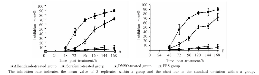
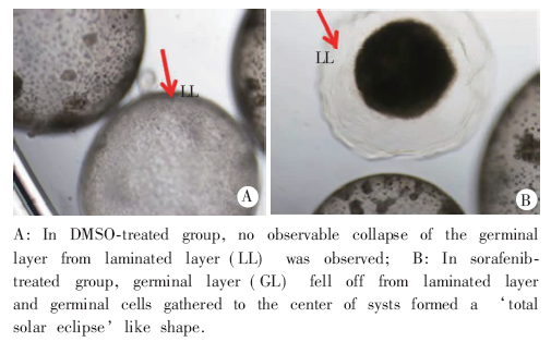
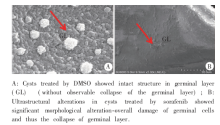
| [1] | Qi YF, Wu WP.Progress on the epidemiology of echinococcosis[J]. Zhongguo Ji Sheng Chong Xue Yu Ji Sheng Chong Bing Za Zhi, 2013, 31(2): 143-148. | | [2] | Cadavid Restrepo AM, Yang YR, McManus DP, et al. The landscape epidemiology of echinococcosis[J]. Infect Dis Poverty, 2016, 5: 13. | | [3] | Xiao N, Yao JW, Ding W, et al. Priorities for research and control of cestode zoonoses in Asia[J]. Infect Dis Poverty, 2013, 2: 16. | | [4] | Wang Q, Huang Y, Huang L, et al. Review of risk factors for human echinococcosis prevalence on the Qinghai-Tibet Plateau, China: a prospective for control options[J]. Infect Dis Poverty, 2014, 3: 3. | | [5] | Wang Q, Yu WJ, Zhong B, et al. Seasonal pattern of Echinococcus re-infection in owned dogs in Tibetan communities of Sichuan, China and its implications for control[J]. Infect Dis Poverty, 2016, 5: 60. | | [6] | Brehm K.The role of evolutionarily conserved signalling systems in Echinococcus multilocularis development and host-parasite interaction[J]. Med Microbiol Immunol, 2010, 199(3): 247-259. | | [7] | Dissous C, Ahier A, Khayath N.Protein tyrosine kinases as new potential targets against human schistosomiasis[J]. Bioessays, 2007, 29(12): 1281-1288. | | [8] | Hemer S, Brehm K.In vitro efficacy of the anticancer drug imatinib on Echinococcus multilocularis larvae[J]. Int J Antimicrob Agents, 2012, 40(5): 458-462. | | [9] | Brumlik MJ, Nkhoma S, Kious MJ, et al. Human p38 mitogen-activated protein kinase inhibitor drugs inhibit Plasmodium falciparum replication[J]. Exp Parasitol, 2011, 128(2): 170-175. | | [10] | Junghae M, Raynes JG.Activation of p38 mitogen-activated protein kinase attenuates Leishmania donovani infection in macrophages[J]. Infect Immun, 2002, 70(9): 5026-5035. | | [11] | Wei S, Marches F, Daniel B, et al. Pyridinylimidazole p38 mitogen-activated protein kinase inhibitors block intracellular Toxoplasma gondii replication[J]. Int J Parasitol, 2002, 32(8): 969-977. | | [12] | Lv H, Li S, Zhang J, et al. In vitro effects of SB202190 on Echinococcus granulosus[J]. Korean J Parasitol, 2013, 51(2): 255-258. | | [13] | Gelmedin V, Caballero-Gamiz R, Brehm K.Characterization and inhibition of a p38-like mitogen-activated protein kinase (MAPK) from Echinococcus multilocularis: antiparasitic activities of p38 MAPK inhibitors[J]. Biochem Pharmacol, 2008, 76(9): 1068-1081. | | [14] | Umemiya-Shirafuji R, Tanaka T, Boldbaatar D, et al. Akt is an essential player in regulating cell/organ growth at the adult stage in the hard tick Haemaphysalis longicornis[J]. Insect Biochem Mol Biol, 2012, 42(3): 164-173. | | [15] | Cao XZ, Xiang HL, Quan MF, et al. Inhibition of cell growth by BrMC through inactivation of Akt in HER-2/neu-overexpressing breast cancer cells[J]. Oncol Lett, 2014, 7(5): 1632-1638. | | [16] | Lathia C, Lettieri J, Cihon F, et al. Lack of effect of ketoconazole-mediated CYP3A inhibition on sorafenib clinical pharmacokinetics[J]. Cancer Chemother Pharmacol, 2006, 57(5): 685-692. | | [17] | Gnoth MJ, Sandmann S, Engel K, et al. In vitro to in vivo comparison of the substrate characteristics of sorafenib tosylate toward P-glycoprotein[J]. Drug Metab Dispos, 2010, 38(8): 1341-1346. | | [18] | Klinkert MQ, Heussler V.The use of anticancer drugs in antiparasitic chemotherapy[J]. Mini Rev Med Chem, 2006, 6(2): 131-143. | | [19] | Stadelmann B, Aeschbacher D, Huber C, et al. Profound activity of the anti-cancer drug bortezomib against Echinococcus multilocularis metacestodes identifies the proteasome as a novel drug target for cestodes[J]. PLoS Negl Trop Dis, 2014, 8(12): e3352. | | [20] | Liance M, Nemati F, Bories C, et al. Experience with doxorubicin-bound polyisohexyl-cyanoacrylate nanoparticles on murine alveolar echinococcosis of the liver[J]. Int J Parasitol, 1993, 23(3): 427-429. | | [21] | Küster T, Lense N, Barna F, et al. A new promising application for highly cytotoxic metal compounds: η6-areneruthenium(Ⅱ) phosphite complexes for the treatment of alveolar echinococcosis[J]. J Med Chem, 2012, 55(9): 4178-4188. |
|

 )
)