
CHINESE JOURNAL OF PARASITOLOGY AND PARASITIC DISEASES ›› 2023, Vol. 41 ›› Issue (6): 760-765.doi: 10.12140/j.issn.1000-7423.2023.06.015
• SHORT COMMUNICATIONS • Previous Articles Next Articles
ZHAO Lei1( ), LI Jia1, MO Gang1, LI Chun1, HUANG Guoyang1, PENG Xiaohong1,2,*()
Received:2023-06-21
Revised:2023-07-18
Online:2023-12-30
Published:2023-12-26
Contact:
* E-mail: Supported by:CLC Number:
ZHAO Lei, LI Jia, MO Gang, LI Chun, HUANG Guoyang, PENG Xiaohong. Effect of Clonorchis sinensis infection on hepatic fibrosis and immune regulation in mice[J]. CHINESE JOURNAL OF PARASITOLOGY AND PARASITIC DISEASES, 2023, 41(6): 760-765.
Add to citation manager EndNote|Ris|BibTeX
URL: https://www.jsczz.cn/EN/10.12140/j.issn.1000-7423.2023.06.015
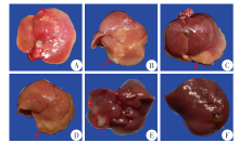

| 小鼠组别 | 感染后时间/周 | 体质量/g | 肝湿重/g | 脾湿重/mg | 脾指数/mg·(10 g)-1 |
|---|---|---|---|---|---|
| 感染组 | 1 | 22.55 ± 0.96 | 1.20 ± 0.12 | 173.3 ± 28.1 | 7.69 ± 1.22 |
| 2 | 23.38 ± 1.22 | 1.43 ± 0.25 | 210.0 ± 42.9 | 8.72 ± 1.05 | |
| 4 | 25.40 ± 0.85 | 1.76 ± 0.66 | 240.0 ± 101.7 | 9.43 ± 3.88 | |
| 8 | 25.97 ± 1.56 | 1.56 ± 0.36 | 181.7 ± 45.8 | 6.95 ± 1.49 | |
| 16 | 25.99 ± 2.23 | 1.20 ± 0.15 | 118.8 ± 15.5 | 4.59 ± 0.62 | |
| 对照组 | 1 | 22.70 ± 1.32 | 1.02 ± 0.35 | 088.6 ± 6.9 | 3.98 ± 0.36 |
| 2 | 23.12 ± 0.88 | 1.03 ± 0.03 | 087.1 ± 7.6 | 3.75 ± 0.38 | |
| 4 | 25.64 ± 1.13 | 1.02 ± 0.03 | 106.0 ± 18.2 | 4.16 ± 0.85 | |
| 8 | 24.80 ± 0.80 | 1.03 ± 0.05 | 091.7 ± 9.8 | 3.69 ± 0.41 | |
| 16 | 27.41 ± 1.34 | 1.13 ± 0.09 | 091.3 ± 9.9 | 3.35 ± 0.26 |
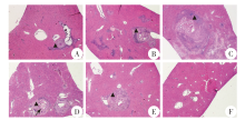


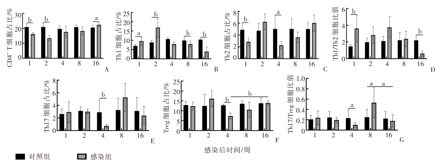
A:TNF;B:IL-6;C:IL-2;D:IFN-γ;E:IL-4;F:IL-10;G:IL-17A。a:P < 0.05,b:P < 0.01。

| [1] |
Qian MB, Utzinger J, Keiser J, et al. Clonorchiasis[J]. Lancet, 2016, 387(10020): 800-810.
doi: 10.1016/S0140-6736(15)60313-0 |
| [2] | Zhao TT, Fang YY, Lai YS. Assessment of the burden of clonorchiasis and its temporal changes in China[J]. Chin J Schisto Control. 2021, 33(2): 162-168. (in Chinese) |
| (赵婷婷, 方悦怡, 赖颖斯. 中国华支睾吸虫病疾病负担估算及其变化趋势分析[J]. 中国血吸虫病防治杂志, 2021, 33(2): 162-168.) | |
| [3] |
Xin HL, Yang YC, Jiang ZH, et al. An investigation of human clonorchiasis prevalence in an endemic county in Guangxi Zhuang Autonomous Region, China, 2016[J]. Food Waterborne Parasitol, 2021, 22: e00109.
doi: 10.1016/j.fawpar.2020.e00109 |
| [4] | Zeng XM, Jiang ZH, Shen JQ, et al. Detection and analysis of fecal egg and serum IgG antibody in 146 suspected cases of Clonorchis sinensis infection in Guangxi[J]. Chin J Parasitol Parasit Dis, 2019, 37(6): 730-732. (in Chinese) |
| (曾雪梅, 蒋智华, 申继清, 等. 广西146例华支睾吸虫疑似感染者粪样虫卵和血清IgG抗体的检测分析[J]. 中国寄生虫学与寄生虫病杂志, 2019, 37(6): 730-732.) | |
| [5] | Huang GH, Zhang B, Lai HT, et al. Analysis on the prevalence of Clonorchis sinensis infection in Lingshan County, Guangxi from 2016 to 2019[J]. Chin J Parasitol Parasit Dis, 2022, 40(5): 673-676. (in Chinese) |
| (黄光华, 张波, 赖海涛, 等. 2016—2019年广西灵山县人群华支睾吸虫感染情况分析[J]. 中国寄生虫学与寄生虫病杂志, 2022, 40(5): 673-676.) | |
| [6] |
Bai XL, Wang N, Zhou J, et al. DX5+ NKT cells’ increase was correlated with liver damage in FVB mice not in BALB/c mice infected by Clonorchis sinensis[J]. Parasite Immunol, 2021, 43(1): e12796.
doi: 10.1111/pim.v43.1 |
| [7] |
Wang N, Bai X, Jin XM, et al. The dynamics of select cellular responses and cytokine expression profiles in mice infected with juvenile Clonorchis sinensis[J]. Acta Trop, 2021, 217: 105852.
doi: 10.1016/j.actatropica.2021.105852 |
| [8] |
Hung KS, Lee TH, Chou WY, et al. Interleukin-10 gene therapy reverses thioacetamide-induced liver fibrosis in mice[J]. Biochem Biophys Res Commun, 2005, 336(1): 324-331.
doi: 10.1016/j.bbrc.2005.08.085 |
| [9] |
Zhou WC, Zhang QB, Qiao L. Pathogenesis of liver cirrhosis[J]. World J Gastroenterol, 2014, 20(23): 7312-7324.
doi: 10.3748/wjg.v20.i23.7312 |
| [10] |
Zhang MJ, Zhang S. T cells in fibrosis and fibrotic diseases[J]. Front Immunol, 2020, 11: 1142.
doi: 10.3389/fimmu.2020.01142 pmid: 32676074 |
| [11] |
Glimcher LH, Murphy KM. Lineage commitment in the immune system: the T helper lymphocyte grows up[J]. Genes Dev, 2000, 14(14): 1693-1711.
doi: 10.1101/gad.14.14.1693 |
| [12] | Elson CO, Cong Y, Brandwein S, et al. Experimental models to study molecular mechanisms underlying intestinal inflammation[J]. Ann N Y Acad Sci, 1998(859): 85-95. |
| [13] |
Lee GR. The balance of Th17 versus Treg cells in autoimmunity[J]. Int J Mol Sci, 2018, 19(3): 730.
doi: 10.3390/ijms19030730 |
| [14] |
Göschl L, Scheinecker C, Bonelli M. Treg cells in autoimmunity: from identification to Treg-based therapies[J]. Semin Immunopathol, 2019, 41(3): 301-314.
doi: 10.1007/s00281-019-00741-8 pmid: 30953162 |
| [15] |
Hanley CJ, Waise S, Ellis MJ, et al. Single-cell analysis reveals prognostic fibroblast subpopulations linked to molecular and immunological subtypes of lung cancer[J]. Nat Commun, 2023, 14(1): 387.
doi: 10.1038/s41467-023-35832-6 pmid: 36720863 |
| [16] | Zhao L, Mo G, Li J, et al. Establishment of a model of hepatic bile duct fibrosis in BALB/c mice infected with Clonorchis sinensis[J]. J Pathog Biol, 2022, 17(10): 1160-1163. (in Chinese) |
| (赵磊, 莫刚, 李佳, 等. 华支睾吸虫感染BALB/c小鼠肝胆管纤维化模型的建立[J]. 中国病原生物学杂志, 2022, 17(10): 1160-1163.) | |
| [17] |
Zhao L, Shi MC, Zhou LN, et al. Clonorchis sinensis adult-derived proteins elicit Th2 immune responses by regulating dendritic cells via mannose receptor[J]. PLoS Negl Trop Dis, 2018, 12(3): e0006251.
doi: 10.1371/journal.pntd.0006251 |
| [18] |
Taylor AE, Carey AN, Kudira R, et al. Interleukin 2 promotes hepatic regulatory T cell responses and protects from biliary fibrosis in murine sclerosing cholangitis[J]. Hepatology, 2018, 68(5): 1905-1921.
doi: 10.1002/hep.30061 pmid: 29698570 |
| [19] |
Yan C, Zhang BB, Hua H, et al. The dynamics of Treg/Th17 and the imbalance of Treg/Th17 in Clonorchis sinensis-infected mice[J]. PLoS One, 2015, 10(11): e0143217.
doi: 10.1371/journal.pone.0143217 |
| [20] |
Dirchwolf M, Podhorzer A, Marino M, et al. Immune dysfunction in cirrhosis: distinct cytokines phenotypes according to cirrhosis severity[J]. Cytokine, 2016, 77: 14-25.
doi: 10.1016/j.cyto.2015.10.006 pmid: 26517154 |
| [21] |
Rey I, Effendi-Ys R. Association between serum IL-6, IL-10, IL-12, and IL-23 levels and severity of liver cirrhosis[J]. Med Arch, 2021, 75(3): 199-203.
doi: 10.5455/medarh.2021.75.199-203 pmid: 34483450 |
| [22] | Velavan TP, Ojurongbe O. Regulatory T cells and parasites[J]. J Biomed Biotechnol, 2011, 2011: 520940. |
| [23] |
Ma X, Hua J, Mohamood AR, et al. A high-fat diet and regulatory T cells influence susceptibility to endotoxin-induced liver injury[J]. Hepatology, 2007, 46(5): 1519-1529.
pmid: 17661402 |
| [1] | LI Xiaoqin, LAI Yashi, CHEN Yu, LV Jiahui, WEI Shuai, ZHANG Lilin, HE Shanshan, SHI Yunliang, LI Yanwen. Ultrastructural observation on excystment of metacercaria of Clonorchis sinensis [J]. CHINESE JOURNAL OF PARASITOLOGY AND PARASITIC DISEASES, 2023, 41(5): 601-608. |
| [2] | XU Yin, LIU Ting, XU Hui, ZENG Xiaojun, LAN Weiming, GONG Zhihong, DAI Kunjiao, QIU Tingting, HAO Xian, XIE Shuying. Establishment and application of PCR-CRISPR/Cas12a-based detection method for Clonorchis sinensis metacercaria [J]. CHINESE JOURNAL OF PARASITOLOGY AND PARASITIC DISEASES, 2023, 41(4): 421-426. |
| [3] | JIANG Xiao-feng, SHEN Yu-juan. Research progress on liver fibrosis caused by Echinococcus infection [J]. CHINESE JOURNAL OF PARASITOLOGY AND PARASITIC DISEASES, 2022, 40(5): 656-660. |
| [4] | HUANG Guang-hua, ZHANG Bo, LAI Hai-tao, Lv Guo-li, LIN Yuan, TANG Wen-qian, WAN Xiao-ling, JIANG Zhi-hua, LIU Jian. Analysis on the prevalence of Clonorchis sinensis infection in Lingshan County, Guangxi from 2016 to 2019 [J]. CHINESE JOURNAL OF PARASITOLOGY AND PARASITIC DISEASES, 2022, 40(5): 673-676. |
| [5] | WANG Ting, YANG Qing-li, LENG Jing, LI Bao-ying, SHEN Ji-qing, DAI Yue. Expression of the high mobility group box 1 homologous protein of Clonorchis sinensis and its effect on nuclear transcription factor-κB activation in mouse macrophages [J]. CHINESE JOURNAL OF PARASITOLOGY AND PARASITIC DISEASES, 2022, 40(3): 305-308. |
| [6] | TANG Lei, YUAN Shuang, YIN Shi-hui, GE Tao, GE Jing-xue, XING Zhi-feng. Investigation on the prevalence of Clonorchis sinensis infection in human population of Heilongjiang Province, 2015 [J]. CHINESE JOURNAL OF PARASITOLOGY AND PARASITIC DISEASES, 2021, 39(6): 741-745. |
| [7] | CHEN Bao-jian, XIE Han-guo, XIE Xian-liang, JIANG Dian-wei, CHEN Yun-hong, GAO Lan-lin. Investigation of Clonorchis sinensis infection in the national surveillance site of Pucheng County, Fujian Province during 2016—2020 [J]. CHINESE JOURNAL OF PARASITOLOGY AND PARASITIC DISEASES, 2021, 39(5): 716-719. |
| [8] | LIU Rong, WEN Li-yong. New progress in basic and clinical research of advanced schistosomiasis [J]. CHINESE JOURNAL OF PARASITOLOGY AND PARASITIC DISEASES, 2021, 39(4): 429-436. |
| [9] | LI An-mei, HUANG Yu-ting, YAO Dan-cheng, ZHU Ai-ya, LI Yang, SHI Wei-fang, DAI Jia-rui, GENG Yan, SHE Dan-ya, ZHANG Nian-heng. Investigation and analysis on the prevalence of Clonorchis sinensis infection in Guizhou Province in 2015 [J]. CHINESE JOURNAL OF PARASITOLOGY AND PARASITIC DISEASES, 2021, 39(1): 125-128. |
| [10] | YUAN Chang-hong, LAN Ming-xing, ZHU Ting-jun, WANG Mei, CHEN Zhe, HU Li-feng, HUANG Qi, JIANG Wei-sheng. Analysis on the status of human infection in national surveillance sites for clonorchiasis in Xinfeng County during 2016-2019 [J]. CHINESE JOURNAL OF PARASITOLOGY AND PARASITIC DISEASES, 2020, 38(5): 561-564. |
| [11] | HUANG Qi, JIANG Wei-sheng, YAN Yue-kang, CHEN Zhe, XIONG Yan-feng, YANG Yu-hua, WANG Shou-shan. Investigation on the status of Clonorchis sinensis infection in Xinfeng County, Jiangxi Province, China [J]. CHINESE JOURNAL OF PARASITOLOGY AND PARASITIC DISEASES, 2020, 38(5): 670-673. |
| [12] | Xiao LI, Jiang-chang WANG, Lei GUAN, Qi GU, Ji-ze DONG, Chen-yun WU, Min YAN, Zhao-jun WANG. Advances in the pathogenesis of cholangiocarcinoma caused by Clonorchis sinensis [J]. CHINESE JOURNAL OF PARASITOLOGY AND PARASITIC DISEASES, 2020, 38(2): 250-254. |
| [13] | Wei-qiang LUO, Xue-liang ZHANG, Zhi-shan ZHOU, Xiu-hong FAN, Xing-ru LI, jie LI. A survey of Clonorchis sinensis and soil-transmitted nematode infections in Qingxin District of Qingyuan City, Guangdong Province [J]. CHINESE JOURNAL OF PARASITOLOGY AND PARASITIC DISEASES, 2019, 37(3): 372-375. |
| [14] | Xin LIU, Yong-fen QI, Yan-rong YU. Effects of soluble egg antigens on hepatic stellate cells in the progression of schistosomiasis-associated liver fibrosis [J]. CHINESE JOURNAL OF PARASITOLOGY AND PARASITIC DISEASES, 2019, 37(2): 218-222. |
| [15] | Meng XU, Wen HUANG, Shen OU, Jian-hai YIN, Sheng-kui CAO, Li-yu MENG, Jian-ping CAO, Yu-juan SHEN. Infection status and molecular identification of Clonorchis sinensis in human population of Tengxian County, Guangxi [J]. CHINESE JOURNAL OF PARASITOLOGY AND PARASITIC DISEASES, 2019, 37(1): 28-32. |
| Viewed | ||||||
|
Full text |
|
|||||
|
Abstract |
|
|||||