
CHINESE JOURNAL OF PARASITOLOGY AND PARASITIC DISEASES ›› 2023, Vol. 41 ›› Issue (5): 601-608.doi: 10.12140/j.issn.1000-7423.2023.05.012
• ORIGINAL ARTICLES • Previous Articles Next Articles
LI Xiaoqin1,2( ), LAI Yashi1, CHEN Yu3, LV Jiahui1, WEI Shuai1,4, ZHANG Lilin1, HE Shanshan1,2, SHI Yunliang1,2, LI Yanwen1,2,*()
Received:2023-03-27
Revised:2023-08-19
Online:2023-10-30
Published:2023-11-06
Contact:
*E-mail: Supported by:CLC Number:
LI Xiaoqin, LAI Yashi, CHEN Yu, LV Jiahui, WEI Shuai, ZHANG Lilin, HE Shanshan, SHI Yunliang, LI Yanwen. Ultrastructural observation on excystment of metacercaria of Clonorchis sinensis[J]. CHINESE JOURNAL OF PARASITOLOGY AND PARASITIC DISEASES, 2023, 41(5): 601-608.

Fig. 1
SEM observation of excystment of C. sinensis A-D: Local swelling or shrinkage observed in the metacercariae (A-C × 1 000, D × 1 440); E-F: Separation of varying degrees is seen between the inner and outer layers of the cyst wall (× 1 500); G-H: The cyst wall exhibits signs of wrinkling and erosive changes (G × 1 000, H × 2 340); i: Enlarged view of figure H (× 4 810); J: The cyst wall has a long tear with smooth edges (× 496); K: The metacercaria is enveloped by the cyst wall, with its anterior spines piercing through the wall (× 1 550); L: The dorsal view of the exysted metacercaria reveals a rough surface with distinct transverse ridges and widespread spines (× 1 500); M: The ventral view of the decapsulated metacestode shows spines covering almost the entire surface, except for a small posterior region. The ventral sucker is noticeably larger than the oral sucker, with its diameter nearly matching the transverse diameter of the body. The inner wall of the ventral sucker displays a longitudinal sphincter muscle structure (× 1 200); N: The inner wall of the oral sucker presents a multi-layered annular ridge-like structure (× 6 500); O: Spines surrounding the ventral sucker have blunt tips, while those further away from the sucker gradually become slender and pointed, with their tips oriented posteriorly (× 4 500); P-Q: The excretory sac is located in the middle-posterior part of the metacercaria and is filled with spherical excretory granules of varying sizes (P × 1 800, Q × 1 500).
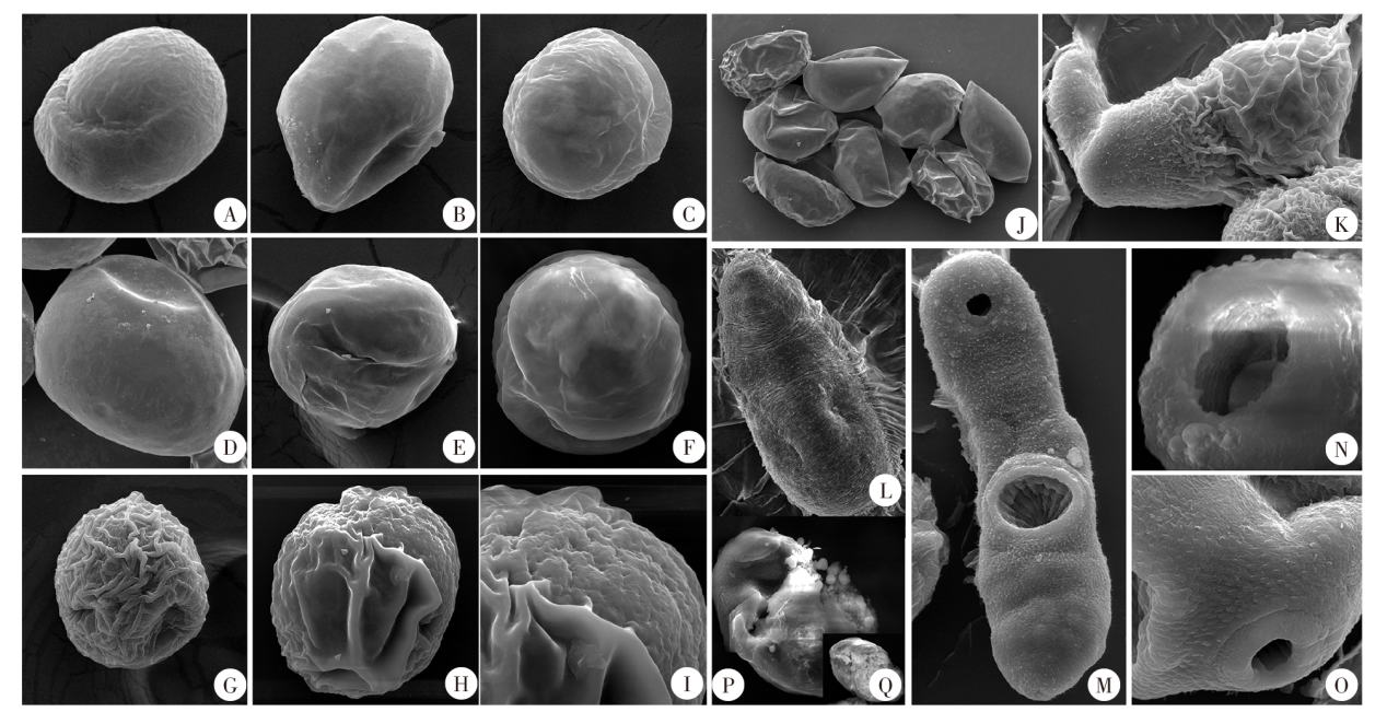

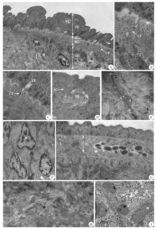
Fig. 2
TEM observation of the structure of excysted metacercaria of Clonorchis sinensis A: The basic structure of the body wall of the excysted metacercariae is divided from the outside to the inside into the tegumental layer (TL), basement layer (BL), muscular layer (ML), and tegumentary cells (TC). Features include spines (S), mitochondrion (M), secretory body (SB), cytoplasm tubules (CT), and cell nucleus (N) (× 8 000); B: Enlarged view of figure A shows the cytoplasmic tubules (CT) in the muscular layer. One can observe mitochondria and secretory granules (× 20 000); C: Vesicles (V) forming at one end of the cytoplasmic tubules, responsible for transporting substances (× 20 000); D: Enlarged view of figure A, depicting large vesicles (V) located within the matrix of the tegumental layer. These vesicles contain elongated secretory granules (SG) (× 40 000); E: Well-developed circular muscles (CM) and longitudinal muscles (LM) are interwoven (× 40 000); F: Deep muscular tissues are found between the tegumental cells, with visible cell nuclei (N) (× 20 000); G: The structure of the basal layer (BL) is loose; in some areas, its width exceeds that of the tegumental layer. It contains secretory vesicles with high electron density (SB) (× 20 000); H: The intercellular matrix is filled with disordered tubular and vesicular structures and mitochondria (× 8 000); I: The mitochondria (M) are tubular and ridge-shaped (× 50 000).
| [1] |
Bouvard V, Baan R, Straif K, et al. A review of human carcinogens: part B- biological agents[J]. Lancet Oncol, 2009, 10(4): 321-322.
doi: 10.1016/S1470-2045(09)70096-8 |
| [2] | National institute of parasitic diseases, Chinese center for disease control and prevention. Report on the national survey of important human parasitic diseases in China (2015)[M]. Beijing: People’s Medical Publishing House, 2018. (in Chinese) |
| (中国疾病预防控制中心寄生虫病预防控制所. 2015年全国人体重点寄生虫病现状调查报告[M]. 北京: 人民卫生出版社, 2018.) | |
| [3] | Huang Y, Zhang GH, Liang MQ, et al. Analysis on the infection status of Clonorchis sinensis among the population in Wuming County, Guangxi[J]. J Appl Prev Med, 2021, 27(3): 197-200. (in Chinese) |
| (黄勇, 张国汉, 梁美群, 等. 广西武鸣县人群华支睾吸虫感染情况分析[J]. 应用预防医学, 2021, 27(3): 197-200.) | |
| [4] | Zeng XM, Liang JM, Wei LL, et al. Survey on infection status of Clonorchis sinensis, Nanning City, 2016—2020[J]. Prev Med Tribune, 2022, 28(3): 169-171. (in Chinese) |
| (曾雪梅, 梁江明, 韦利玲, 等. 2016—2020年南宁市华支睾吸虫感染状况调查[J]. 预防医学论坛, 2022, 28(3): 169-171.) | |
| [5] | Fried B. Metacercarial excystment of trematodes[J]. Adv Parasitol, 1994, 33: 91-144. |
| [6] |
Saxton T, Fried B. An update on metacercarial excystment of trematodes[J]. Parasitol Res, 2009, 105(5): 1185-1191.
doi: 10.1007/s00436-009-1561-3 pmid: 19609562 |
| [7] | Ohyama F. Effects of acid pepsin pretreatment, bile acids and reductants on the excystation of Clonorchis sinensis (Trematoda: Opisthorchiidae) metacercariae in vitro[J]. Parasitol Int, 1998, 47(1): 29-39. |
| [8] |
Chung YB, Kong Y, Joo IJ, et al. Excystment of Paragonimus westermani metacercariae by endogenous cysteine protease[J]. J Parasitol, 1995, 81(2): 137-142.
pmid: 7707186 |
| [9] |
Li SY, Chung YB, Chung BS, et al. The involvement of the cysteine proteases of Clonorchis sinensis metacercariae in excystment[J]. Parasitol Res, 2004, 93(1): 36-40.
doi: 10.1007/s00436-004-1097-5 |
| [10] |
Kaewpitoon N, Laha T, Kaewkes S, et al. Characterization of cysteine proteases from the carcinogenic liver fluke, Opisthorchis viverrini[J]. Parasitol Res, 2008, 102(4): 757-764.
doi: 10.1007/s00436-007-0831-1 pmid: 18092178 |
| [11] | Zheng B, Yang YC, Lu ZC, et al. Establishment of indoor micro-ecological environment for life cycle of Clonorchis sinensis[J]. Chin J Zoonoses, 2019, 35(1): 51-53. (in Chinese) |
| (郑宝, 杨益超, 卢作超, 等. 华支睾吸虫生活史室内微生态建立[J]. 中国人兽共患病学报, 2019, 35(1): 51-53.) | |
| [12] |
Irwin SWB. In vitro excystment of the metacercaria of Maritrema arenaria (Digenea∶Microphallidae)[J]. Int J Parasitol, 1983, 13(2): 191-196.
doi: 10.1016/0020-7519(83)90011-5 |
| [13] |
Li YW, Hu XC, Liu XQ, et al. Molecular cloning and analysis of stage and tissue-specific expression of cathepsin L-like protease from Clonorchis sinensis[J]. Parasitol Res, 2009, 105(2): 447-452.
doi: 10.1007/s00436-009-1406-0 |
| [14] |
Li YW, Huang Y, Hu XC, et al. 41.5-kDa cathepsin L protease from Clonorchis sinensis: expression, characterization, and serological reactivity of one excretory-secretory antigen[J]. Parasitol Res, 2012, 111(2): 673-680.
doi: 10.1007/s00436-012-2885-y |
| [15] |
McDowall AA, James BL. The functional morphology of the circumoral spines of Timoniella imbutiforme (Molin, 1859) Brooks, 1980 (Digenea∶Acanthostomidae)[J]. Int J Parasitol, 1988, 18(4): 523-530.
doi: 10.1016/0020-7519(88)90017-3 |
| [16] | Ye B, Tong XH. Scanning electron microscopic observations on Clonorchis sisnensis juveniles and adults[J]. Chin J Zoonoses, 1996, (6): 17-19. (in Chinese) |
| (叶彬, 童新华. 华支睾吸虫童虫体表发育的扫描电镜观察[J]. 中国人兽共患病杂志, 1996(6): 17-19.) | |
| [17] |
Fujino T, Ishii Y, Choi DW. The ultrastructural characterization of the tegument of Clonorchis sinensis (Cobbold, 1875) cercaria[J]. Z Parasitenkd, 1979, 60(1): 65-76.
doi: 10.1007/BF00928972 |
| [18] | Lee SH, Hong ST, Seo BS. A study on the fine tegumental structures of the metacercaria and juvenile stages of Clonorchis sinensis[J]. Kisaengchunghak Chapchi, 1982, 20(2): 123-132. |
| [19] |
Zhou XX, Xie F, Wang L, et al. The function and clinical application of extracellular vesicles in innate immune regulation[J]. Cell Mol Immunol, 2020, 17(4): 323-334.
doi: 10.1038/s41423-020-0391-1 pmid: 32203193 |
| [20] |
Chaiyadet S, Sotillo J, Smout M, et al. Carcinogenic liver fluke secretes extracellular vesicles that promote cholangiocytes to adopt a tumorigenic phenotype[J]. J Infect Dis, 2015, 212(10): 1636-1645.
doi: 10.1093/infdis/jiv291 pmid: 25985904 |
| [21] |
Simonsen PE, Vennervald BJ, Birch-Andersen A. Echinostoma caproni in mice: ultrastructural studies on the formation of immune complexes on the surface of an intestinal trematode[J]. Int J Parasitol, 1990, 20(7): 935-941.
pmid: 2276867 |
| [22] |
Christensen NO, Knudsen J, Andreassen J. Echinostoma revolutum: resistance to secondary and superimposed infections in mice[J]. Exp Parasitol, 1986, 61(3): 311-318.
pmid: 3709749 |
| [23] |
Hanna RE. Fasciola hepatica : autoradiography of protein synthesis, transport, and secretion by the tegument[J]. Exp Parasitol, 1980, 50(3): 297-304.
pmid: 7428908 |
| [24] | Shi YL, Wan XL Jiang ZH, et al. Scanning electron microscopic and transmission electron microscopic observations of the tegument structure of adult Clonorchis sinensis[J]. Chin J Parasitol Parasit Dis, 2018, 36(2): 184-186. (in Chinese) |
| (石云良, 万孝玲, 蒋智华, 等. 华支睾吸虫成虫体被结构扫描电镜和透射电镜观察[J]. 中国寄生虫学与寄生虫病杂志, 2018, 36(2): 184-186.) | |
| [25] |
Køie M, Nansen P, Christensen NO. Stereoscan studies of rediae, cercariae, cysts, excysted metacercariae and migratory stages of Fasciola hepatica[J]. Z Parasitenkd, 1977, 54(3): 289-297.
doi: 10.1007/BF00390120 |
| [26] |
Hanna RE. Fasciola hepatica: glycocalyx replacement in the juvenile as a possible mechanism for protection against host immunity[J]. Exp Parasitol, 1980, 50(1): 103-114.
pmid: 7389854 |
| [27] |
Threadgold LT, Gallagher SS. Electron microscope studies of Fasciola hepatica: Ⅰ- the ultrastructure and interrelationship of the parenchymal cells[J]. Parasitology, 1966, 56(2): 299-304.
pmid: 5962402 |
| [28] |
Gallagjer SS, Threadgold LT. Electron-microscope studies of Fasciola hepatica: Ⅱ-the interrelationship of the parenchyma with other organ systems[J]. Parasitology, 1967, 57(4): 627-632.
pmid: 5583380 |
| [29] |
Dalton JP, Skelly P, Halton DW. Role of the tegument and gut in nutrient uptake by parasitic platyhelminths[J]. Can J Zool, 2004, 82(2): 211-232.
doi: 10.1139/z03-213 |
| [30] | He M. Cloning expression, histological localization and immunological characteristic study of cysteine protease from Clonorchis sinensis[D]. Nanning: Guangxi Medical University, 2018: 29-32). (in Chinese) |
| (何勉. 华支睾吸虫半胱氨酸蛋白酶克隆表达、组织定位及免疫学特性研究[D]. 南宁: 广西医科大学, 2018: 29-32). |
| [1] | XU Yin, LIU Ting, XU Hui, ZENG Xiaojun, LAN Weiming, GONG Zhihong, DAI Kunjiao, QIU Tingting, HAO Xian, XIE Shuying. Establishment and application of PCR-CRISPR/Cas12a-based detection method for Clonorchis sinensis metacercaria [J]. CHINESE JOURNAL OF PARASITOLOGY AND PARASITIC DISEASES, 2023, 41(4): 421-426. |
| [2] | HUANG Guang-hua, ZHANG Bo, LAI Hai-tao, Lv Guo-li, LIN Yuan, TANG Wen-qian, WAN Xiao-ling, JIANG Zhi-hua, LIU Jian. Analysis on the prevalence of Clonorchis sinensis infection in Lingshan County, Guangxi from 2016 to 2019 [J]. CHINESE JOURNAL OF PARASITOLOGY AND PARASITIC DISEASES, 2022, 40(5): 673-676. |
| [3] | WANG Ting, YANG Qing-li, LENG Jing, LI Bao-ying, SHEN Ji-qing, DAI Yue. Expression of the high mobility group box 1 homologous protein of Clonorchis sinensis and its effect on nuclear transcription factor-κB activation in mouse macrophages [J]. CHINESE JOURNAL OF PARASITOLOGY AND PARASITIC DISEASES, 2022, 40(3): 305-308. |
| [4] | TANG Lei, YUAN Shuang, YIN Shi-hui, GE Tao, GE Jing-xue, XING Zhi-feng. Investigation on the prevalence of Clonorchis sinensis infection in human population of Heilongjiang Province, 2015 [J]. CHINESE JOURNAL OF PARASITOLOGY AND PARASITIC DISEASES, 2021, 39(6): 741-745. |
| [5] | CHEN Bao-jian, XIE Han-guo, XIE Xian-liang, JIANG Dian-wei, CHEN Yun-hong, GAO Lan-lin. Investigation of Clonorchis sinensis infection in the national surveillance site of Pucheng County, Fujian Province during 2016—2020 [J]. CHINESE JOURNAL OF PARASITOLOGY AND PARASITIC DISEASES, 2021, 39(5): 716-719. |
| [6] | LI An-mei, HUANG Yu-ting, YAO Dan-cheng, ZHU Ai-ya, LI Yang, SHI Wei-fang, DAI Jia-rui, GENG Yan, SHE Dan-ya, ZHANG Nian-heng. Investigation and analysis on the prevalence of Clonorchis sinensis infection in Guizhou Province in 2015 [J]. CHINESE JOURNAL OF PARASITOLOGY AND PARASITIC DISEASES, 2021, 39(1): 125-128. |
| [7] | YUAN Chang-hong, LAN Ming-xing, ZHU Ting-jun, WANG Mei, CHEN Zhe, HU Li-feng, HUANG Qi, JIANG Wei-sheng. Analysis on the status of human infection in national surveillance sites for clonorchiasis in Xinfeng County during 2016-2019 [J]. CHINESE JOURNAL OF PARASITOLOGY AND PARASITIC DISEASES, 2020, 38(5): 561-564. |
| [8] | HUANG Qi, JIANG Wei-sheng, YAN Yue-kang, CHEN Zhe, XIONG Yan-feng, YANG Yu-hua, WANG Shou-shan. Investigation on the status of Clonorchis sinensis infection in Xinfeng County, Jiangxi Province, China [J]. CHINESE JOURNAL OF PARASITOLOGY AND PARASITIC DISEASES, 2020, 38(5): 670-673. |
| [9] | Xiao LI, Jiang-chang WANG, Lei GUAN, Qi GU, Ji-ze DONG, Chen-yun WU, Min YAN, Zhao-jun WANG. Advances in the pathogenesis of cholangiocarcinoma caused by Clonorchis sinensis [J]. CHINESE JOURNAL OF PARASITOLOGY AND PARASITIC DISEASES, 2020, 38(2): 250-254. |
| [10] | Wei-qiang LUO, Xue-liang ZHANG, Zhi-shan ZHOU, Xiu-hong FAN, Xing-ru LI, jie LI. A survey of Clonorchis sinensis and soil-transmitted nematode infections in Qingxin District of Qingyuan City, Guangdong Province [J]. CHINESE JOURNAL OF PARASITOLOGY AND PARASITIC DISEASES, 2019, 37(3): 372-375. |
| [11] | Meng XU, Wen HUANG, Shen OU, Jian-hai YIN, Sheng-kui CAO, Li-yu MENG, Jian-ping CAO, Yu-juan SHEN. Infection status and molecular identification of Clonorchis sinensis in human population of Tengxian County, Guangxi [J]. CHINESE JOURNAL OF PARASITOLOGY AND PARASITIC DISEASES, 2019, 37(1): 28-32. |
| [12] | Bin JIANG, Yi TAO, Li-li XU, Le-le HUO, Yu-fen WEI, Ni LIU, Meng YIN, Hao-bing ZHANG. Pharmacokinetics of tribendimidine in rat plasma and bile [J]. CHINESE JOURNAL OF PARASITOLOGY AND PARASITIC DISEASES, 2018, 36(5): 449-454. |
| [13] | Yun-liang SHI, Xiao-ling WAN, Zhi-hua JIANG, Xiao-jing CHENG, Yi-chao YANG. Scanning electron microscopic and transmission electron microscopic observations of the tegument structure of adult Clonorchis sinensis [J]. CHINESE JOURNAL OF PARASITOLOGY AND PARASITIC DISEASES, 2018, 36(2): 184-186. |
| [14] | Zhi-hua JIANG, Qing-li YANG, Yi-chao YANG. Development of cytochrome c oxidase subunit 1 gene-based duplex PCR for identifying Clonorchis sinensis and Haplorchis taichui [J]. CHINESE JOURNAL OF PARASITOLOGY AND PARASITIC DISEASES, 2018, 36(2): 187-189. |
| [15] | Min-xia GU, Yao-jun YU, Jin-chuan ZHANG, Bin WANG, Yi-jiang YU, Cong-han REN. Current status and influencing factors of Clonorchis sinensis infection in rural areas of Ninghai County in Zhejiang Province [J]. CHINESE JOURNAL OF PARASITOLOGY AND PARASITIC DISEASES, 2017, 35(6): 585-587. |
| Viewed | ||||||||||||||||||||||||||||||||||||||||||||||||||
|
Full text 348
|
|
|||||||||||||||||||||||||||||||||||||||||||||||||
|
Abstract 285
|
|
|||||||||||||||||||||||||||||||||||||||||||||||||