中国寄生虫学与寄生虫病杂志 ›› 2026, Vol. 44 ›› Issue (1): 85-93.doi: 10.12140/j.issn.1000-7423.2026.01.013

• 论著 • 上一篇    下一篇

孕期刚地弓形虫感染致滋养层细胞LILRB4表达变化及对妊娠的影响

张帆1()(), 牟汝涛2, 张振东1, 刘现兵1, 张海霞1, 胡雪梅1, 李志丹1,*()()   

  1. 1 滨州医学院免疫学教研室山东烟台 264003
    2 烟台市烟台山医院介入医学科山东烟台 264003
  • 收稿日期:2025-11-12 修回日期:2025-12-23 出版日期:2026-02-28 发布日期:2026-02-14
  • 通讯作者: 李志丹(ORCID:0009-0000-6790-4045),女,博士,讲师,从事生殖免疫与感染免疫相关研究。E-mail:lizhidandemingzi@163.com
  • 作者简介:张帆(ORCID:0009-0004-7639-9641),女,硕士研究生,从事生殖免疫与感染免疫相关研究。E-mail:164156088@qq.com
  • 基金资助:
    山东省医药卫生科技项目(202502070799);山东省自然科学基金(ZR2021QH039)

Changes of LILRB4 expression on trophoblast cells and its impact on pregnancy caused by Toxoplasma gondii infection

ZHANG Fan1()(), MOU Rutao2, ZHANG Zhendong1, LIU Xianbing1, ZHANG Haixia1, HU Xuemei1, LI Zhidan1,*()()   

  1. 1 Department of Immunology, Binzhou Medical University, Yantai 264003, Shandong, China
    2 Department of Interventional Medicine, Yantai Mountain Hospital, Yantai 264003, Shandong, China
  • Received:2025-11-12 Revised:2025-12-23 Online:2026-02-28 Published:2026-02-14
  • Contact: E-mail: lizhidandemingzi@163.com
  • Supported by:
    Shandong Provincial Medical and Health Science and Technology Project(202502070799);Natural Science Foundation of Shandong Province(ZR2021QH039)

摘要:

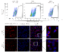
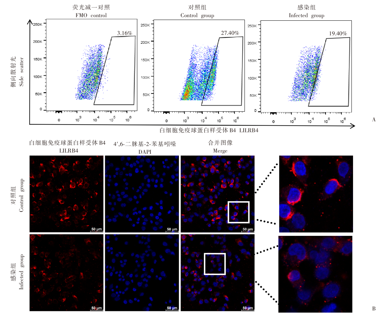
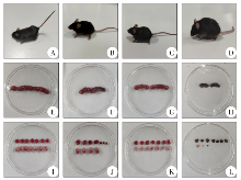
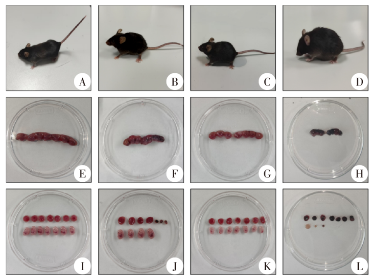
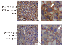
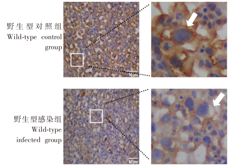
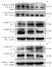
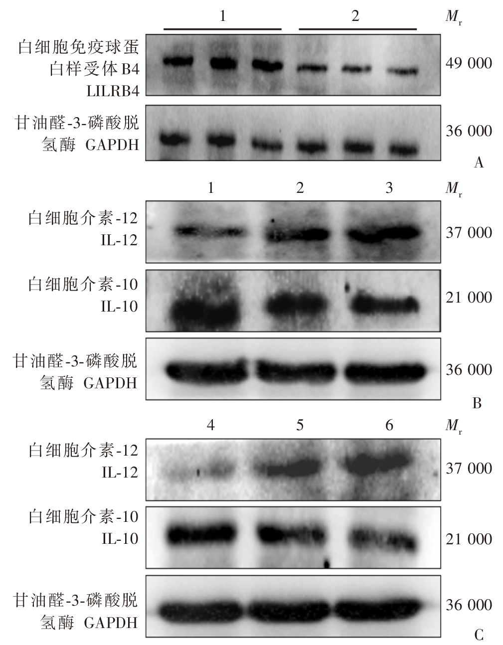
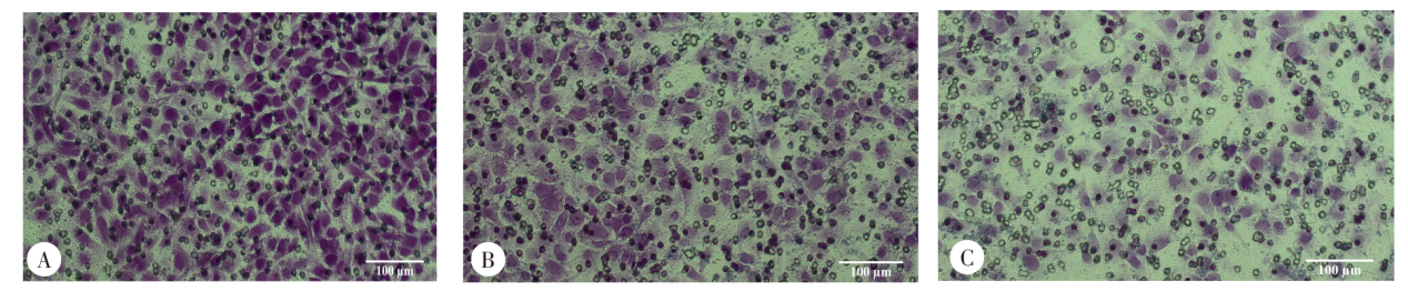
目的 探讨孕期刚地弓形虫感染后滋养层细胞表面白细胞免疫球蛋白样受体B4(LILRB4)的表达水平变化,以及对滋养层细胞功能及妊娠的影响。 方法 将人原代滋养层细胞培养于细胞培养皿中(1 × 10⁷个/皿),分为对照组和感染组。感染组按细胞与弓形虫1:1感染,加入别藻蓝蛋白(APC)标记的抗人LILRB4单克隆抗体,固定破膜后加入别藻蓝蛋白偶联花青7染料(APC-Cy7)标记的抗人细胞角蛋白7(CK7)单克隆抗体和Alexa Fluor 488标记抗人波形蛋白单克隆抗体,流式细胞术检测LILRB4表达情况。取对数生长期的HTR-8/SVneo细胞接种于24孔板中(2 × 105个/孔),分为对照组和感染组。感染组按细胞与弓形虫1:1感染,加入鼠抗人LILRB4单克隆抗体(1:200)、Elab Fluor 647标记的羊抗鼠IgG抗体(1:200)孵育,封片后使用激光共聚焦显微镜拍照,Image J软件分析LILRB4蛋白表达的荧光强度。C57BL/6J野生型雌鼠40只与雄鼠20只、LILRB4-/-雌鼠40只与雄鼠20只分别按雌雄比2:1随机合笼过夜,次晨发现阴栓的雌鼠为孕0 d。野生型孕鼠随机分为野生型对照组和野生型感染组,LILRB4-/-孕鼠随机分为LILRB4-/-对照组和LILRB4-/-感染组,每组6只。孕8 d,野生型感染组和LILRB4-/-感染组孕鼠腹腔注射300个弓形虫速殖子,对照组注射等量生理盐水。孕14 d,解剖各组孕鼠子宫,观察胎盘、胎鼠发育情况。免疫组化检测野生型对照组和野生型感染组小鼠胎盘组织中LILRB4蛋白表达,Image J软件分析LILRB4蛋白阳性表达情况。蛋白质免疫印迹(Western blotting)检测野生型对照组和野生型感染组小鼠胎盘组织中LILRB4蛋白相对表达水平,以及野生型对照组、野生型感染组和LILRB4-/-感染组小鼠胎盘组织中白细胞介素-10(IL-10)和IL-12蛋白的相对表达水平。将HTR-8/SVneo细胞接种于细胞培养皿中(1 × 107个/皿),分为对照组、感染组和LILRB4阻断加感染组,LILRB4阻断加感染组加入LILRB4中和抗体预处理2 h,感染组和LILRB4阻断加感染组按细胞与弓形虫1:1感染,Western blotting检测3组HTR-8/SVneo细胞中IL-10和IL-12蛋白的相对表达水平。Transwell法检测弓形虫感染后HTR-8/SVneo细胞侵袭功能。所有数据均采用GraphPad Prism 9.0软件统计分析,两组比较采用独立样本t检验,多组比较采用单因素方差分析和Tukey事后检验。 结果 流式细胞术结果显示,对照组人原代滋养层细胞中LILRB4阳性细胞比例为(26.10 ± 1.99)%,高于感染组的(18.60 ± 1.13)%(t = 15.00,P < 0.01)。免疫荧光结果显示,感染组LILRB4蛋白的平均荧光强度为122.56 ± 5.24,弱于对照组的149.27 ± 3.50(t = 5.36,P < 0.05)。野生型对照组和LILRB4-/-对照组孕鼠毛发有光泽,精神正常,胎盘和胎鼠发育良好;野生型感染组和LILRB4-/-感染组孕鼠萎靡不振、毛发蓬松粗糙,胎盘缺血且胎鼠发育不良。野生型感染组胎盘和胎鼠体质量分别为(54.82 ± 7.12)和(140.59 ± 3.19)mg,低于野生型对照组的(72.51 ± 1.11)和(201.03 ± 17.37)mg(t = 4.25、5.92,P < 0.05、0.01);LILRB4-/-感染组胎盘和胎鼠体质量分别为(41.24 ± 2.80)和(68.25 ± 11.55)mg,均低于野生型感染组(t = 3.07、10.45,P < 0.05、0.01)。免疫组化结果显示,野生型对照组小鼠胎盘组织内高表达LILRB4蛋白,主要集中在细胞膜,野生型感染组LILRB4蛋白表达较少。野生型感染组LILRB4蛋白阳性表达率为(16.13 ± 2.55)%,低于野生型对照组的(36.64 ± 6.62)%(t = 5.00,P < 0.01)。Western blotting结果显示,野生型对照组小鼠胎盘内LILRB4蛋白相对表达水平为1.15 ± 0.05,高于野生型感染组的0.78 ± 0.10(t = 5.40,P < 0.05)。野生型感染组小鼠胎盘组织内IL-10蛋白相对表达水平为0.93 ± 0.09,低于野生型对照组的1.28 ± 0.16(Tukey事后检验,P < 0.05);LILRB4-/-感染组小鼠胎盘组织内IL-10蛋白相对表达水平为0.61 ± 0.10,低于野生型感染组(Tukey事后检验,P < 0.05)。野生型感染组小鼠胎盘组织内IL-12蛋白相对表达水平为1.08 ± 0.11,高于野生型对照组的0.55 ± 0.18(Tukey事后检验,P < 0.05);LILRB4-/-感染组小鼠胎盘内IL-12蛋白相对表达水平为1.67 ± 0.29,高于野生型感染组(Tukey事后检验,P < 0.05)。感染组HTR-8/SVneo细胞内IL-10蛋白相对表达水平为0.85 ± 0.05,低于对照组的1.15 ± 0.06(Tukey事后检验,P < 0.05);LILRB4阻断加感染组IL-10蛋白相对表达水平为0.72 ± 0.04,低于感染组(Tukey事后检验,P < 0.05)。感染组HTR-8/SVneo细胞内IL-12蛋白相对表达水平为0.89 ± 0.10,高于对照组的0.52 ± 0.14(Tukey事后检验,P < 0.05);LILRB4阻断加感染组IL-12蛋白相对表达水平为1.21 ± 0.04,高于感染组的0.89 ± 0.10(Tukey事后检验,P < 0.05)。感染组HTR-8/SVneo细胞侵袭细胞数为(178 ± 21)个,低于对照组的(278 ± 18)个(t = 45.60,P < 0.01),LILRB4阻断加感染组侵袭细胞数为(119 ± 9)个,较感染组侵袭细胞数量进一步减少(t = 5.50,P < 0.05)。 结论 弓形虫感染可致人滋养层细胞和小鼠胎盘组织LILRB4蛋白表达水平显著下调,LILRB4下调可促进IL-12表达,抑制IL-10产生,减弱滋养层细胞侵袭功能。

关键词: 刚地弓形虫, 滋养层细胞, 白细胞免疫球蛋白样受体B4, 细胞因子, 不良妊娠

Abstract:

Objective To investigate the changes in the expression of leukocyte immunoglobulin-like receptor subfamily B member 4 (LILRB4) on the surface of trophoblast cells following Toxoplasma gondii infection during pregnancy, and to examine its effects on trophoblast cell functions and pregnancy outcomes. Methods Human primary trophoblast cells were incubated in cell culture dishes at a density of 1 × 10⁷ cells per dish and divided into control and infected groups. Cells in the infected group were infected with T. gondii at a ratio of 1:1, incubated with APC-conjugated anti-human LILRB4 monoclonal antibody, fixed, permeabilized, incubated with APC-Cy7-conjugated anti-human cytokeratin 7 (CK7) monoclonal antibody and Alexa Fluor 488-conjugated anti-human vimentin monoclonal antibody, and the expression of LILRB4 was detected by flow cytometry. HTR-8/SVneo cells in the logarithmic growth phase were seeded into 24-well plates at a density of 2 × 10⁵ cells per well and divided into control and infected groups. Cells in the infected group were infected with T. gondii at a ratio of 1:1, incubated with mouse anti-human LILRB4 monoclonal antibody (1:200 dilution) and Elab Fluor 647-conjugated goat anti-mouse IgG monoclonal antibody (1:200 dilution) and enveloped. Images were photographed with a confocal laser scanning microscope, and the immunofluorescence intensity of LILRB4 protein was analyzed using the Image J software. Forty female and 20 male wild-type mice of the C57BL/6J strain and 40 female and 20 male LILRB4-/- mice were randomly caged overnight, and presence of copulatory plugs in female mice on morning of the following day was considered gestational day 0. Wild-type pregnant mice were randomly assigned to wild-type control and infected groups, while LILRB4-/- pregnant mice were randomly divided into LILRB4-/- control and infected groups, with 6 mice in each group. Pregnant mice in wild-type and LILRB4-/- infected groups were intraperitoneally injected with 300 T. gondii tachyzoites on day 8 of gestation, while animals in the control groups received the same volume of physiological saline. On day 14 of gestation, all mice were sacrificed and their uteri were dissected to evaluate placental and fetal development. The expression of LILRB4 protein was detected using immunohistochemistry in mouse placental tissues in wild-type control and infected groups, and the percentage of positive LILRB4 protein expression was analyzed with the software Image J. The relative expression of LILRB4 protein was determined using Western blotting in mouse placental tissues in wild-type control and infected groups. Western blotting was used to detect the relative protein expression levels of interleukin-10 (IL-10) and IL-12 in mouse placental tissues of wild-type control group, wild-type infected group, and LILRB4-/- infected group. HTR-8/SVneo cells were seeded into cell culture dishes at a density of 1 × 10⁷ cells per dish and divided into control, infected, and LILRB4 blockade and infected group. Cells in the LILRB4 blockade and infected group were pretreated with LILRB4 neutralizing antibody for 2 hours, while cells in the infected and LILRB4 blockade and infected groups were infected with T. gondii at a ratio of 1:1. The relative expression of IL-10 and IL-12 proteins was quantified using Western blotting in HTR-8/SVneo cells in control, infected and LILRB4 blockade and infected groups. The invasive ability of HTR-8/SVneo cells was evaluated using Transwell assay following T. gondii infected. All statistical analyses were performed using the software GraphPad Prism 9.0. Difference of means between groups was tested for statistical significance with independent-sample t-test, and multiple-group comparisons were conducted with one-way analysis of variance (ANOVA) followed by Tukey’s post hoc test. Results Flow cytometry detected a higher percentage of LILRB4-positive human primary trophoblast cells in the control group than in the infected group [(26.10 ± 1.99)% vs. (18.60 ± 1.13)%; t = 15.00, P < 0.01], and immunofluorescence staining revealed that the mean fluorescence intensity of LILRB4 protein was lower in the infected group than in the control group [(122.56 ± 5.24) vs. (149.27 ± 3.50); t = 5.36, P < 0.05]. Pregnant mice in the wild-type and LILRB4-/- control groups presented shiny fur, normal mental state, and well placental and fetal development, while pregnant mice in the wild-type and LILRB4-/- infected groups exhibited remarkable low spirits, shaggy and rough fur, placental ischemia, and poor fetal growth. The mouse placental [(54.82 ± 7.12) mg vs. (72.51 ± 1.11) mg; t = 4.25, P < 0.05] and fetal weights [(140.59 ± 3.19) mg vs. (201.03 ± 17.37) mg; t = 5.92, P < 0.01] were lower in the wild-type infected group than in the wild-type control group, and the mouse placental [(41.24 ± 2.80) mg vs. (54.82 ± 7.12) mg; t = 3.07, P < 0.05] and fetal weights [(68.25 ± 11.55) mg vs. (140.59 ± 3.19) mg; t = 10.45, P < 0.01] were lower in the LILRB4-/- infected group than in the wild-type infected group. Immunohistochemical staining showed that the LILRB4 protein was highly positive in mouse placental tissues in the wild-type control group, with positive LILRB4 protein expression primarily found in cell membranes, and low LILRB4 protein expression was found in the wild-type infected group. The proportion of positive LILRB4 protein expression was lower in the wild-type infected group than in the wild-type control group [(16.13 ± 2.55)% vs. (36.64 ± 6.62)%; t = 5.00, P < 0.01]. Western blotting showed that the relative protein expression of LILRB4 in mouse placental tissues was higher in the wild-type control group than in the wild-type infected group [(1.15 ± 0.05) vs. (0.78 ± 0.10); t = 5.40, P < 0.05], and the relative IL-10 protein expression was lower in mouse placental tissues in the wild-type infected group than in the wild-type control group [(0.93 ± 0.09) vs. (1.28 ± 0.16); Tukey’s post hoc test, P < 0.05], and lower in the LILRB4-/- infected group than in the wild-type infected group [(0.61 ± 0.10) vs. (0.93 ± 0.09); Tukey’s post hoc test, P < 0.05]. The relative IL-12 protein expression was higher in mouse placental tissues in the wild-type infected group than in the wild-type control group [(1.08 ± 0.11) vs. (0.55 ± 0.18); Tukey’s post hoc test, P < 0.05], and higher relative IL-12 protein expression was detected in the LILRB4-/- infected group than in the wild-type infected group [(1.67 ± 0.29) vs. (1.08 ± 0.11); Tukey’s post hoc test, P < 0.05]. The relative IL-10 protein expression was lower in HTR-8/SVneo cells in the infected group than in the control group [(0.85 ± 0.05) vs. (1.15 ± 0.06); Tukey’s post hoc test, P < 0.05], and lower in the LILRB4 blockade and infected group (0.72 ± 0.04) than in the infected group (Tukey’s post hoc test, P < 0.05), and the relative IL-12 protein expression was higher in HTR-8/SVneo cells in the infected group than in the control group [(0.89 ± 0.10) vs. (0.52 ± 0.14); Tukey’s post hoc test, P < 0.05], and higher in the LILRB4 blockade and infected group than in the infected group [(1.21 ± 0.04) vs. (0.89 ± 0.10); Tukey’s post hoc test, P < 0.05]. In addition, the counts of invasive HTR-8/SVneo cells were lower in the infected group than in the control group [(178 ± 21) vs. (278 ± 18); t = 45.60, P < 0.01], and lower in the LILRB4 blockade and infected group (119 ± 9) than in the infected group (t = 5.50, P < 0.05). Conclusion T. gondii infection may significantly downregulate LILRB4 protein expression in human trophoblast cells and mouse placental tissues, and downregulation of LILRB4 promotes IL-12 expression and inhibits IL-10 production, thereby attenuating the invasive ability of trophoblast cells.

Key words: Toxoplasma gondii, Trophoblast cells, LILRB4, Cytokine, Adverse pregnancy

中图分类号: