中国寄生虫学与寄生虫病杂志 ›› 2025, Vol. 43 ›› Issue (5): 635-642.doi: 10.12140/j.issn.1000-7423.2025.05.006

• 论著 • 上一篇    下一篇

刚地弓形虫慢性感染诱导小胶质细胞焦亡加剧神经炎症的机制

陈柯旭1()(), 孙彦鑫2, 洪欣雨1, 任立芹1, 李晓冉2, 潘伟3, 张玉梅1,*()()   

  1. 1 滨州医学院病原生物学教研室山东 烟台 264003
    2 滨州医学院第二临床医学院山东 烟台 264003
    3 徐州医科大学病原生物学和免疫学教研室江苏省免疫与代谢重点实验室江苏 徐州 221004
  • 收稿日期:2025-05-21 修回日期:2025-06-03 出版日期:2025-10-30 发布日期:2025-10-23
  • 通讯作者: *张玉梅(ORCID:0000-0001-5089-1225),女,博士,教授,从事弓形虫致脑病的机制研究。E-mail:meiyuzh@163.com
  • 作者简介:陈柯旭(ORCID:0009-0001-2147-7620),女,硕士研究生,从事小胶质细胞焦亡与弓形虫神经炎症的机制研究。E-mail:gg17612973914@163.com
  • 基金资助:
    山东省自然科学基金(ZR2021MH310);大学生创新创业训练计划省级项目(S202410440091);滨州医学院校级教学改革与研究项目(JYKTMS202234)

Mechanism of neuroinflammation exacerbated by microglial pyroptosis induced by chronic Toxoplasma gondii infection

CHEN Kexu1()(), SUN Yanxin2, HONG Xinyu1, REN Liqin1, LI Xiaoran2, PAN Wei3, ZHANG Yumei1,*()()   

  1. 1 Department of Pathogenic Biology, Binzhou Medical University, Yantai 264003, Shandong, China
    2 The Second School of Clinical Medicine, Binzhou Medical University, Yantai 264003, Shandong, China
    3 Department of Pathogen Biology and Immunology, Xuzhou Medical University, Jiangsu Key Laboratory of Immunity and Metabolism, Xuzhou 221004, Jiangsu, China
  • Received:2025-05-21 Revised:2025-06-03 Online:2025-10-30 Published:2025-10-23
  • Contact: *E-mail: meiyuzh@163.com
  • Supported by:
    Natural Science Foundation of Shandong Province(ZR2021MH310);Provincial Project of College Students’Innovation and Entrepreneurship Training Program(S202410440091);Teaching Reform and Research Project of Binzhou Medical University(JYKTMS202234)

摘要:

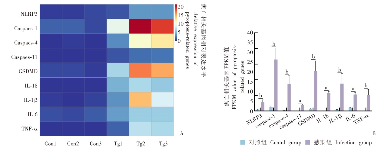
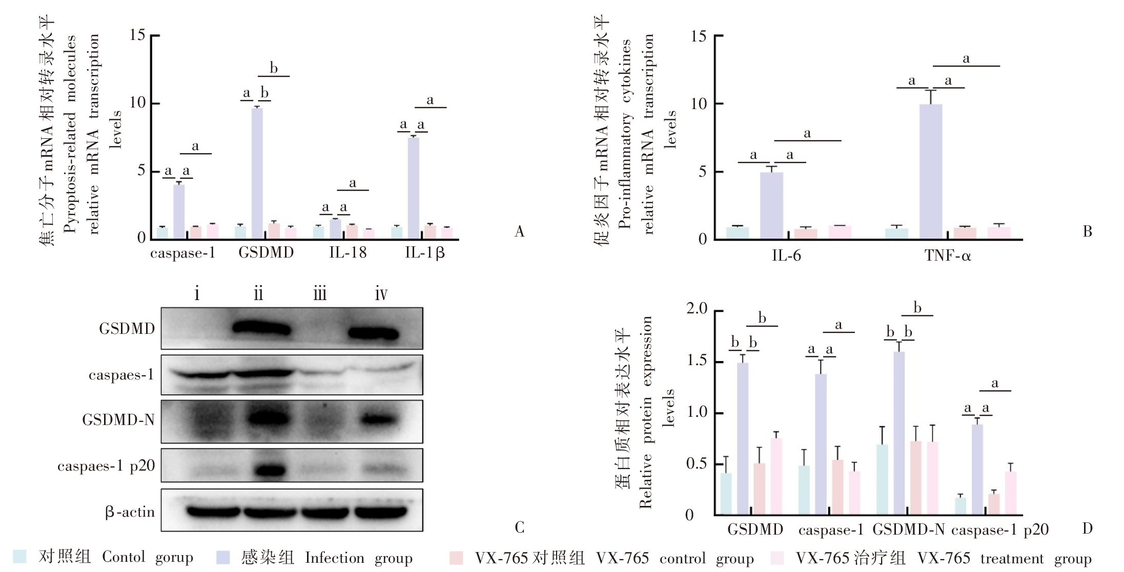
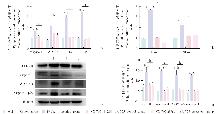
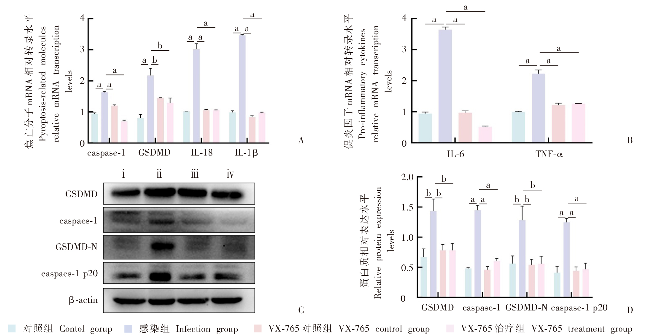
目的 探讨刚地弓形虫慢性感染通过诱导小胶质细胞焦亡加重神经炎症的机制。方法 将20只C57BL/6小鼠随机分为对照组和弓形虫感染组,每组10只,感染组每鼠经口灌胃10个弓形虫Chinese 1型Wh6虫株(TgCtwh6)包囊(悬于200 μl PBS),对照组经口灌胃等量PBS。感染后第6周,收集小鼠脑皮质,通过转录组测序筛选焦亡相关基因的差异表达谱。将60只C57BL/6小鼠随机分为对照组、感染组、VX-765对照组和VX-765治疗组,每组15只,感染组和VX-765治疗组每鼠经口灌胃10个TgCtwh6包囊。感染后第4周起,VX-765对照组和VX-765治疗组每2天腹腔注射1次半胱天冬酶1(caspase-1)抑制剂VX-765(50 mg/kg),共注射7次,感染后第8周收集各组小鼠脑皮质。将小鼠小胶质细胞(BV2细胞)分为对照组、感染组、VX-765对照组和VX-765治疗组,每组5 × 105个细胞,感染组和VX-765治疗组加入等量TgCtwh6速殖子感染,VX-765对照组和VX-765治疗组加入20 μmol/L VX-765处理,培养24 h后收集各组细胞。提取各组小鼠脑皮质和BV2细胞RNA,采用实时荧光定量逆转录PCR(RT-qPCR)检测焦亡分子caspase-1、消皮素D(GSDMD)和促炎因子白细胞介素-18(IL-18)、IL-1β、IL-6、肿瘤坏死因子-α(TNF-α)的表达水平。提取各组小鼠脑皮质和BV2细胞蛋白,采用Western blotting检测caspase-1、caspase-1 p20亚基(caspase-1 p20)、GSDMD、GSDMD N端片段(GSDMD-N)的表达水平。两组间比较采用独立样本t检验,多组间比较采用单因素方差分析和Tukey事后检验。结果 转录组测序显示,感染组小鼠脑皮质中焦亡基因NOD样受体热蛋白结构域相关蛋白3(NLRP3)、caspase-1、caspase-4、GSDMD、IL-1β、IL-18等表达上调,其中caspase-1、GSDMD mRNA相对转录水平(16.48 ± 6.40、12.80 ± 5.62)与对照组(1.33 ± 0.42、0.59 ± 0.20)差异有统计学意义(t = 4.09、3.76,P < 0.05)。RT-qPCR结果显示,感染组小鼠脑皮质中caspase-1、GSDMD、IL-18、IL-1β、IL-6和TNF-α的mRNA相对转录水平分别为4.04 ± 0.38、9.67 ± 0.27、1.49 ± 0.16、7.50 ± 0.27、4.96 ± 0.79、9.97 ± 1.77,均高于对照组的0.88 ± 0.18、1.00 ± 0.26、0.97 ± 0.16、0.94 ± 0.21、0.92 ± 0.22、0.82 ± 0.42(Tukey事后检验,均P < 0.01);VX-765治疗组小鼠caspase-1、GSDMD、IL-18、IL-1β、IL-6和TNF-α的mRNA相对转录水平分别为1.13 ± 0.13、0.87 ± 0.25、0.77 ± 0.05、0.89 ± 0.11、1.03 ± 0.05、0.93 ± 0.43,均较感染组明显下降(Tukey事后检验,均P < 0.01)。Western blotting结果显示,感染组小鼠脑皮质中GSDMD、caspase-1、GSDMD-N、caspase-1 p20的相对表达量分别为1.49 ± 0.14、1.38 ± 0.24、1.60 ± 0.17、0.89 ± 0.11,均高于对照组的0.41 ± 0.29、0.50 ± 0.29、0.70 ± 0.30、0.17 ± 0.06(Tukey事后检验,均P < 0.05);VX-765治疗组小鼠皮质中GSDMD、caspase-1、GSDMD-N、caspase-1 p20的相对表达量分别为0.76 ± 0.11、0.43 ± 0.15、0.72 ± 0.29、0.43 ± 0.14,均较感染组明显减少(Tukey事后检验,均P < 0.05)。RT-qPCR结果显示,感染组BV2细胞中caspase-1、GSDMD、IL-18、IL-1β、IL-6和TNF-α的mRNA相对转录水平分别为1.64 ± 0.03、2.17 ± 0.40、3.01 ± 0.31、3.47 ± 0.05、3.64 ± 0.15、2.23 ± 0.20,均高于对照组的0.94 ± 0.05、0.81 ± 0.21、1.02 ± 0.01、0.99 ± 0.08、0.94 ± 0.09、0.99 ± 0.03(Tukey事后检验,均P < 0.01);VX-765治疗组细胞caspase-1、GSDMD、IL-18、IL-1β、IL-6和TNF-α的mRNA相对转录水平分别为0.70 ± 0.06、1.29 ± 0.27、1.07 ± 0.01、0.98 ± 0.03、0.52 ± 0.03、1.26 ± 0.03,均较感染组明显降低(Tukey事后检验,均P < 0.05)。Western blotting结果显示,感染组BV2细胞中GSDMD、caspase-1、GSDMD-N、caspase-1 p20的相对表达量分别为1.43 ± 0.34、1.45 ± 0.14、1.29 ± 0.40、1.25 ± 0.11,均高于对照组的0.67 ± 0.23、0.48 ± 0.02、0.56 ± 0.22、0.41 ± 0.18(Tukey事后检验,均P < 0.05);VX-765治疗组细胞中GSDMD、caspase-1、GSDMD-N、caspase-1 p20相对表达量分别为0.78 ± 0.20、0.61 ± 0.07、0.56 ± 0.22、0.47 ± 0.17,均较感染组明显减少(Tukey事后检验,均P < 0.05)。结论 弓形虫TgCtwh6慢性感染诱导小胶质细胞发生焦亡,促进促炎因子释放,进而加重神经炎症反应,而VX-765抑制剂能有效抑制焦亡及炎症反应。

关键词: 刚地弓形虫, 小胶质细胞, 细胞焦亡, 神经炎症

Abstract:

Objective To unravel the mechanism of aggravation of neuroinflammation induced by microglial pyroptosis following chronic infection with the Toxoplasma gondii Chinese 1 genotype Wh6 strain (TgCtwh6). Methods Twenty C57BL/6 mice were divided into a control group and an infection group randomly, of 10 mice each group. Mice in the infection group were each administered by oral gavage with 10 TgCtwh6 strain cysts suspended in 200 μl PBS to establish a chronic T. gondii infection model, while mice in the control group received an equal volume of PBS via oral gavage. Mouse cerebral cortex was collected, and the differential expression profiles of pyroptosis-related genes were screened in the mouse cerebral cortex using transcriptome sequencing 6 weeks post-infection. Sixty C57BL/6 mice were divided into a control group, an infection group, a VX-765 control group, and a VX-765 treatment group randomly, of 15 mice each group. Mice in the infection group and VX-765 treatment group were each administered by oral gavage with 10 TgCtwh6 strain cysts. Mice in the VX-765 control group and VX-765 treatment groups were intraperitoneally injected with the caspase-1 inhibitor VX-765 (at a dose of 50 mg/kg) once every 2 days since 4 weeks post-infection for 7 injections, and mouse cerebral cortex was collected from each group 8 weeks post-infection. BV2 mouse microglial cells were assigned to a control group, an infection group, a VX-765 control group, and a VX-765 treatment group, of 5 × 10⁵ cells each group. Cells in the infection group and VX-765 treatment group were infected with an equal amount of TgCtwh6 strain tachyzoites, while cells in the VX-765 control and VX-765 treatment groups were treated with 20 μmol/L VX-765. Cells were harvested 24 h following culture. RNA was extracted from mouse cerebral cortex and BV2 cells, and the relative RNA expression of pyroptosis-related genes caspase-1 and gasdermin D (GSDMD) and pro-inflammatory cytokines interleukin-18 (IL-18), IL-1β, IL-6, tumor necrosis factor-α (TNF-α) were quantified using real-time quantitative reverse transcription PCR (RT-qPCR) assay. Protein was extracted from mouse cerebral cortex and BV2 cells, and the expression of caspase-1, caspase-1 p20 subunit (caspase-1 p20), GSDMD, and GSDMD N-terminal fragment (GSDMD-N) were determined using Western blotting assay. Comparisons of means between groups were done using independent sample t-test, and multi-group comparisons were conducted with one-way analysis of variance (ANOVA) and Tukey’s post hoc test. Results Transcriptome sequencing revealed upregulation of pyroptosis-related genes NOD-like receptor thermal protein domain associated protein 3 (NLRP3), caspase-1, caspase-4, GSDMD, IL-1β and IL-18 expression in the mouse cerebral cortex in the infection group, with relatively higher caspase-1 (16.48 ± 6.40 vs. 1.33 ± 0.42; t = 4.09, P < 0.05) and GSDMD (12.80 ± 5.62 vs. 0.59 ± 0.20; t = 3.76, P < 0.05) expression in the infection group than in the control group. RT-qPCR assay quantified relatively higher mRNA levels of caspase-1 (4.04 ± 0.38 vs. 0.88 ± 0.18), GSDMD (9.67 ± 0.27 vs. 1.00 ± 0.26), IL-18 (1.49 ± 0.16 vs. 0.97 ± 0.16), IL-1β (7.50 ± 0.27 vs. 0.94 ± 0.21), IL-6 (4.96 ± 0.79 vs. 0.92 ± 0.22) and TNF-α (9.97 ± 1.77 vs. 0.82 ± 0.42) in the mouse cerebral cortex in the infection group than in the control group [Tukey’s Honest Significant Difference test (Tukey’s HSD test), all P < 0.01], and lower relative mRNA expression of caspase-1 (1.13 ± 0.13), GSDMD (0.87 ± 0.25), IL-18 (0.77 ± 0.05), IL-1β (0.89 ± 0.11), IL-6 (1.03 ± 0.05), and TNF-α (0.93 ± 0.43) in the VX-765 treatment group than in the infection group (Tukey’s HSD test, all P < 0.05). Western blotting assay determined higher relative expression of GSDMD (1.49 ± 0.14 vs. 0.41 ± 0.29), caspase-1 (1.38 ± 0.24 vs. 0.50 ± 0.29), GSDMD-N (1.60 ± 0.17 vs. 0.70 ± 0.30), and caspase-1 p20 (0.89 ± 0.11 vs. 0.17 ± 0.06) in the mouse cerebral cortex in the infection group than in the control group (Tukey’s HSD test, all P < 0.05), and lower relative expression of GSDMD (0.76 ± 0.11), caspase-1 (0.43 ± 0.15), GSDMD-N (0.72 ± 0.29), and caspase-1 p20 (0.43 ± 0.14) in the VX-765 treatment group than in the infection group (Tukey’s HSD test, all P < 0.05). RT-qPCR assay detected relatively higher mRNA expression of caspase-1 (1.64 ± 0.03 vs. 0.94 ± 0.05), GSDMD (2.17 ± 0.40 vs. 0.81 ± 0.21), IL-18 (3.01 ± 0.31 vs. 1.02 ± 0.01), IL-1β (3.47 ± 0.05 vs. 0.99 ± 0.08), IL-6 (3.64 ± 0.15 vs. 0.94 ± 0.09), and TNF-α (2.23 ± 0.20 vs. 0.99 ± 0.03) in BV2 cells in the infection group than in the control group (Tukey’s HSD test, all P < 0.01), and significantly lower mRNA expression of caspase-1 (0.70 ± 0.06), GSDMD (1.29 ± 0.27), IL-18 (1.07 ± 0.01), IL-1β (0.98 ± 0.03), IL-6 (0.52 ± 0.03), and TNF-α (1.26 ± 0.03) in the VX-765 treatment group than in the infection group (Tukey’s HSD test, all P < 0.05). Western blotting assay determined higher relative expression of GSDMD (1.43 ± 0.34 vs. 0.67 ± 0.23), caspase-1 (1.45 ± 0.14 vs. 0.48 ± 0.02), GSDMD-N (1.29 ± 0.40 vs. 0.56 ± 0.22), and caspase-1 p20 (1.25 ± 0.11 vs. 0.41 ± 0.18) in BV2 cells in the infection group than in the control group (Tukey’s HSD test, all P < 0.05), and lower relative expression of GSDMD (0.78 ± 0.20), caspase-1 (0.61 ± 0.07), GSDMD-N (0.56 ± 0.22), and caspase-1 p20 (0.47 ± 0.17) in the VX-765 treatment group than in the infection group (Tukey’s HSD test, all P < 0.05). Conclusion Chronic infection with the TgCtwh6 strain induces microglial pyroptosis and promotes the release of pro-inflammatory cytokines, thereby exacerbating neuroinflammatory responses, while VX-765 inhibitor is effective to suppress pyroptosis and inflammatory responses.

Key words: Toxoplasma gondii, Microglia, Pyroptosis, Neuroinflammation

中图分类号: