
CHINESE JOURNAL OF PARASITOLOGY AND PARASITIC DISEASES ›› 2024, Vol. 42 ›› Issue (6): 737-743.doi: 10.12140/j.issn.1000-7423.2024.06.007
• ORIGINAL ARTICLES • Previous Articles Next Articles
LI Zhenwei( ), WANG Cheng, WANG Zhixin, LIU Jinming, ZHAO Qian, WANG Haijiu, XIE Zhi*()
Received:2024-08-22
Revised:2024-10-12
Online:2024-12-30
Published:2025-01-14
Contact:
E-mail: Supported by:CLC Number:
LI Zhenwei, WANG Cheng, WANG Zhixin, LIU Jinming, ZHAO Qian, WANG Haijiu, XIE Zhi. Research on the application of 3D visualization-based technology to guide surgical plan for hepatic alveolar echinococcosis[J]. CHINESE JOURNAL OF PARASITOLOGY AND PARASITIC DISEASES, 2024, 42(6): 737-743.
Add to citation manager EndNote|Ris|BibTeX
URL: https://www.jsczz.cn/EN/10.12140/j.issn.1000-7423.2024.06.007
Table 1
Comparison of basic information between patients with hepatic alveolar echinococcosis who underwent/did not undergo 3D visualization technology before surgery
| 项目Project | 研究组患者(n = 50) Patients in research group (n = 50) | 对照组患者(n = 50) Patients in control group (n = 50) | t/χ2 | P |
|---|---|---|---|---|
| 年龄/岁 Age/Year | 43.15 ± 4.37 | 43.23 ± 4.25 | 0.072 | 0.943 |
| 性别 Gender | 0.05 | 0.823 | ||
| 男 Male | 26(52.00%)a | 24(48.00%)a | ||
| 女 Female | 24(48.00%)a | 26(52.00%)a | ||
| 体质量指数/kg•m-2 BMI/kg•m-2 | 23.35 ± 2.98 | 23.41 ± 3.34 | 0.02 | 0.984 |
| 肝多房棘球蚴边缘浸润带 Marginal infiltrative zone of hepatic Echinococcus multilocularis | 0.363 | 0.547 | ||
| 有侵犯 Infringement | 23(46.00%)a | 24(48.00%)a | ||
| 无侵犯 No infringement | 27(54.00%)a | 26(52.00%)a | ||
| 切除部位 Removal site | 1.958 | 0.581 | ||
| 左半肝切除 Left hemihepatectomy | 13(26.00%)a | 14(28.00%)a | ||
| 左半肝切除 + 肝门棘球蚴切除 Left hemihepatectomy + hepatic portal echinococcectomy | 10(20.00%)a | 13(26.00%)a | ||
| 右半肝切除 Right hemihepatectomy | 10(20.00%)a | 7(14.00%)a | ||
| 右半肝切除 + 肝门棘球蚴切除 Right hemihepatectomy + hepatic portal echinococcectomy | 13(26.00%)a | 12(24.00%)a | ||
| 解剖性肝段切除 Anatomical liver segmentectomy | 4(8.00%)a | 4(8.00%)a | ||
| 病灶直径/cm Diameter of lesion/cm | 8.24 ± 1.16 | 8.37 ± 1.22 | 0.001 | 0.970 |
| 切除肝脏体积/ml Liver resection volume/ml | 2 138.75 ± 157.94 | 2 129.42 ± 148.73 | 0.353 | 0.725 |
| 剩余肝脏体积/ml Remaining liver volume/ml | 1 193.46 ± 85.73 | 1 215.63 ± 91.48 | 0.125 | 0.693 |
Table 2
Comparison of imaging data between patients with hepatic alveolar echinococcosis who underwent/did not undergo 3D visualization technology before surgery
| 项目 Project | 研究组例数(占比/%)(n = 50) No. research group case (Proportion/%) (n = 50) | 对照组例数(占比/%)(n = 50) No. control group case (Proportion/%) (n = 50) | t/χ2 | P |
|---|---|---|---|---|
| CT特征 CT feature | ||||
| 分型 Classification | 0.294 | 0.770 | ||
| 结节型 Nodular type | 16(32.00) | 19(38.00) | ||
| 巨块型 Giant block typae | 22(44.00) | 20(40.00) | ||
| 弥漫型 Diffuse type | 12(24.00) | 11(22.00) | ||
| 病灶大小/cm Disease size/cm | 0.214 | 0.648 | ||
| < 5 | 27(54.00) | 25(50.00) | ||
| ≥ 5 | 23(46.00) | 25(50.00) | ||
| 瘤周强化 Peritumoral enhancement | 0.113 | 0.739 | ||
| 是 Yes | 31(62.00) | 29(58.00) | ||
| 否 No | 19(38.00) | 21(42.00) | ||
| 边缘表现 Edge performance | 0.321 | 0.572 | ||
| 光滑 Smooth | 14(28.00) | 13(26.00) | ||
| 不光滑 Not smooth | 36(72.00) | 37(74.00) | ||
| 瘤周低信号 Low signal around the tumor | 0.182 | 0.671 | ||
| 是 Yes | 26(52.00) | 22(44.00) | ||
| 否 No | 24(48.00) | 28(56.00) | ||
| 强化方式 Strengthening method | 0.328 | 0.567 | ||
| 典型 Typical | 27(54.00) | 25(50.00) | ||
| 不典型 Not typical | 23(46.00) | 25(50.00) | ||
| 增强类型 Enhanced type | 0.42 | 0.518 | ||
| 均匀 Uniformity | 18(36.00) | 16(32.00) | ||
| 不均匀 Uneven | 32(64.00) | 34(68.00) | ||
| MRI | ||||
| T1WI | 0.625 | 0.430 | ||
| 稍低信号 Slightly lower signal | 13(26.00) | 11(22.00) | ||
| 等信号 Middle signal | 20(40.00) | 24(48.00) | ||
| 稍高信号 Slightly high signal | 17(34.00) | 15(30.00) | ||
| T2WI | 0.222 | 0.638 | ||
| 稍低信号 Slightly lower signal | 9(18.00) | 13(26.00) | ||
| 等信号 Middle signal | 22(44.00) | 19(38.00) | ||
| 稍高信号 Slightly high signal | 19(38.00) | 18(36.00) |
Table 3
Comparison of perioperative data between patients with hepatic alveolar echinococcosis who underwent/did not undergo 3D visualization technology before surgery
| 项目 Project | 研究组(n = 50) Research group (n = 50) | 对照组(n = 50) Control group (n = 50) | t/χ2 | P |
|---|---|---|---|---|
| 手术时间/min Surgical duration/min | 228.56 ± 29.47 | 284.32 ± 24.89 | 3.856 | <0.001 |
| 出血量/ml Bleeding volume/ml | 435.14 ± 98.76 | 517.45 ± 88.65 | 6.069 | 0.014 |
| 术后住院时间/d Postoperative hospitalization time/d | 15.27 ± 2.13 | 16.43 ± 2.98 | 3.561 | 0.037 |
| Pringle法全肝阻断时间/min Pringle method whole liver block time/min | 26.65 ± 4.78 | 33.98 ± 3.91 | 5.4 | 0.023 |
| 住院费用/万元 Hospitalization expenses/10 000 yuan | 4.58 ± 0.79 | 4.79 ± 0.92 | 2.627 | 0.01 |
| 医患沟通满意度 Satisfaction with doctor-patient communication | 89.65 ± 9.23 | 81.50 ± 10.49 | 3.812 | <0.001 |
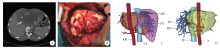
Fig. 1
Traditional 2D CT manifestations, intraoperative findings, and 3D reconstruction model of hepatic alveolar echinococcosis lesions A: Preoperative CT scan revealed a huge alveolar echinococcosis lesion (white arrow); B: Intraoperative lesion (white arrow); C, D: A three-dimensional model automatically generated from two-dimensional CT tomography images (the black arrows indicate huge alveolar echinococcosis lesions). AR: Arteries; HV: Hepatic vein; PV: Portal vein; IV: Inferior vena cava.
Table 4
Preoperative 3D visualization reconstruction data of patients with hepatic alveolar echinococcosis(n = 50)
| 项目 Project | 研究组患者 Patients in research group |
|---|---|
| 肝脏体积/ml Liver volume/ml | 3 354.52 ± 174.96 |
| 棘球蚴病灶体积/ml Volume of Echinococcus lesion/ml | 6 771.95 ± 365.45 |
| 预留肝脏体积/ml Reserved liver volume/ml | 1 203.27 ± 98.23 |
| 预切肝脏体积/ml Pre cut liver volume/ml | 2 154.43 ± 136.52 |
| 残肝比 Residual liver ratio/% | 41.22 ± 6.38 |
| 实际切除肝脏体积/ml Actual volume of liver resection/ml | 2 138.75 ± 157.94 |
| 实际剩余肝脏体积/ml Actual remaining liver volume/ml | 1 193.46 ± 85.73 |
Table 5
Analysis of prognostic factors in patients with hepatic alveolar echinococcosis
| 项目 Project | β | SE | 95% CI | 参数估计 Parameter estimation | OR 95% CI | ||||||
|---|---|---|---|---|---|---|---|---|---|---|---|
| 上限 Upper limits | 下限 Lower limits | Wald χ2 | OR | P | 上限 Upper limits | 下限 Lower limits | |||||
| 肝多房棘球蚴边缘浸润带 Marginal infiltrative zone of hepatic E. multilocularis | 0.987 | 0.456 | 1.645 | 0.328 | 7.890 | 2.682 | 0.005 | 4.756 | 1.378 | ||
| 切除部位 Removal site | 0.123 | 0.100 | 0.321 | -0.075 | 1.485 | 1.130 | 0.223 | 1.382 | 0.885 | ||
| 病灶直径 Diameter of lesion | -0.087 | 0.098 | 0.023 | -0.197 | 0.789 | 0.917 | 0.375 | 1.025 | 0.812 | ||
| 切除肝脏体积 Liver resection volume | 0.045 | 0.067 | 0.157 | -0.067 | 0.456 | 1.046 | 0.500 | 1.158 | 0.934 | ||
| 剩余肝脏体积 Remaining liver volume | 0.234 | 0.156 | 0.545 | -0.077 | 2.345 | 1.263 | 0.126 | 1.645 | 0.928 | ||
| 手术时间 Surgical duration | 0.156 | 0.145 | 0.446 | 0.006 | 1.456 | 1.169 | 0.228 | 1.678 | 1.006 | ||
| 出血量 Bleeding volume | -0.567 | 0.345 | -0.078 | 1.056 | 6.012 | 0.568 | 0.064 | 0.876 | 1.324 | ||
| 术后住院时间 Postoperative hospitalization time | 0.002 | 0.045 | 0.094 | -0.090 | 0.009 | 1.002 | 0.925 | 1.107 | 0.905 | ||
| Pringle法全肝阻断时间 Pringle method whole liver block time | -0.012 | 0.034 | 0.050 | -0.074 | 0.112 | 0.988 | 0.738 | 1.040 | 0.934 | ||
| 住院费用 Hospitalization expenses | 0.056 | 0.067 | 0.189 | -0.077 | 0.632 | 1.057 | 0.427 | 1.221 | 0.923 | ||
| 分型 Classification | 0.023 | 0.089 | 0.191 | -0.145 | 0.063 | 1.023 | 0.802 | 1.130 | 0.889 | ||
| 病灶大小 Disease size | 0.012 | 0.090 | 0.204 | -0.180 | 0.017 | 1.012 | 0.897 | 1.125 | 0.890 | ||
| 瘤周强化 Peritumoral enhancement | 0.045 | 0.067 | 0.177 | -0.087 | 0.456 | 1.046 | 0.500 | 1.155 | 0.937 | ||
| 边缘表现 Edge performance | -0.023 | 0.045 | 0.045 | -0.111 | 0.289 | 0.977 | 0.590 | 1.079 | 0.876 | ||
| 瘤周低信号 Low signal around the tumor | 0.067 | 0.089 | 0.245 | -0.111 | 0.587 | 1.070 | 0.444 | 1.287 | 0.890 | ||
| 强化方式 Strengthening methods | -0.009 | 0.045 | 0.037 | -0.095 | 0.041 | 0.991 | 0.840 | 1.033 | 0.902 | ||
| 增强类型 Enhanced type | 0.045 | 0.089 | 0.223 | -0.137 | 0.267 | 1.046 | 0.605 | 1.200 | 0.891 | ||
| T1WI | 1.038 | 0.423 | 0.447 | 1.273 | 6.023 | 2.824 | 0.062 | 1.564 | 3.571 | ||
| T2WI | 0.468 | 0.213 | -0.449 | 0.776 | 4.817 | 1.596 | 0.162 | 0.638 | 2.173 | ||
| 三维可视化重建 3D visualization reconstruction | -0.378 | 0.192 | -0.794 | -0.122 | 3.876 | 0.685 | 0.025 | 0.452 | 0.885 | ||
|
| [1] | ZULIPIKAER Tusunniyazi, JIANG Tiemin, WEN Hao. Advances in surgical treatment of hepatic alveolar echinococcosis [J]. CHINESE JOURNAL OF PARASITOLOGY AND PARASITIC DISEASES, 2024, 42(6): 783-789. |
| [2] | RAOWAN Tuolehong, ABUDUSALAMU Abulikemu, YANG Lingfei, CHEN Lu, LI Zhao, JIA Fang, SONG Tao. Effect evaluation and factor analysis of ultrasonic manifestations in the diagnosis of hepatic alveolar echinococcosis [J]. CHINESE JOURNAL OF PARASITOLOGY AND PARASITIC DISEASES, 2023, 41(3): 312-318. |
| [3] | AN Xiu-qing, WANG Miao-miao, ZHOU Hong-qian, MENG Kai, CAI Jian-ping, LIU Guang-hui, A Ji-de, YANG Jing-yu. Research progress on microvascular density in hepatic alveolar echinococcosis [J]. CHINESE JOURNAL OF PARASITOLOGY AND PARASITIC DISEASES, 2022, 40(6): 792-797. |
| [4] | ZHANG Ting-ting, DU Qiu-pei, GUO Xin-jian, ZHANG Ling-qiang, WANG Zhi-xin, CHANG Zheng-song, ZHAO Qian, WANG Hai-jiu, HOU Li-zhao. Research progress on vascular invasion of hepatic alveolar echinococcosis [J]. CHINESE JOURNAL OF PARASITOLOGY AND PARASITIC DISEASES, 2022, 40(4): 516-523. |
| [5] | WU Liang-liang, YANG Ling-fei, SONG Tao. Ultrasound and pathological manifestations of lesions in SD rats with hepatic Echinococcus multilocularis infection established by different methods [J]. CHINESE JOURNAL OF PARASITOLOGY AND PARASITIC DISEASES, 2022, 40(4): 549-552. |
| [6] | ZHU Ling-hong, ZHU Lu-min, WANG Bo, YANG Zhi-yong, ZHANG Jing-ni, JI Li, CAI Qi-gang, HAN Xiu-min. Analysis of clinical features of echinococcosis cases [J]. CHINESE JOURNAL OF PARASITOLOGY AND PARASITIC DISEASES, 2021, 39(1): 61-68. |
| [7] | KASIMU Aihaiti, ABUDUSALAMU Aini, TUERGANAILI Aji, SHAO Ying-mei, ZHANG Rui-qing, TALAITI Tuergan, JIANG Tie-min, RAN Bo, ABUDUAINI Abulizi, MIERADILI Aierken, WEN Hao. Analysis of hospital expenses for patients with end-stage hepatic alveolar echinococcosis receiving ex vivo liver resection and autotransplantation [J]. CHINESE JOURNAL OF PARASITOLOGY AND PARASITIC DISEASES, 2020, 38(1): 53-57. |
| [8] | Wei-na ZHANG, Wei GU, Jian-ming JIAO, Jiao LUO. Analysis of clinical features on 15 case of fascioliasis [J]. CHINESE JOURNAL OF PARASITOLOGY AND PARASITIC DISEASES, 2019, 37(2): 207-212. |
| [9] | Xiao-lei XU, Zhi-xin WANG, Zhan WANG, Hai-wen YE, Ming-quan PANG, Ying ZHOU, Hai-jiu WANG, Hai-ning FAN. Treatment of complicated hepatic alveolar echinococcosis: our experience of 98 cases [J]. CHINESE JOURNAL OF PARASITOLOGY AND PARASITIC DISEASES, 2018, 36(6): 552-559. |
| [10] | ABUDUSALAMU Aini1, TUERHONGJIANG Tuxun2, MA Hai-zhang3, ZHANG Heng2, ZHANG Hao1, ABUDUKAIYOUMU Maimaiti4, LI Yu-peng2, SHADIKE Apaer2, LIN Ren-yong5, SHAO Ying-mei1, WEN Hao5*. Changes of Toll-like Receptor mRNA and Related Cytokines in Patients with Hepatic Alveolar Echinococcosis [J]. , 2016, 34(6): 12-542-546. |
| [11] | ZHU Di-wen1,ZHANG Hai-xiao1,REN Wei-xin1 *,XIONG Jin2,XU Xiao-hui2,WEN Hao3. Pathological Changes of after Trans-Portal Vein Chemoembolization Echinococcus multilocularis in the Liver of Infected Rats [J]. , 2014, 32(1): 13-58-61. |
| [12] | FAN Yu-Xiang, LIN Wei-Xin, DI Li-Mu-La-Chi-·Ba-Wu-Dong, GU Dun-Feng, HU Xiao-Dong, ZHANG Hai-Xiao, JI Wei-Zheng, JIANG Chao, WEN Hao. Therapeutic Effect of Hepatic Artery Infusion with Albendazole Microspheres on Hepatic Alveolar Echinococcosis in Rats [J]. , 2011, 29(6): 4-415-418. |
| [13] | TANG Qun-Ke, ZHANG Ying, LI Yong-Shou, YUAN Chun-Ping, ZHANG Dong-Tian. Non-surgical Treatment for Nonresectable Advanced Hepatic Alveolar Echinococcosis [J]. , 2011, 29(1): 11-46-48. |
| Viewed | ||||||
|
Full text |
|
|||||
|
Abstract |
|
|||||