中国寄生虫学与寄生虫病杂志 ›› 2025, Vol. 43 ›› Issue (1): 103-111.doi: 10.12140/j.issn.1000-7423.2025.01.016

• 论著 • 上一篇    下一篇

刚地弓形虫Ⅰ/Ⅲ型ROP16调控TAF15对THP-1细胞影响及机制研究

殷荷1,2(), 马磊3, 党甜甜1,2, 李佳铭1,2, 赵志军1,2,*()   

  1. 1 宁夏医科大学总医院医学实验中心,宁夏 银川 750004
    2 宁夏医学检验临床医学研究中心,宁夏 银川 750004
    3 宁夏大学生命科学学院,宁夏 银川 750004
  • 收稿日期:2024-06-24 修回日期:2024-10-16 出版日期:2025-02-28 发布日期:2025-03-26
  • 通讯作者: 赵志军,男,博士,研究员,从事微生物与感染性疾病的研究。E-mail:z15815z@163.com
  • 作者简介:殷荷,女,硕士,技师,从事弓形虫致病机制的研究。E-mail:1573928868@qq.com
  • 基金资助:
    宁夏自然科学基金(2024AAC03696);宁夏回族自治区重点研发计划(2023BEG02002)

Effect and mechanism of Toxoplasma gondii ROP16 Ⅰ/Ⅲ regulating TAF15 on THP-1 cells

YIN He1,2(), MA Lei3, DANG Tiantian1,2, LI Jiaming1,2, Zhao Zhijun1,2,*()   

  1. 1 General Hospital of Ningxia Medical University, Yinchuan 750004, Ningxia, China
    2 Ningxia Medical Laboratory Clinical Research Center, Yinchuan 750004, Ningxia, China
    3 College of Life Sciences of Ningxia University, Yinchuan 750004, Ningxia, China
  • Received:2024-06-24 Revised:2024-10-16 Online:2025-02-28 Published:2025-03-26
  • Contact: E-mail: z15815z@163.com
  • Supported by:
    Ningxia Natural Science Foundation Project(2024AAC03696);Key Research and Development Program of Ningxia Hui Autonomous Region(2023BEG02002)

摘要:

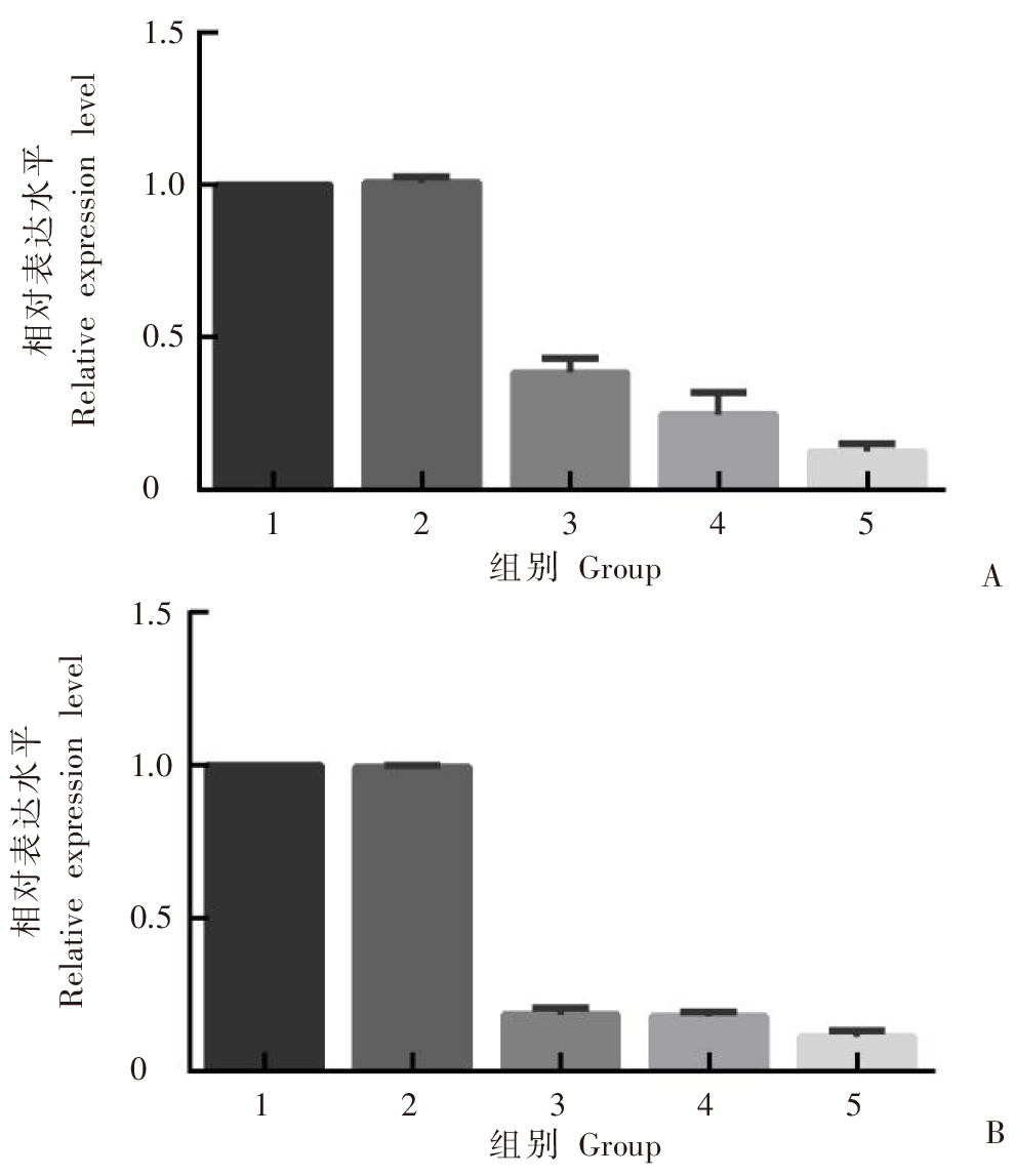
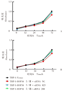
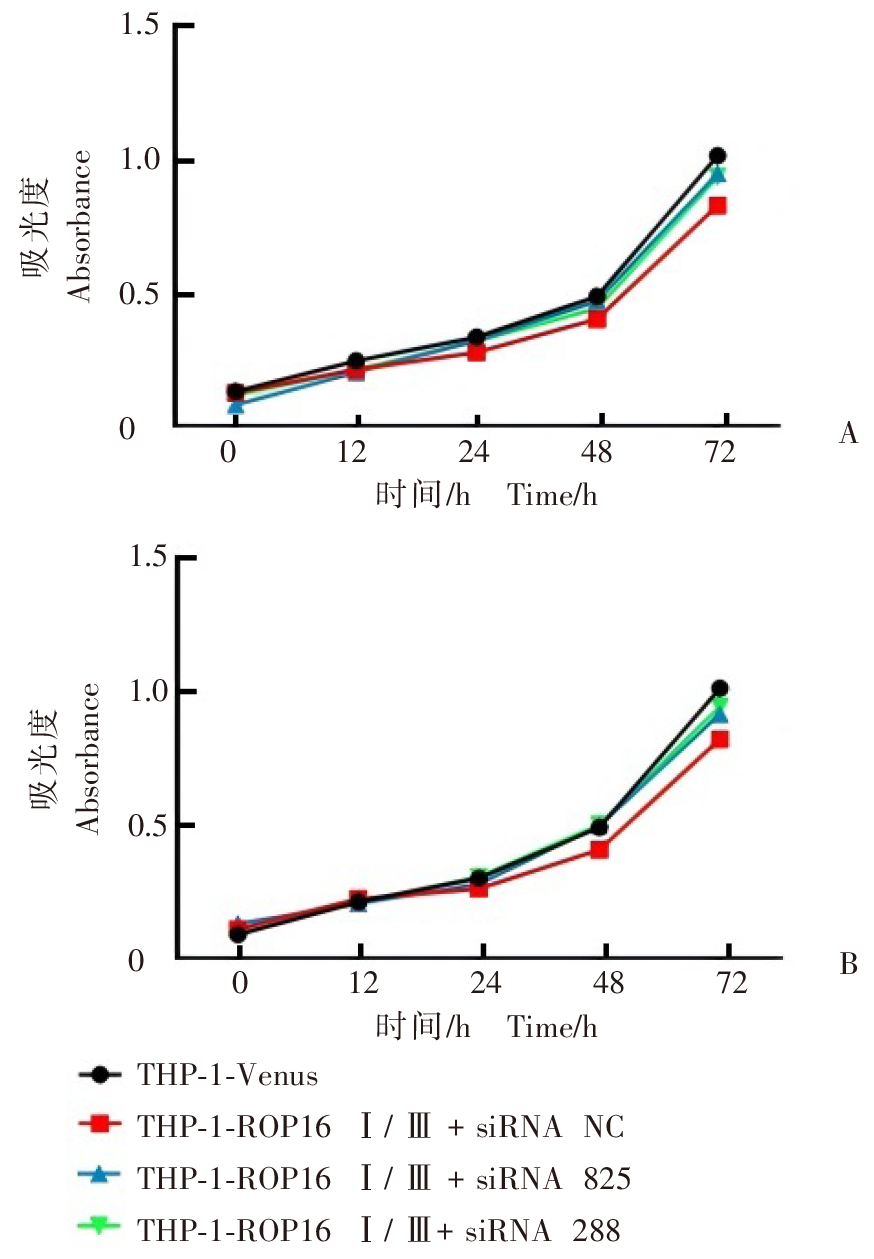
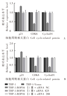
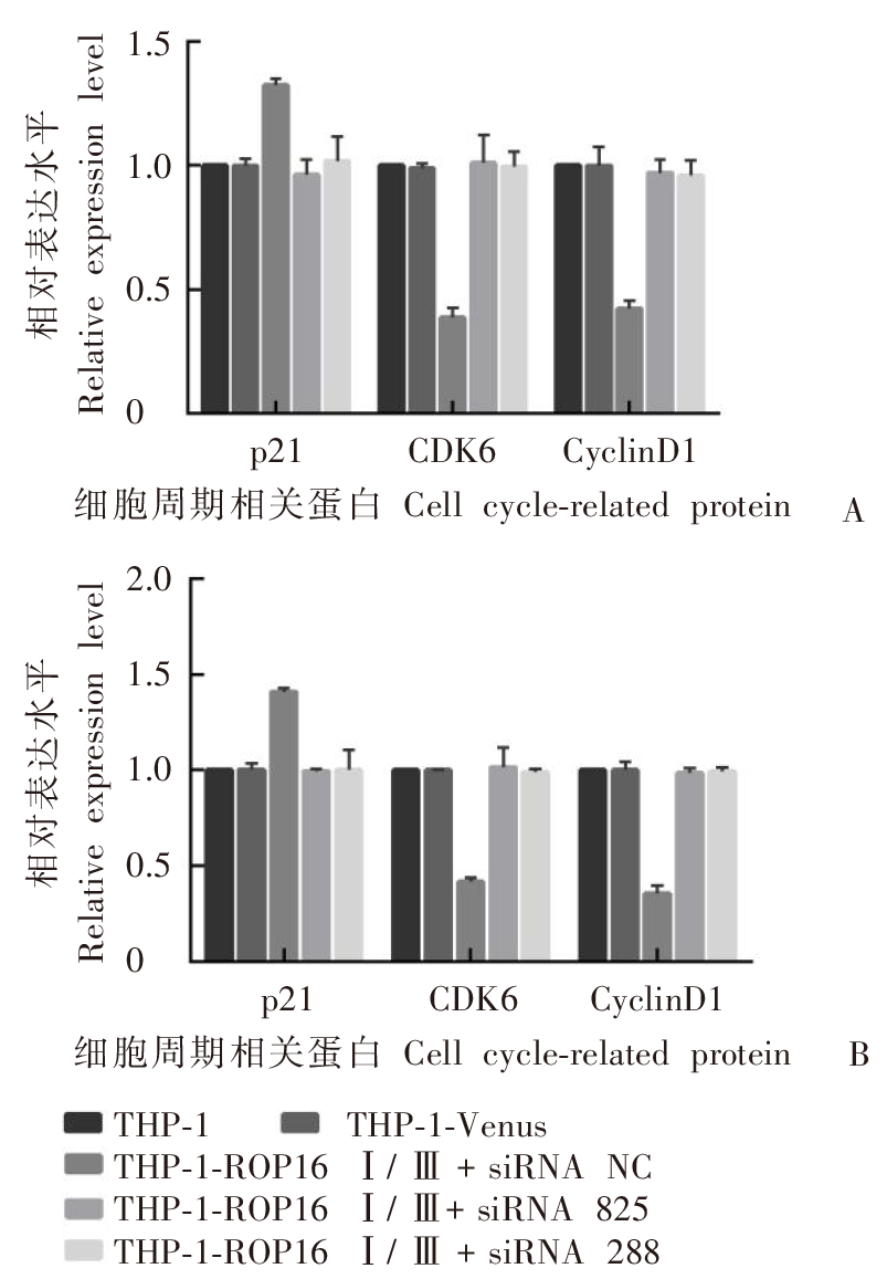
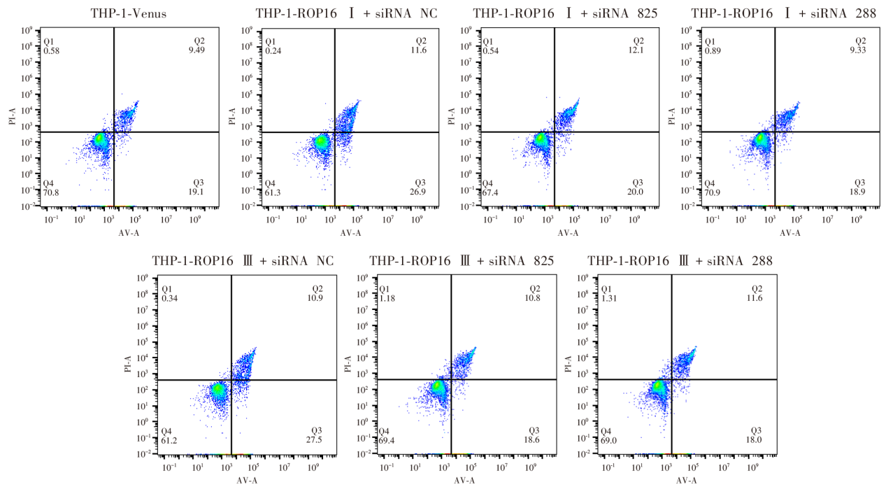
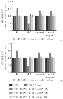
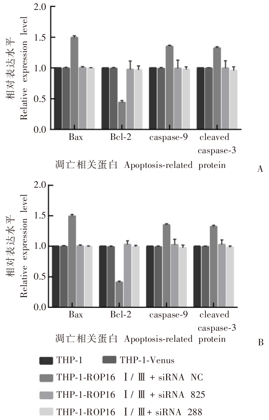
目的 探究刚地弓形虫Ⅰ、Ⅲ型棒状体蛋白16(ROP16)通过调控TATA结合蛋白相关因子15(TAF15)对人单核细胞白血病细胞(THP-1)增殖、凋亡的影响及作用机制。方法 将Ⅰ、Ⅲ型ROP16过表达慢病毒转染至THP-1细胞,构建稳定表达ROP16的细胞株(THP-1-ROP16Ⅰ/Ⅲ),以转染空载体慢病毒的细胞为空载体对照组(THP-1-Venus),未转染细胞为对照组(THP-1),通过实时荧光定量逆转录PCR(RT-qPCR)和蛋白质免疫印迹(Western blotting)验证过表达效果。利用免疫沉淀-质谱联用技术(IP-MS)在THP-1-ROP16Ⅰ/Ⅲ细胞株中预测ROP16的互作蛋白,并通过RT-qPCR和Western blotting检测细胞中互作蛋白TAF15的表达。将3条作用于TAF15基因不同位点的小干扰RNA(siRNA 1215、siRNA 825、siRNA 288)分别干扰THP-1-ROP16Ⅰ/Ⅲ细胞株,分为THP-1-ROP16Ⅰ/Ⅲ + siRNA 1215/825/288组,同时设未干扰对照组(THP-1-ROP16Ⅰ/Ⅲ + siRNA NC),Western blotting验证TAF15沉默效果。通过细胞计数试剂盒8(CCK-8)与流式细胞术检测各组细胞增殖及凋亡情况,Western blotting检测细胞周期依赖性蛋白激酶抑制因子1A(p21)、周期素依赖性激酶6(CDK6)、G1/S特异性周期蛋白(CyclinD1)、B细胞淋巴瘤/白血病-2蛋白(Bcl-2)、Bcl-2相关X蛋白(Bax)、裂解型半胱氨酸蛋白酶-3(cleaved caspase-3)、半胱氨酸蛋白酶-9(caspase-9)及磷酸化信号转导和转录激活因子3(P-STAT3)蛋白的表达。结果 THP-1-ROP16Ⅰ/Ⅲ组ROP16 mRNA的相对转录水平为2 679.427 ± 250.600、2 395.410 ± 325.700,均高于THP-1-Venus组(1.036 ± 0.102)(F = 153.3,P < 0.01);ROP16蛋白的相对表达水平为4.526 ± 0.020、5.457 ± 0.250,均高于THP-1-Venus组(1.688 ± 0.653)(F = 76.4,P < 0.01)。TAF15为Ⅰ、Ⅲ型ROP16的互作蛋白,THP-1-ROP16Ⅰ/Ⅲ组TAF15 mRNA的相对转录水平为6.027 ± 0.313、5.567 ± 0.088,均高于THP-1-Venus组(0.985 ± 0.027)(F = 869.4,P < 0.01);TAF15蛋白的相对表达水平为1.789 ± 0.145、1.593 ± 0.029,均高于THP-1-Venus组(1.010 ± 0.365)(F = 50.6,P < 0.01)。TAF15 siRNA转染48 h后,THP-1-ROP16Ⅰ + siRNA 1215/825/288组TAF15蛋白的相对表达水平分别为0.384 ± 0.047、0.246 ± 0.072、0.125 ± 0.026,均低于THP-1-Venus组(1.007 ± 0.019)(F = 313.1,P < 0.01);THP-1-ROP16Ⅲ + siRNA 1215/825/288组细胞TAF15蛋白的相对表达水平分别为0.186 ± 0.020、0.180 ± 0.015、0.112 ± 0.019,均低于THP-1-Venus组(0.995 ± 0.052)(F = 3046.0,P < 0.01)。THP-1-ROP16Ⅰ/Ⅲ + siRNA NC组细胞CCK-8实验A450值分别为0.803 ± 0.015、0.813 ± 0.011,低于THP-1-Venus组(0.997 ± 0.010、0.995 ± 0.016)(t = 19.2、24.0,均P < 0.01);THP-1-ROP16Ⅰ/Ⅲ + siRNA 825/288组A450值分别为0.986 ± 0.010、0.983 ± 0.004,0.980 ± 0.006、0.984 ± 0.010(F = 3.5、2.9,均P > 0.05)。THP-1-ROP16Ⅰ/Ⅲ + siRNA NC组细胞凋亡率分别为(38.19 ± 0.45)%、(38.06 ± 0.84)%,高于THP-1-Venus组的(28.41 ± 0.69)%(t = 20.5、17.7,均P < 0.01);THP-1-ROP16Ⅰ/Ⅲ + siRNA 825/288组分别为(30.03 ± 1.83)%、(28.78 ± 0.72)%,(29.33 ± 0.80)%、(28.94 ± 0.58)%(F = 1.5、0.4,均P > 0.05)。THP-1-ROP16Ⅰ + siRNA NC组p21、Bax、caspase-9、cleaved caspase-3及P-STAT3蛋白的相对表达水平分别为1.322 ± 0.027、1.493 ± 0.030、1.349 ± 0.021、1.324 ± 0.020、10.500 ± 1.005,均高于THP-1-Venus组(1.000 ± 0.026、0.996 ± 0.016、0.989 ± 0.019、0.994 ± 0.010、1.000 ± 0.001)(t = 14.8、25.4、22.3、25.0、15.6,均P < 0.01);CDK6、CyclinD1及Bcl-2蛋白的相对表达水平分别为0.387 ± 0.040、0.424 ± 0.030、0.438 ± 0.035,均低于THP-1-Venus组(0.989 ± 0.018、1.000 ± 0.074、0.991 ± 0.016)(t = 23.6、12.4、25.0,均P < 0.01)。THP-1-ROP16Ⅲ + siRNA NC组p21、Bax、caspase-9、cleaved caspase-3及P-STAT3蛋白的相对表达水平分别为1.409 ± 0.020、1.493 ± 0.030、1.349 ± 0.021、1.324 ± 0.020、16.210 ± 0.664,均高于THP-1-Venus组(1.004 ± 0.032、0.996 ± 0.015、0.989 ± 0.019、0.994 ± 0.010、1.000 ± 0.001)(t = 18.7、25.4、22.3、25.0、39.7,均P < 0.01);CDK6、CyclinD1及Bcl-2蛋白的相对表达水平分别为0.418 ± 0.021、0.357 ± 0.040、0.411 ± 0.019,均低于THP-1-Venus组(1.000 ± 0.001、1.001 ± 0.042、0.991 ± 0.016)(t = 47.7、19.1、40.7,均P < 0.01)。结论 弓形虫Ⅰ、Ⅲ型ROP16蛋白可通过促进TAF15的表达抑制THP-1细胞增殖、促进细胞凋亡,其作用机制可能与抑制细胞内STAT3信号通路的活化有关。

关键词: 刚地弓形虫, 棒状体蛋白16, TATA结合蛋白相关因子15, THP-1细胞

Abstract:

Objective To investigate the effect and mechanisms of Toxoplasma gondii type Ⅰ and Ⅲ rhoptry protein 16 (ROP16) on the proliferation and apoptosis of human monocytic leukemia THP-1 cells via TATA-binding protein-associated factor 15 (TAF15). Methods THP-1 cells were transfected with entiviruses overexpressing T. gondii type Ⅰ and Ⅲ ROP16 to generate cell lines that stably expressed ROP16 (THP-1-ROP16 Ⅰ/Ⅲ), and cell transfected with lentiviruses containing empty vectors (THP-1-Venus) served as an empty vector control, while non-transfected cells (THP-1) served as controls. The efficiency of overexpression was checked using quantitative real-time PCR (RT-qPCR) assay and Western blotting. The proteins interacting with ROP16 were identified using immunoprecipitation-mass spectrometry (IP-MS) in THP-1-ROP16 Ⅰ/Ⅲ cell lines, and the expression of the ROP16-interacting protein TAF15 was quantified using RT-qPCR and Western blotting assays. Three siRNA targeting different sites of TAF15 gene (siRNA 1215, siRNA 825, siRNA 288) were used to interfere with THP-1-ROP16 Ⅰ/Ⅲ cell lines and divided into THP-1-ROP16 Ⅰ/Ⅲ + siRNA 1215/825/288 groups, while an undisturbed control group (THP-1-ROP16 Ⅰ/Ⅲ + siRNA NC) was set up and the silencing efficiency of TAF15 was checked using Western blotting. In addition, the cell proliferation and apoptosis was measured using cell counting kit-8 (CCK-8) assay and flow cytometry, and the expression of cyclin-dependent kinase inhibitor 1A (p21), cyclin-dependent kinase 6 (CDK6), G1/S-specific cyclin (CyclinD1), B-cell lymphoma/leukemia-2 protein (Bcl-2), Bcl-2-associated X protein (Bax), cleaved caspase-3, caspase-9 and phosphorylated signal transducer and activator of transcription 3 (P-STAT3) was determined using Western blotting. Results The relative ROP16 mRNA expression was 2 679.427 ± 250.600 in the THP-1-ROP16 Ⅰ group and 2 395.410 ± 325.700 in the THP-1-ROP16 Ⅲ group, which was both higher than in the THP-1-Venus group (1.036 ± 0.102) (F = 153.3, P < 0.01), and the relative ROP16 protein expression was higher in the THP-1-ROP16 Ⅰ group (4.526 ± 0.020) and THP-1-ROP16 Ⅲ group (5.457 ± 0.250) than in the THP-1 Venus group (1.688 ± 0.653) (F = 76.4, P < 0.01). TAF15 was identified as a protein interacting with type Ⅰ and Ⅲ ROP16, and the relative TAF15 mRNA and protein expression was both higher in the THP-1 ROP16 Ⅰ group (6.027 ± 0.313 and 1.789 ± 0.145) and THP-1 ROP16 Ⅲ group (5.567 ± 0.088 and 1.593 ± 0.029) than in the THP-1 Venus group (0.985 ± 0.027 and 1.010 ± 0.365) (F = 869.4 and 50.6, P < 0.01). The relative TAF15 protein expression was 0.384 ± 0.047, 0.246 ± 0.072, and 0.125 ± 0.026 in the THP-1 ROP16 Ⅰ + siRNA 1215 group, the THP-1 ROP16 Ⅰ + siRNA 825 group, and the THP-1 ROP16 Ⅰ + siRNA 288 group 48 hours post-transfection with TAF15 siRNA, which was all lower than in the THP-1-Venus group (1.007 ± 0.019) (F = 313.1, P < 0.01), and the relative TAF15 protein expression was 0.186 ± 0.020, 0.180 ± 0.015, and 0.112 ± 0.019 in the THP-1 ROP16 Ⅲ + siRNA 1215 group, the THP-1 ROP16 Ⅲ + siRNA 825 group, and the THP-1 ROP16 Ⅲ + siRNA 288 group 48 hours post-transfection with TAF15 siRNA, which was all lower than in the THP-1 Venus group (0.995 ± 0.052) (F = 3 046.0, P < 0.01). CCK-8 assay measured the A450 values of 0.803 ± 0.015 and 0.813 ± 0.011 in the THP-1-ROP16 Ⅰ + siRNA NC group and the THP-1-ROP16 Ⅲ + siRNA NC group, which were both lower than in the THP-1 Venus group (0.997 ± 0.010 and 0.995 ± 0.016) (t = 19.2 and 24.0, both P < 0.01). A450 values of THP-1-ROP16 Ⅰ/Ⅲ + siRNA 825/288 groups were 0.986 ± 0.010, 0.983 ± 0.004; 0.980 ± 0.006, 0.984 ± 0.010 (F = 3.5, 2.9; both P > 0.05), respectively. The apoptotic rates of THP-1 cells were (38.19 ± 0.45)% in the THP-1-ROP16 Ⅰ + siRNA NC group and (38.06 ± 0.84)% in the THP-1-ROP16 Ⅲ + siRNA NC group, which were both higher than in the THP-1-Venus group [(28.41 ± 0.69)% ] (t = 20.5 and 17.7; both P < 0.01). The apoptotic rates of THP-1-ROP16 Ⅰ/Ⅲ + siRNA 825/288 group were (30.03 ± 1.83)%, (28.78 ± 0.72)%; (29.33 ± 0.80)%, (28.94 ± 0.58)% (F = 1.5, 0.4,both P > 0.05). The relative expression of p21, Bax, Caspase-9, Cleaved Caspase-3, and P-STAT3 proteins was 1.322 ± 0.027, 1.493 ± 0.030, 1.349 ± 0.021, 1.324 ± 0.020, and 10.500 ± 1.005 in the THP-1-ROP16 Ⅰ + siRNA NC group, which was all higher than in the THP-1-Venus group (1.000 ± 0.026, 0.996 ± 0.016, 0.989 ± 0.019, 0.994 ± 0.010 and 1.000 ± 0.001) (t = 14.8, 25.4, 22.3, 25.0 and 15.6; all P < 0.01), and the relative CDK6, CyclinD1, and Bcl-2 protein expression was 0.387 ± 0.040, 0.424 ± 0.030, and 0.438 ± 0.035 in the THP-1-ROP16 Ⅰ + siRNA NC group, which was all lower than in the THP-1-Venus group (0.989 ± 0.018, 1.000 ± 0.074 and 0.991 ± 0.016) (t = 23.6, 12.4 and 25.0; all P < 0.01). The relative expression of p21, Bax, caspase-9, cleaved caspase-3, and P-STAT3 proteins was 1.409 ± 0.020, 1.493 ± 0.030, 1.349 ± 0.021, 1.324 ± 0.020, and 16.210 ± 0.664 in the THP-1-ROP16 Ⅲ + siRNA NC group, which was all higher than in the THP-1-Venus group (1.004 ± 0.032, 0.996 ± 0.015, 0.989 ± 0.019, 0.994 ± 0.010 and 1.000 ± 0.001) (t = 18.7, 25.4, 22.3, 25.0 and 39.7; all P < 0.01), and the relative expression of CDK6, CyclinD1, and Bcl-2 proteins was 0.418 ± 0.021, 0.357 ± 0.040, and 0.411 ± 0.019 in the THP-1-ROP16 Ⅲ + siRNA NC group, which was all lower than in the THP-1-Venus group (1.000 ± 0.001, 1.001 ± 0.042 and 0.991 ± 0.016) (t = 47.7, 19.1 and 40.7; all P < 0.01). Conclusion T. gondii type Ⅰ and Ⅲ ROP16 proteins inhibit THP-1 cell proliferation and promote cell apoptosis through promoting TAF15 expression, which may be associated with inhibition of activation of the STAT3 signaling pathway in THP-1 cells.

Key words: Toxoplasma gondii, ROP16 protein, TAF15, THP-1 cell

中图分类号: