中国寄生虫学与寄生虫病杂志 ›› 2025, Vol. 43 ›› Issue (3): 329-334.doi: 10.12140/j.issn.1000-7423.2025.03.004

• 论著 • 上一篇    下一篇

穿心莲内酯抑制刚地弓形虫增殖作用的研究

柳润春1,2,3()(), 邹伟浩1,2,3, 郑书雨1,2,3, 吴蔚玲1,2,3, 彭鸿娟1,2,3,*()()   

  1. 1 南方医科大学公共卫生学院病原生物学系,广东 广州 510515
    2 广东省热带病研究重点实验室,广东 广州 510515
    3 华南传染病防治教育部重点实验室(南方医科大学),广东 广州 510515
  • 收稿日期:2025-03-05 修回日期:2025-04-28 出版日期:2025-06-30 发布日期:2025-06-27
  • 通讯作者: 彭鸿娟(0000-0002-9345-8218),女,博士,教授,从事寄生虫感染与免疫研究。E-mail:hongjuan@smu.edu.cn
  • 作者简介:柳润春(0009-0003-6889-7352),男,硕士研究生,从事寄生虫感染与免疫研究。E-mail:1732934516@qq.com
  • 基金资助:
    国家自然科学基金(82330072);国家自然科学基金(82272364);广东省自然科学基金(2023A1515011733);广东省自然科学基金(2024A1515011327)

Investigating the inhibitory effect of andrographolide on Toxoplasma gondii proliferation

LIU Runchun1,2,3()(), ZOU Weihao1,2,3, ZHENG Shuyu1,2,3, WU Weiling1,2,3, PENG Hongjuan1,2,3,*()()   

  1. 1 Department of Pathogen Biology, School of Public Health, Southern Medical University, Guangzhou 510515, Guangdong, China
    2 Guangdong Provincial Key Laboratory of Tropical Disease Research, Guangzhou 510515, Guangdong, China
    3 Key Laboratory of the Ministry of Education for Prevention and Control for Infectious Diseases of South China, Guangzhou 510515, Guangdong, China
  • Received:2025-03-05 Revised:2025-04-28 Online:2025-06-30 Published:2025-06-27
  • Contact: E-mail: hongjuan@smu.edu.cn
  • Supported by:
    National Natural Science Foundation of China(82330072);National Natural Science Foundation of China(82272364);Guangdong Provincial Natural Science Foundation(2023A1515011733);Guangdong Provincial Natural Science Foundation(2024A1515011327)

摘要:

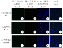
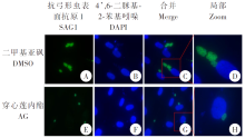
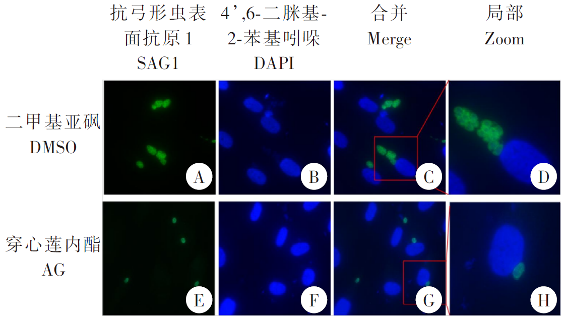
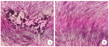
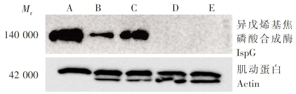
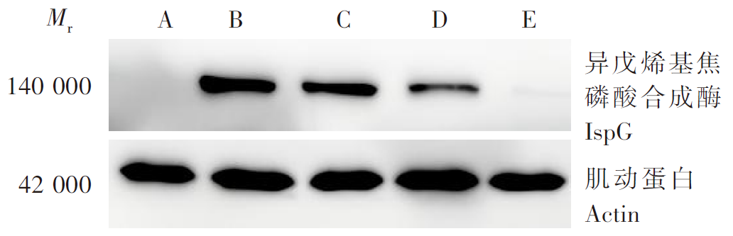
目的 探讨穿心莲内酯(AG)对刚地弓形虫的抑制作用及作用靶点。 方法 在人包皮成纤维细胞(HFF)中加入0、5、10、20、40、80、120、160、320 μmol/L AG和10 μl细胞计数试剂盒-8(CCK-8)溶液,检测各组细胞的吸光度(A450值),绘制增殖曲线,筛选对细胞无明显毒性的浓度用于后续研究。将弓形虫感染的HFF细胞分为二甲基亚砜(DMSO)组、AG组和乙胺嘧啶(PYR)组,分别加入兔源抗弓形虫表面抗原1抗体,绿色荧光标记的羊抗兔IgG抗体(1:1 000),红色荧光标记的羊抗兔IgG抗体(1:1 000),光学显微镜下观察弓形虫入侵情况,并计算入侵率。将HFF细胞分为DMSO组和AG组,加入绿色荧光标记的羊抗鼠IgG抗体(1:1 000),光学显微镜下观察弓形虫增殖情况。将HFF细胞分为DMSO组、AG组,分别加入100 μl DMSO、40 μmol/L AG,显微镜下观察HFF细胞形成的空斑面积。采用表面等离子体共振成像(SPRi)技术筛选互作蛋白,药物亲和响应靶标稳定性实验验证异戊烯基焦磷酸合成酶(IspG)蛋白与AG的互作,蛋白质免疫印迹(Western blotting)检测IspG蛋白变化情况。实时荧光定量PCR(qPCR)检测互作蛋白基因mRNA相对转录水平。使用GraphPad Prism 8.0.2软件进行统计学分析。 结果 增殖实验结果显示,在0、5、10、20、40 μmol/L AG中HFF细胞相对活力分别为100.00%、107.45%、100.66%、109.21%和90.94%,细胞活力维持在较高水平,无明显毒性;在80、120、160、320 μmol/L AG中相对活力分别为57.83%、34.16%、48.25%和30.75%,毒性明显(F = 14.96,P < 0.01)。选择40 μmol/L浓度用于后续研究。间接免疫荧光结果显示,AG组和PYR组弓形虫入侵率分别为(8.06 ± 2.40)%和(6.36 ± 1.79)%,均低于DMSO组的(42.49 ± 9.75)%(F = 35.88,P < 0.01)。DMSO组平均每个纳虫泡的弓形虫数量约为(5.78 ± 0.94)个,高于AG组的(1.40 ± 0.12)个(t = 7.98,P < 0.01)。空斑实验结果显示,AG组和DMSO组的空斑形成面积分别为0 μm²和(3 210 ± 1 840)μm²,差异有统计学意义(t = 19.03,P < 0.01)。SPRi鉴定AG与弓形虫的互作蛋白结果显示,蛋白质谱评分较高的弓形虫蛋白分子为核糖体RNA加工蛋白、ATP合酶α亚基、IspG蛋白和START结构域蛋白,分别为2.19、4.01、4.01和2.12分,分子间具有高度的相关性。药物亲和响应靶标稳定性实验结果显示,10 μmol/L AG组的IspG蛋白相对表达水平为0.25 ± 0.01,高于对照组的0.12 ± 0.01(F = 294.2,P < 0.01)。qPCR结果显示,40 μmol/L AG组IspG mRNA的相对转录水平为4.903 ± 1.546,高于DMSO组的1.19 ± 0.20(t = 4.123,P < 0.05)。Western blotting检测结果显示,IspG蛋白在0、10、20和40 μmol/L AG组的相对表达水平分别为0.57 ± 0.01、0.52 ± 0.02、0.24 ± 0.05和0.03 ± 0.01,呈现明显的浓度依赖性下降趋势(F = 313.4,P < 0.01)。 结论 AG通过靶向IspG蛋白,抑制弓形虫的入侵和增殖,具有显著的抗弓形虫活性。

关键词: 刚地弓形虫, 穿心莲内酯, 异戊烯基焦磷酸合成酶, 甲基赤藓醇磷酸途径

Abstract:

Objective To investigate the inhibitory effect of andrographolide (AG) on Toxoplasma gondii and unravel its potential therapeutic mechanisms. Methods Human foreskin fibroblasts (HFFs) were treated with 0, 5, 10, 20, 40, 80, 120, 160, or 320 μmol/L AG and 10 μl CCK-8 solutions. The absorbance (A450 value) was measured in HFFs following treatment and cell proliferation curves were plotted. Screening for concentrations that were not significantly toxic to cells for subsequent studies. For invasion assays, HFFs were divided into dimethyl sulfoxide (DMSO) group, AG group, and pyrimethamine (PYR) group, which were incubated in SAG1 antibody, green fluorescence-labeled goat anti-rabbit IgG antibody (1:1 000), and red fluorescence-labeled goat anti-rabbit IgG antibody (1:1 000), respectively; and then, T. gondii invasion was observed under an optical microscope, and the invasive rate was calculated. For proliferation assays, HFFs were divided into DMSO and AG groups, treated with green fluorescence-labeled goat anti-mouse IgG antibody (1:1 000), and T. gondii proliferation was observed under an optical microscope. For plaque assays, HFFs were divided into DMSO and AG groups, which were treated with 100 μl DMSO and 40 μmol/L AG, respectively, and the size of plaques were observed under an optical microscope. Interacting proteins were screened using surface plasmon resonance imaging (SPRi), and the interaction of isopentenyl diphosphate isomerase (IspG) gene with AG was validated with drug affinity responsive target stability assays. The IspG protein expression was determined using Western blotting assay, and the relative mRNA levels of interacting protein-coding genes were quantified using real time quantitative reverse transcription PCR (qPCR) assay. All statistical analyses were performed using the software GraphPad Prism 8.0.2. Results The results of proliferation experiments showed that the relative viability of HFF cells in 0, 5, 10, 20, and 40 μmol/L AG was 100.00%, 107.45%, 100.66%, 109.21%, and 90.94%, respectively, and the cell viability was maintained at the same level without obvious toxicity, and the relative viabilities of HFFs were 57.83%, 34.16%, 48.25%, and 30.75% following treatment with AG at doses of 80, 120, 160 and 320 μmol/L, appearing a remarkable toxicity (F = 14.96, P < 0.01). Indirect immunofluorescence assay revealed lower invasion rates in AG [(8.06 ± 2.40) %] and PYR groups [(6.36 ± 1.79) %] than in the DMSO group [(42.49 ± 9.75) %] (F = 35.88, P < 0.01), and the number of T. gondii parasitophorous vacuoles was higher in the DMSO group [(5.78 ± 0.94) parasites/vacuole] than in the AG group [(1.40 ± 0.12) parasites/vacuole] (t = 7.98, P < 0.01). Plaque assays showed a higher plaque size in the AG group (0 μm2) than in the DMSO group [(3 210 ± 1 840) μm2] (t = 19.03, P < 0.01). SPRi identified that the AG-interacting T. gondii proteins with the highest mass spectrometry scores included ribosomal RNA processing protein (2.19), ATP synthase α subunit (4.01), IspG (4.01), and START domain protein (2.12), appearing high molecular relevance. Drug affinity responsive target stability assays showed higher IspG expression in HFFs treated with 10 μmol/L AG (0.25 ± 0.01) than in controls (0.12 ± 0.01) (F = 294.2, P < 0.01). qPCR assay quantified higher relative IspG mRNA expression (4.903 ± 1.546) in HFFs treated with 40 μmol/L AG than in those treated with DMSO (1.19 ± 0.20) (t = 4.123, P < 0.05), and Western blotting determined that the IspG protein expression appeared a tendency towards a concentration-dependent manner, with relative IspG protein expression of 0.57 ± 0.01, 0.52 ± 0.02, 0.24 ± 0.05, and 0.03 ± 0.01 in HFFs treated with 0 (DMSO), 10, 20, and 40 μmol/L AG, respectively (F = 313.4, P < 0.01). Conclusion AG inhibits T. gondii invasion and proliferation through targeting IspG, appearing a remarkable anti-T. gondii activity.

Key words: Toxoplasma gondii, Andrographolide, Isopentenyl diphosphate isomerase, Methylerythritol phosphate pathway

中图分类号: