中国寄生虫学与寄生虫病杂志 ›› 2023, Vol. 41 ›› Issue (4): 520-523.doi: 10.12140/j.issn.1000-7423.2023.04.022

• 病例报告 • 上一篇    下一篇

贵州省儿童多房棘球蚴病1例

朱爱娅1(), 王旭2, 王江友3, 王颖4, 李杨1,*(), 宋珊5, 耿燕1, 兰子尧1, 戴佳芮1   

  1. 1 贵州省疾病预防控制中心,贵阳 550004
    2 中国疾病预防控制中心寄生虫病预防控制所(国家热带病研究中心),国家卫生健康委员会寄生虫病原与媒介生物学重点实验室,世界卫生组织热带病合作中心,国家级热带病国际联合研究中心,上海 200025
    3 紫云县疾病预防控制中心,贵州紫云 550899
    4 安顺市疾病预防控制中心,贵州安顺 561099
    5 赤水市疾病预防控制中心,贵州赤水 563299
  • 收稿日期:2023-02-01 修回日期:2023-05-31 出版日期:2023-08-30 发布日期:2023-09-06
  • 通讯作者: *李杨(1968-),男,本科,主任医师,从事寄生虫病防治工作。E-mail:835235727@qq.com
  • 作者简介:朱爱娅(1978-),女,硕士,主任医师,从事寄生虫病防治工作。E-mail:zhuaiya520@126.com
  • 基金资助:
    贵州省人才团队项目;上海市卫生健康委员会科研项目(20214Y0213)

A child case of alveolar echinococcosis in Guizhou Province

ZHU Aiya1(), WANG Xu2, WANG Jiangyou3, WANG Ying4, LI Yang1,*(), SONG Shan5, GENG Yan1, LAN Ziyao1, DAI Jiarui1   

  1. 1 Guizhou Center for Disease Control and Prevention, Guiyang 550004, China
    2 National Institute of Parasitic Diseases, Chinese Center for Disease Control and Prevention (Chinese Center for Tropical Diseases Research); NHC Key Laboratory of Parasite and Vector Biology; WHO Collaborating Center for Tropical Diseases, National Center for International Research on Tropical Diseases, Shanghai 200025, China
    3 Ziyun County Center for Disease Control and Prevention, ZiYun 550899, Guizhou, China
    4 Anshun Center for Disease Control and Prevention, Anshun 561099, Guizhou, China
    5 Chishui Center for Disease Control and Prevention, Chishui 563299, Guizhou, China
  • Received:2023-02-01 Revised:2023-05-31 Online:2023-08-30 Published:2023-09-06
  • Contact: *E-mail: 835235727@qq.com
  • Supported by:
    Guizhou Provincial Talent Team Project;Research Project of Shanghai Municipal Health Commission(20214Y0213)

摘要:

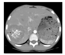
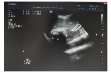
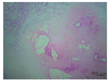
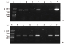
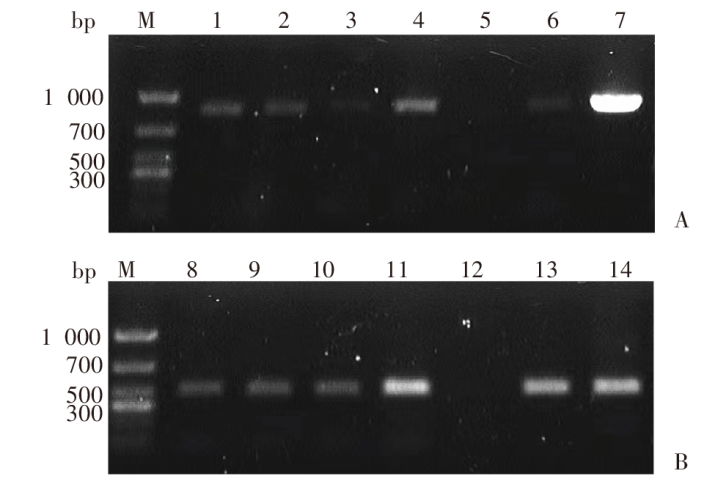
患儿,男,9岁,学生,贵州省安顺市紫云县人。2021年8月起,患儿常感右上腹疼痛不适。2022年9月10日以“肝占位病变:疑似肝脓肿并钙化或肿瘤”收治于中国贵航集团三0二医院。流行病学调查结果显示,患儿无流行区旅居史,其父母长期从事牲畜(犬)宰杀职业,部分犬只从西藏自治区、四川省等棘球蚴病流行区引入,患儿常与待宰犬只接触。入院查体:全身皮肤巩膜无黄染,右上腹稍有压痛,肝脏包块未触及。血常规示:嗜酸粒细胞百分比20.1%。肝功能检测结果示,γ谷氨酰转移酶36 U/L。上腹部增强CT显示,肝右后叶见团块状混杂密度影,呈分叶状,内见多发斑点状、迂曲蚯蚓状高密度钙化影,呈簇状分布;增强扫描病灶见多发网格样分隔影,病灶边缘及分隔呈轻度持续强化。腹部彩色超声示,肝右后叶可见类椭圆形不均匀强回声团。9月14日行腹腔镜下肝右后叶切除术,见肝右后叶代偿性增大,Ⅵ、Ⅶ段可见乳白色肿块,约6 cm × 7 cm × 5 cm,表面凹凸不平,质地硬。术后病理切片结果示,肝组织炎性坏死。术后复查,血常规示嗜酸粒细胞百分比正常;上腹部CT提示术后改变,余未见明显异常,遂出院。11月10日至贵州医科大学附属医院复查,血清学检测结果示棘球绦虫抗体阳性。11月29日病灶组织DNA经中国疾病预防控制中心寄生虫病预防控制所(国家热带病研究中心)检测,PCR扩增出棘球绦虫线粒体细胞色素c氧化酶亚基1(cox1)和烟酰胺腺嘌呤二核苷酸脱氢酶亚基1(nad1)基因片段,分别与多房棘球绦虫的cox1基因(参考序列登录号AB477010)和nad1基因(参考序列登录号AJ132907)的序列相似性高达99.42%和99.59%,确诊为多房棘球蚴病。予患儿阿苯达唑15 mg/(kg·d),早晚餐后分服,继续治疗6个月。定期回访。

关键词: 多房棘球蚴病, 儿童, 贵州省

Abstract:

The patient is a 9-year-old boy, a student from Ziyun County, Anshun City, Guizhou Province. He has experienced constant pain and discomfort in his upper right abdomen since August 2021. He was admitted to the 302 Hospital of the China Guihang Group on September 10, 2022 with a "liver occupying lesion: suspected liver abscess with calcification or tumor". The epidemiological investigation indicated that the boy has no history of travelling to epidemic areas. His parents have been engaged in livestock (dogs) slaughter for a long time, and some dogs were purchased from Echinococcosis epidemic areas such as Tibet Autonomous Region and Sichuan Province. The patient often came into contact with those dogs. The admission examination showed no yellowing of the skin and sclera throughout the body, slight tenderness in the right upper abdomen, and no palpable liver mass. Blood cell count showed that the percentage of eosinophils was 20.1%. The liver function testing results were showed γ Glutamic transferase was 36 U/L. The enhanced CT scan of the upper abdomen showed a patchy mixed-density shadow in the right posterior lobe of the liver, which is lobulated. Multiple patchy and tortuous earthworm-like high-density calcification shadows distributed in clusters. Multiple grid-like septal shadows were observed in the enhanced scan at the lesion, with mild continuous enhancement at the edges and septal areas. The abdominal color ultrasound showed an elliptical-shaped, uneven, strong echogenic mass in the right posterior lobe of the liver. Laparoscopic resection of the right posterior lobe of the liver was performed on September 14th, and compensatory enlargement of the right posterior lobe was observed. A milky white mass was seen in segments Ⅵ and Ⅶ, approximately 6 cm × 7 cm × 5 cm, with uneven surface and hard texture. The postoperative pathological section results showed inflammatory necrosis of the liver tissue. Postoperative blood cell count showed a normal of eosinophil percentage. Postoperative CT reexamination of the upper abdomen revealed postoperative changes, and no other obvious abnormalities were found. Therefore, the child was discharged. On November 10th, the patient went to the Affiliated Hospital of Guizhou Medical University for review, and the serum test indicated that Echinococcus antibody was positive. The cytochrome c oxidase subunit 1 (cox1) and nicotinamide adenine dinucleotide dehydrogenase subunit 1 (nad1) gene fragments of Echinococcus were amplified by PCR from the lesion tissue DNA by National Institute of Parasitic Diseases, Chinese Center for Disease Control and Prevention (Chinese Center for Tropical Diseases Research) on November 29th. The amplified cox1 and nad1 gene fragments have high sequence similarity of 99.42% and 99.59% with the cox1 gene (reference sequence registration number AB477010) and nad1 gene (reference sequence registration number AJ132907) of E. multilocularis, respectively. The diagnosis of echinococcosis multilocularis was confirmed. The patient was administered with 15 mg/(kg·d) orally albendazole, which was taken separately after breakfast and dinner for a course of 6 months and followed-up the patient regularly.

Key words: Alveolar echinococcosis, Child, Guizhou Province

中图分类号: