中国寄生虫学与寄生虫病杂志 ›› 2023, Vol. 41 ›› Issue (2): 253-256.doi: 10.12140/j.issn.1000-7423.2023.02.023

• 病例报告 • 上一篇    下一篇

儿童肺细粒棘球蚴病1例

路伟民1,2(), 杨小涛1, 朱瑛1, 张鸿3, 李霁伟4, 王艳春1,*()   

  1. 1 昆明市儿童医院感染二科,云南昆明 650034
    2 大理大学临床医学院,云南大理 671000
    3 昆明市儿童医院胸外科,云南昆明 650034
    4 昆明市儿童医院病理科,云南昆明 650034
  • 收稿日期:2022-06-13 修回日期:2022-07-04 出版日期:2023-04-24 发布日期:2023-04-24
  • 通讯作者: 王艳春
  • 作者简介:路伟民(1992-),男,硕士研究生,从事儿童感染性疾病研究。E-mail:1227659374@qq.com

A child case of pulmonary cystic echinococcosis

LU Weimin1,2(), YANG Xiaotao1, ZHU Ying1, ZHANG Hong3, LI Jiwei4, WANG Yanchun1,*()   

  1. 1 The 2nd Department of Infectious Disease, Kunming Children’s Hospital, Kunming 650034, Yunnan, China
    2 Medical College of Dali University, Dali 671000, Yunnan, China
    3 Department of Thoracic Surgery, Kunming Children’s Hospital, Kunming 650034, Yunnan, China
    4 Department of Pathology, Kunming Children’s Hospital, Kunming 650034, Yunnan, China
  • Received:2022-06-13 Revised:2022-07-04 Online:2023-04-24 Published:2023-04-24
  • Contact: WANG Yanchun
  • Supported by:
    “Ten Hundred Thousand” Project of Kunming Municipal Health Commission(2020-SW (province))

摘要:

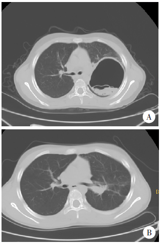
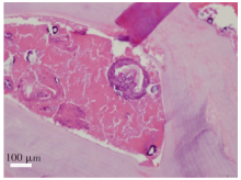
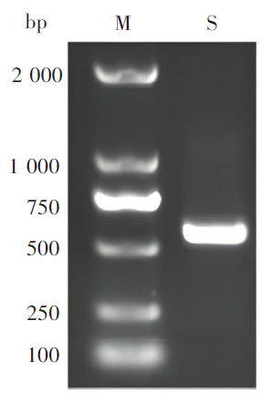
患儿,男,12岁,汉族,大理祥云县人。2020年8月2日出现阵咳、有痰,口服阿莫西林胶囊3 d后无减轻;8月5日至当地卫生室予阿莫西林克拉维酸钾、炎琥宁、溴己新输液治疗3 d,仍咳嗽;8月11日起发热,并出现口周青紫、呼吸稍促;8月11日转至祥云县医院,血常规示白细胞、嗜酸粒细胞升高,胸部CT提示左肺上叶脓肿形成可能,诊断为“肺脓肿、重症肺炎”,予万古霉素静滴3 d,患儿咳嗽减轻,无气促,仍发热;8月14日转至昆明市儿童医院,以“肺炎、肺脓肿”收住院。流行病学调查结果显示,患儿无外出旅居史,常与家养犬接触。实验室检查示嗜酸粒细胞增高,血清棘球蚴抗体IgG阳性;胸部CT示双肺感染并左肺上叶空腔形成,见“水上浮莲征”,临床诊断为肺细粒棘球蚴病。予左肺上叶包囊剥离术,术后肺组织病理检查鉴定为细粒棘球蚴感染。包囊组织DNA经PCR扩增后测序,结果显示与细粒棘球绦虫线粒体细胞色素氧化酶亚单位2(cox2)基因序列一致性高达99.24%。术后口服阿苯达唑[10 mg/(kg•d),2次/d,每4周为1个疗程,间歇1周,连用3个疗程],体温恢复正常,咳嗽明显减轻。术后6个月复查,胸部CT示左肺上叶空腔样病灶明显缩小,邻近渗出灶较前局部密实,部分吸收。术后定期随访18个月,无复发。

关键词: 肺细粒棘球蚴病, 儿童, 云南

Abstract:

The patient was a 12-year-old boy of Han ethnic group who lived in Xiangyun County, Dali. The patient complained of wet cough on August 2, 2020, and took amoxicillin capsules for 3 days, without relief. Then he was brought to the local clinic and received amoxicilin/Clavulanate potassium, potassium sodium dehydroandroan drographolide succinate for injection and bromhexine hydrochloride for 3 days. The patient was subsequently referred to the Department of Pediatrics of the Xiangyun County People’s Hospital due to fever, shortness of breath and cyanosis on August 11, 2020. The blood routine test showed elevated white blood cells and eosinophils. The chest CT scan showed a large cystic mass with a thick wall in the left lobe of the lung. The patient was misdiagnosis as lung abscess and severe pneumonia, received intravenous vancomycin for 3 days, but also in fever. The patient was admitted to Kunming Children’s Hospital with diagnosed as pneumonia and lung abscess on August 14, 2020. The patient, who was an inhabitant of a rural area, had a history of prolonged exposed to domestic dogs and goats. Laboratory investigations showed marked eosinophilia and positive Echinococcus IgG antibody. The chest CT scan showed infiltration in the lung and a large cystic mass in the left upper lobe of the lung with curvilinear septa within the lesion. The Water-Lily sign was reported. The patient was therefore diagnosed with pulmonary cystic echinococcosis. Removal of cyst in the left upper lobe of the patient’s left lung. The pathological biopsy identified E. granulosus parasite in the lung tissue. The total DNA was extracted from the paraffin-embedded samples of the hydatid cyst, and the molecular analysis was performed. The amplified PCR products were purified and subjected to direct sequencing. The results showed 99.2% sequence identity to the cox2 gene of mitochondrial of E. granulosus. The patient was administrated with albendazole at oral dose of 10 mg/(kg•d), twice a day for 3 months. The body temperature of the patient returned to normal and without cough. A postoperative chest CT scan showed the cavity in the left upper lobe of the lung was significantly reduced and sufficient expansion of the lung in the sixth mouth. Recurrence was not observed for more than 18 months of follow-up.

Key words: Pulmonary cystic echinococcosis, Children, Yunnan

中图分类号: