
中国寄生虫学与寄生虫病杂志 ›› 2022, Vol. 40 ›› Issue (4): 516-523.doi: 10.12140/j.issn.1000-7423.2022.04.016
张婷婷1,2,4( ), 杜秋沛1, 郭新建3, 张灵强2,4, 王志鑫2,4, 常正松1,3, 赵乾2,4, 王海久2,4, 侯立朝2,4,*()
收稿日期:2021-09-23
修回日期:2022-01-04
出版日期:2022-08-30
发布日期:2022-09-07
通讯作者:
侯立朝
作者简介:张婷婷(1995-),女,硕士研究生,从事肝胆胰相关疾病研究,E-mail: 705042690@qq.com
基金资助:
ZHANG Ting-ting1,2,4(), DU Qiu-pei1, GUO Xin-jian3, ZHANG Ling-qiang2,4, WANG Zhi-xin2,4, CHANG Zheng-song1,3, ZHAO Qian2,4, WANG Hai-jiu2,4, HOU Li-zhao2,4,*()
Received:2021-09-23
Revised:2022-01-04
Online:2022-08-30
Published:2022-09-07
Contact:
HOU Li-zhao
Supported by:摘要:
肝多房棘球蚴病病灶对人体重要的脉管系统(血管、胆管、淋巴结)是否侵犯及其侵犯程度,直接影响患者的手术治疗方式。目前,临床对于血管侵犯、胆管侵犯及淋巴结转移的病理状态、检查指征及治疗方式的关系尚不明确。本文综合国内外最新研究进展,对肝多房棘球蚴病的增殖与转移机制,病灶的病理分区,及脉管侵犯的肉眼、影像、显微镜下的特点进行深入阐述,以期推进肝多房棘球蚴病脉管侵犯诊断与治疗等研究的发展。
中图分类号:
张婷婷, 杜秋沛, 郭新建, 张灵强, 王志鑫, 常正松, 赵乾, 王海久, 侯立朝. 肝多房棘球蚴病脉管侵犯的研究进展[J]. 中国寄生虫学与寄生虫病杂志, 2022, 40(4): 516-523.
ZHANG Ting-ting, DU Qiu-pei, GUO Xin-jian, ZHANG Ling-qiang, WANG Zhi-xin, CHANG Zheng-song, ZHAO Qian, WANG Hai-jiu, HOU Li-zhao. Research progress on vascular invasion of hepatic alveolar echinococcosis[J]. Chinese Journal of Parasitology and Parasitic Diseases, 2022, 40(4): 516-523.
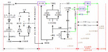
图1
多房棘球蚴的传播途径、增殖转移机制、病灶分区及分层概括图
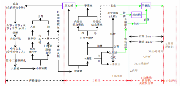
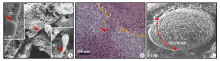
图2
多房棘球蚴囊泡生发层的生发细胞释放与游离[9⇓-11] A:电镜下的突起,末端逐渐膨大;B:光镜下的突起(红色箭头示),呈指状向外延伸;C:电镜下的“溶囊”现象,宿主免疫细胞附着于多房棘球蚴囊泡表面。TU:管状结构/实心延伸;BE:囊状扩大/膨大;E:嗜酸粒细胞;L:淋巴细胞;M:巨噬细胞;N:中性粒细胞;P:浆细胞。
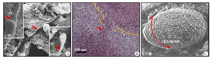
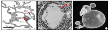
图3
多房棘球蚴囊泡的出芽性增殖与脱落[12⇓-14] A:幼虫囊泡示意图,囊泡向内/外出芽性增殖;B:光镜下的腔隙壁,外源性芽殖突破腔隙壁;C:电镜下的外源性出芽性增殖,子囊泡脱落。ENV:内源性幼虫囊泡;EXV:外源性幼虫囊泡;FLW:纤维细胞腔隙壁;EB:外源性出芽性增殖;PV:初级囊泡;SV:次级囊泡。
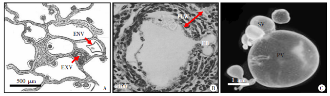
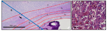
图4
光镜下多房棘球蚴病灶分区及分层 A:光镜下的肝多房棘球蚴病灶分区及分层,可分为3区、5层; B:光镜下的炎症细胞带区,分布着大量宿主免疫细胞。I :病灶区; II:边缘带区; :正常肝组织区; a:角皮层; 1:坏死区层; 2:内环层; 3a/b: 内/外纤维环层; 4:外环层; 5:周围肝,组织层(黑色箭头为淋巴细胞浸润)。
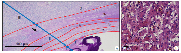
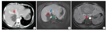
图5
肝多房棘球蚴病灶侵犯血管影像观[21-22] A:CT影像图,肝右静脉(红色箭头)受病灶推压而狭窄;B:MRI影像图,肝右静脉(红色箭头)被病灶局部包绕、肝中静脉(蓝色箭头)被病灶截断、下腔静脉(绿色箭头)未见显示;C:MRI影像图,肝右动脉(红色箭头)受病灶推压而出现移位、变形但未出现明显狭窄,管壁显影清楚。
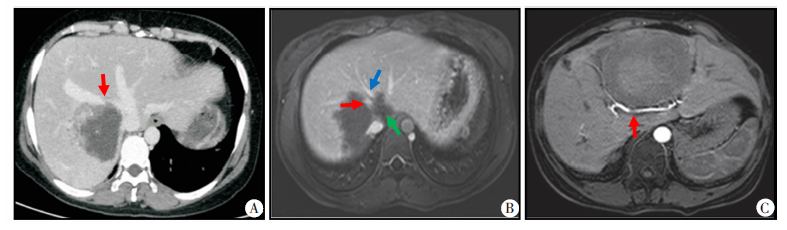
表1
多房棘球蚴病灶分区及分层
| 分区 | 分层 | 特点 |
|---|---|---|
| Ⅰ区:病灶 | a:角皮层 | 类同心圆叠层的粉皮,无细胞结构 |
| 第1层:坏死区 | 角皮层周围广泛的无结构坏死区 | |
| Ⅱ区:边缘带/ 浸润带/ 炎症反应带 | 第2层:内环 | 不规则,由上皮样细胞和中性粒细胞形成 |
| 第3a层:内纤维环 | 长条形纤维化区域,由纤维胶质形成 | |
| 第4层:外环 | 炎症细胞带区,由大量淋巴细胞、嗜酸粒细胞、巨噬细胞、中性粒细胞、浆细胞等炎症细胞及少量反应组织细胞组成( | |
| 第3b层:外纤维环 | 额外的纤维性边缘,可见淋巴细胞浸润,部分可无该层 | |
| Ⅲ区:正常肝组织 | 第5层:周围肝组织 | 邻近肝组织,肝细胞气球样变,周围纤维组织增生、淤血、胆汁瘀滞,汇管区炎症细胞浸润 |

图6
血管(下腔静脉)受侵肉眼观示意图(A)和实图(B)[23]
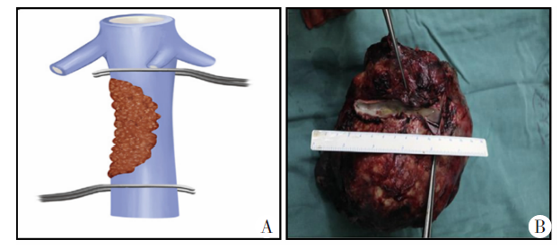
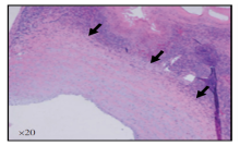
图7
肝多房棘球蚴病灶侵犯下腔静脉显微观(黑色箭头)[22]

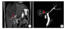
图8
肝多房棘球蚴病灶侵犯肝右胆管(红色箭头)影像观[26] A:MRI下,肝右胆管受病灶推压而狭窄;B:磁共振胆胰管成像,肝右胆管受压狭窄。


图9
肝多房棘球蚴病灶侵犯胆管显微观[26] 黑色箭头:多房棘球蚴病灶及边缘带区域;红色箭头:胆管黏膜
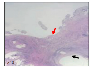
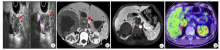
图10
肝多房棘球蚴病淋巴结转移(红色箭头)影像观[28⇓-30] A:超声下,转移的淋巴结无血流信号;B:CT下,转移的淋巴结未见强化;C:MRI下,转移的淋巴结未见强化;D:PET-CT下,转移的胰周淋巴结呈高摄取。


图11
肝多房棘球蚴病淋巴结转移肉眼观[28]
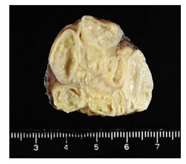

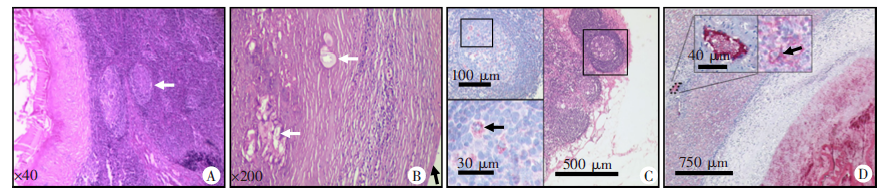
图12
肝多房棘球蚴病淋巴结转移显微观[30⇓-32] A:HE染色,转移淋巴结中央区可见淋巴小结(白色箭头);B:HE染色,转移淋巴结周边区可见淋巴结被膜(黑色箭头)、多房棘球蚴囊泡(白色箭头);C:Em2G11单克隆抗体染色,转移淋巴结可见SPEMS颗粒(黑色箭头);D:Em2G11单克隆抗体染色,病灶周围肝窦内可见SPEMS颗粒(黑色箭头)
| [1] | Huang H, Zhang Sk. Research progress on mechanisms of infiltration and metastasis for the alveolar echinococcosis[J]. Chin J Zoonoses, 2016. 32(7): 670-673, 678. (in Chinese) |
| ( 黄红, 张淑坤. 多房棘球蚴病的浸润和转移机制研究进展[J]. 中国人兽共患病学报, 2016, 32(7): 670-673, 678.) | |
| [2] |
Yu XK, Zhang L, Ma WJ, et al. An overview of hepatic echinococcosis and the characteristic CT and MRI imaging manifestations[J]. Infect Drug Resist, 2021, 14: 4447-4455.
doi: 10.2147/IDR.S331957 |
| [3] | Chen Y, Chen YF, Chen XP. The surgical theory and clinical application of hepatic perivascular space[J]. J Abdom Surg, 2021, 34(2): 88-91. (in Chinese) |
| ( 陈姚, 陈义发, 陈孝平. 肝血管旁间隙外科理念及其临床应用[J]. 腹部外科, 2021, 34(2): 88-91.) | |
| [4] | Shen S. Expert consensus on diagnosis and treatment of alveolar hepatic echinococcosis (2020 Edition)[J]. Chin J Bases Clin Gen Surg, 2020, 27(1): 13-17. (in Chinese) |
| 沈舒. 泡型肝包虫病诊疗专家共识(2020版)[J]. 中国普外基础与临床杂志, 2020, 27(1): 13-17.) | |
| [5] | Yang XW. Expert consensus on diagnosis and treatment of complex hepatic alveolar echinococcosis (2020 Edition)[J]. Chin J Bases Clin Gen Surg, 2020, 27(1): 18-23. (in Chinese) |
| 杨先伟. 复杂肝泡型包虫病诊疗专家共识(2020版)[J]. 中国普外基础与临床杂志, 2020, 27(1): 18-23.) | |
| [6] | Shao YM, Jiang TM, TuErGanAiLi·AJ, et al. Radical and quasi-radical surgery for end-stage hepatic alveolar echinococcosis[J]. Chin J Digest Surg, 2011, 10(4): 296-298. (in Chinese) |
| ( 邵英梅, 蒋铁民, 吐尔干艾力·阿吉, 等. 根治性及准根治性手术治疗终末期肝泡型包虫病[J]. 中华消化外科杂志, 2011, 10(4): 296-298.) | |
| [7] | Wen H, Vuitton L, Tuxun T, et al. Echinococcosis: advances in the 21st Century[J]. Clin Microbiol Rev, 2019, 32(2): e00075-e00018. |
| [8] | Zheng CJ, Xue CZ, Han S, et al. National alveolar echinococcosis distribution: China, 2012—2016[J]. China CDC Wkly, 2020, 2(1): 1-7. |
| [9] |
Mehlhorn H, Eckert J, Thompson RC. Proliferation and metastases formation of larval Echinococcus multilocularis. Ⅱ. Ultrastructural investigations[J]. Z Parasitenkd, 1983, 69(6): 749-763.
doi: 10.1007/BF00927424 |
| [10] | Maimaiti WSL. Effects of different surgical margins on postoperative recurrence of hepatic alveolar echinococcosis[D]. Urumqi: Xinjiang Medical University, 2018: 4-30. (in Chinese) |
| ( 买买提·瓦司力. 不同的手术切缘对肝泡型包虫病术后复发的影响[D]. 乌鲁木齐: 新疆医科大学, 2018: 4-30.) | |
| [11] |
Ali-Khan Z, Siboo R, Gomersall M, et al. Cystolytic events and the possible role of germinal cells in metastasis in chronic alveolar hydatidosis[J]. Ann Trop Med Parasitol, 1983, 77(5): 497-512.
doi: 10.1080/00034983.1983.11811742 |
| [12] |
Rausch R. Studies on the helminth fauna of Alaska XX. the histogenesis of the alveolar larva of Echinococcus species[J]. J Infect Dis, 1954, 94(2): 178-186.
doi: 10.1093/infdis/94.2.178 |
| [13] |
Ali-Khan Z, Siboo R. Pathogenesis and host response in subcutaneous alveolar hydatidosis[J]. Z Parasitenkd, 1980, 62(3): 241-254.
doi: 10.1007/BF00926565 |
| [14] |
Gottstein B, Hemphill A. Echinococcus multilocularis: the parasite-host interplay[J]. Exp Parasitol, 2008, 119(4): 447-452.
doi: 10.1016/j.exppara.2008.03.002 pmid: 18410929 |
| [15] |
Grimm J, Beck A, Nell J, et al. Combining computed tomography and histology leads to an evolutionary concept of hepatic alveolar echinococcosis[J]. Pathogens, 2020, 9(8): 634.
doi: 10.3390/pathogens9080634 |
| [16] | Liver cancer branch of China Association for the promotion of international exchanges in health care. Expert consensus on hepatocellular carcinoma complicated with vascular invasion (discussion draft)[J]. Electron J Liver Tumor, 2015, 2(3): 1-11. (in Chinese) |
| 中国医疗保健国际交流促进会肝脏肿瘤分会. 肝细胞癌合并血管侵犯专家共识(讨论稿)[J]. 肝癌电子杂志, 2015, 2(3): 1-11.) | |
| [17] | Cong WM, Bu H, Chen J, et al. Evidence-based practice guidelines for the standardized pathological diagnosis of primary liver cancer(2015 edition)[J]. Med J Chin People’s Liberation Army, 2015, 40(11): 865-872. (in Chinese) |
| 丛文铭, 步宏, 陈杰, 等. 原发性肝癌规范化病理诊断指南(2015年版)[J]. 解放军医学杂志, 2015, 40(11): 865-872.) | |
| [18] |
San Norberto EM, Fuente R, Taylor JH, et al. Endovascular management of inferior vena cava invasion by hepatic hydatid cyst[J]. J Vasc Interv Radiol, 2015, 26(1): 144-146.
doi: 10.1016/j.jvir.2014.10.003 pmid: 25541456 |
| [19] |
Kaynak K, Köksal C, Kazimoĝlu K, et al. Vascular echinococcosis[J]. Asian Cardiovasc Thorac Ann, 2002, 10(3): 259-261.
doi: 10.1177/021849230201000317 |
| [20] | Du QP, Wang ZX, Ren L, et al. A case report of obstructive jaundice caused by rupture of hepatic Echinococcus granulosus cyst into biliary tract[J]. Chin J Parasitol Parasit Dis, 2021, 39(1): 99-100. (in Chinese) |
| ( 杜秋沛, 王志鑫, 任利, 等. 肝细粒棘球蚴包囊破入胆道致梗阻性黄疸1例[J]. 中国寄生虫学与寄生虫病杂志, 2021, 39(1): 99-100.) | |
| [21] | Yang XF, Bao HH, Cao JY, et al. Study on the characteristics of "venophilic vessels" in hepatic alveolar echinococcosis[J]. J Practical Radiol, 2018, 34(4): 541-544, 567. (in Chinese) |
| ( 杨晓菲, 鲍海华, 曹佳媛, 等. 肝泡型包虫病"嗜静脉血管"特点研究[J]. 实用放射学杂志, 2018, 34(4): 541-544, 567.) | |
| [22] | Yang XF, Kang YL, Qiao YJ, et al. Magnetic resonance imaging evaluation of characteristics of vascular invasion in intermediate and advanced hepatic alveolar echinococcosis[J]. Exp Ther Med, 2019, 17(5): 4197-4204. |
| [23] |
Shen S, Kong J, Zhao J, et al. Outcomes of different surgical resection techniques for end-stage hepatic alveolar echinococcosis with inferior vena cava invasion[J]. HPB (Oxford), 2019, 21(9): 1219-1229.
doi: 10.1016/j.hpb.2018.10.023 |
| [24] | WHO informal working group on echinococcosis. Guidelines for treatment of cystic and alveolar echinococcosis in humans[J]. Bull WHO, 1996, 74(3): 231-242. |
| [25] | Ayifuhan AH, Cao J, Tuerganaili, et al. Surgical treatment of hepatic alveolar echinococcosis: an analysis of 43 cases[J]. Chin J Hepatobiliary Surg, 2011, 17(3): 213-215. (in Chinese) |
| ( 阿依甫汗·阿汗, 曹峻, 吐尔干艾力. 等. 肝泡型包虫病的手术治疗:附43例病例分析[J]. 中华肝胆外科杂志, 2011, 17(3): 213-215.) | |
| [26] | Kang YL, Bao HH, Wang LY. The value of MRI in differential diagnosis of hilar biliary invasion and hilar cholangiocarcinoma in echinococcosis[J]. J Clin Radiol, 2019, 38(10): 1862-1866. (in Chinese) |
| ( 康莹丽, 鲍海华, 王理祎. MRI对泡型包虫病肝门部胆管侵犯与肝门部胆管癌鉴别诊断的价值[J]. 临床放射学杂志, 2019, 38(10): 1862-1866.) | |
| [27] | Kang YL. MRI evaluation of biliary tract damage in hepatic alveolar echinococcosis[D]. Xining: Qinghai University, 2019: 1-27. (in Chinese) |
| 康莹丽. 肝泡型包虫病胆道损害的MRI评价[D]. 西宁: 青海大学, 2019: 1-27.). | |
| [28] |
Amano T, Hayashi S, Nishida T, et al. Alveolar echinococcosis mimicking a hepatic neoplasm with lymph node metastasis: a case report[J]. Case Rep Gastroenterol, 2018, 12(3): 587-596.
doi: 10.1159/000492461 |
| [29] | Gao HY, Li WX, Sun ZX, et al. CT and MRI in growth feature evaluation for the children hepatic alveolar echinococcosis[J]. J Clin Radiol, 2017, 36(8): 1170-1173. (in Chinese) |
| ( 高会艳, 李伟霞, 孙再兴, 等. CT、 MRI对儿童肝泡状棘球蚴病生长特性的评价[J]. 临床放射学杂志, 2017, 36(8): 1170-1173.) | |
| [30] | Pu P, Liu L, Chen ZX, et al. Imaging findings and pathological analysis of lymph node metastasis in alveolar echinococcosis[J]. J Xinjiang Med Univ, 2019, 42(5): 647-651. (in Chinese) |
| ( 蒲鹏, 刘丽, 陈增雄, 等. 泡状棘球蚴淋巴结转移的影像表现及病理分析[J]. 新疆医科大学学报, 2019, 42(5): 647-651.) | |
| [31] |
Hillenbrand A, Beck A, Kratzer W, et al. Impact of affected lymph nodes on long-term outcome after surgical therapy of alveolar echinococcosis[J]. Langen Arch Surg, 2018, 403(5): 655-662.
doi: 10.1007/s00423-018-1687-9 |
| [32] | Barth TF, Herrmann TS, Tappe D, et al. Sensitive and specific immunohistochemical diagnosis of human alveolar echinococcosis with the monoclonal antibody Em2G11[J]. PLoS Negl Trop Dis, 2012, 6(10): e1877. |
| [33] |
Buttenschoen K, Kern P, Reuter S, et al. Hepatic infestation of Echinococcus multilocularis with extension to regional lymph nodes[J]. Langen Arch Surg, 2009, 394(4): 699-704.
doi: 10.1007/s00423-009-0481-0 |
| [34] |
Morine Y, Shimada M. The value of systematic lymph node dissection for intrahepatic cholangiocarcinoma from the viewpoint of liver lymphatics[J]. J Gastroenterol, 2015, 50(9): 913-27.
doi: 10.1007/s00535-015-1071-2 pmid: 25833009 |
| [35] | Xu K, Wang HJ, Zhang L, et al. Research progress on the mechanisms underlying the impairment of host hepatocytes by Echinococcus multilocularis[J]. Chin J Parasitol Parasit Dis, 2021, 39(2): 256-260. (in Chinese) |
| ( 徐凯, 王海久, 张丽, 等. 多房棘球蚴对宿主肝细胞损害机制的研究进展[J]. 中国寄生虫学与寄生虫杂志, 2021, 39(2): 256-260.) | |
| [36] | Liang HJ, Qin SK, Shen F, et al. CSCO expert consensus on diagnosis and treatment of biliary system tumors (2019 Edition)[J]. Chin Clin Oncol, 2019, 24(9): 828-838. (in Chinese) |
| 梁后杰, 秦叔逵, 沈锋. CSCO胆道系统肿瘤诊断治疗专家共识(2019年版)[J]. 临床肿瘤学杂志, 2019, 24(9): 828-838.) | |
| [37] | Xu K, Huang LL, Wu CL, et al. Research progress on the experimental treatment of Echinococcus infection by using Chinese traditional medicine[J]. Chin J Parasitol Parasit Dis, 2021, 39(5): 710-715. (in Chinese) |
| ( 徐凯, 黄璐璐, 吴传玲, 等. 中药实验治疗棘球蚴病的研究进展[J]. 中国寄生虫学与寄生虫病杂志, 2021, 39(5): 710-715.) |
| [1] | 娆琬·托勒洪, 阿不都撒拉木·阿不力克木, 杨凌菲, 陈璐, 李钊, 贾芳, 宋涛. 超声表现诊断肝多房棘球蚴病的效果评价及因素分析[J]. 中国寄生虫学与寄生虫病杂志, 2023, 41(3): 312-318. |
| [2] | 安秀青, 王苗苗, 周鸿乾, 孟凯, 蔡剑平, 刘光辉, 阿吉德, 杨金煜. 肝多房棘球蚴病微血管密度的研究进展[J]. 中国寄生虫学与寄生虫病杂志, 2022, 40(6): 792-797. |
| [3] | 吴亮亮, 杨凌菲, 宋涛. 不同方式建立肝多房棘球蚴感染SD大鼠模型病灶的超声及病理表现[J]. 中国寄生虫学与寄生虫病杂志, 2022, 40(4): 549-552. |
| [4] | 喀斯木·艾海提, 阿卜杜萨拉木·艾尼, 吐尔干艾力·阿吉, 邵英梅, 张瑞青, 塔来提·吐尔干, 蒋铁民, 冉博, 阿卜杜艾尼·啊卜力孜, 米尔阿迪力·艾尔肯, 温浩. 离体肝切除和自体肝移植术治疗终末期肝多房棘球蚴病的住院医疗费用分析[J]. 中国寄生虫学与寄生虫病杂志, 2020, 38(1): 53-57. |
| [5] | 许晓磊, 王志鑫, 王展, 叶海雯, 庞明泉, 周灜, 王海久, 樊海宁. 98例复杂性肝多房棘球蚴病外科治疗方案的比较分析[J]. 中国寄生虫学与寄生虫病杂志, 2018, 36(6): 552-559. |
| [6] | 阿卜杜萨拉木·艾尼1,吐尔洪江·吐逊2,马海长3,张恒2,张皓1,阿卜杜凯尤木·麦麦提4,李玉鹏2,沙地克·阿帕尔2,林仁勇5,邵英梅1,温浩5*. Toll样受体mRNA和相关细胞因子在肝多房棘球蚴病患者体内的变化[J]. 中国寄生虫学与寄生虫病杂志, 2016, 34(6): 12-542-546. |
| 阅读次数 | ||||||
|
全文 |
|
|||||
|
摘要 |
|
|||||