| 1 | Ryan U, Paparini A, Monis P, et al. It's official-Cryptosporidium is a gregarine: what are the implications for the water industry[J]. Water Res, 2016, 105: 305-313. | | 2 | McDonald V, Korbel DS, Barakat FM, et al. Innate immune responses against Cryptosporidium parvum infection[J]. Parasite Immunol, 2013, 35(2): 55-64. | | 3 | Kawai T, Akira S. The role of pattern-recognition receptors in innate immunity: update on Toll-like receptors[J]. Nat Immunol, 2010, 11(5): 373-384. | | 4 | 鱼艳荣, 齐永芬. Toll样受体在蠕虫感染中的作用[J]. 中国寄生虫学与寄生虫病杂志, 2010, 28(5): 377-381. | | 5 | Yang Z, Fu Y, Gong P, et al. Bovine TLR2 and TLR4 mediate Cryptosporidium parvum recognition in bovine intestinal epithelial cells[J]. Microb Pathog, 2015, 85: 29-34. | | 6 | Steinman RM, Adams JC, Cohn ZA. Identification of a novel cell type in peripheral lymphoid organs of mice. IV. Identification and distribution in mouse spleen[J]. J Exp Med, 1975, 141(4): 804-820. | | 7 | Tsai JJ, Wang HC, Chiu CL, et al. The effect of Dermatophagoides pteronyssinus group 7 allergen (Der p 7) on dendritic cells and its role in T cell polarization[J]. Immunobiology, 2016, 221(11): 1319-1328. | | 8 | 姜晶, 赵权, 杨桂连. 树突状细胞在蠕虫感染免疫应答中的作用[J]. 中国寄生虫学与寄生虫病杂志, 2015, 33(2): 147-150. | | 9 | Lundie RJ, Young LJ, Davey GM, et al. Blood-stage Plasmodium berghei infection leads to short-lived parasite-associated antigen presentation by dendritic cells[J]. Eur J Immunol, 2010, 40(6): 1674-1681. | | [10] | 曲凯歌, 赵权, 姜晶, 等. 树突状细胞在寄生虫感染免疫中的作用[J]. 中国寄生虫学与寄生虫病杂志, 2014, 32(2): 152-156. | | [11] | Liu J, He C, Zhou H, et al. Effects of TLR4 on β2-glycoprotein I-induced bone marrow-derived dendritic cells maturation[J]. Cell Immunol, 2014, 290(2): 226-232. | | [12] | 刘晓霞, 朱明, 徐琦, 等. 寄生虫感染对树突状细胞亚群的影响[J]. 中国人兽共患病学报, 2012, 28(10): 1020-1024. | | [13] | Bedi B, McNair NN, Mead JR. Dendritic cells play a role in host susceptibility to Cryptosporidium parvum infection[J]. Immunol Lett, 2014, 158(1/2): 42-51. | | [14] | Poncini CV, Giménez G, Pontillo CA, et al. Central role of extracellular signal-regulated kinase and Toll-like receptor 4 in IL-10 production in regulatory dendritic cells induced by Trypanosoma cruzi[J]. Mol Immunol, 2010, 47(11-12): 1981-1988. | | [15] | Inaba K, Inaba M, Romani N, et al. Generation of large numbers of dendritic cells from mouse bone marrow cultures supplemented with granulocyte/macrophage colony-stimulating factor[J]. J Exp Med, 1992, 176(6): 1693-1702. | | [16] | 李培英, 廖圣法, 陆凤林, 等. 安徽省奶牛寄生蠕虫感染情况调查[J]. 安徽农业科学, 1999, 27(2): 77-79. | | [17] | Perez-Cordon G, Yang G, Zhou B, et al. Interaction of Cryptosporidium parvum with mouse dendritic cells leads to their activation and parasite transportation to mesenteric lymph nodes[J]. Pathog Dis, 2014, 70(1): 17-27. | | [18] | Xiao L, Escalante L, Yang C, et al. Phylogenetic analysis of Cryptosporidium parasites based on the small-subunit rRNA gene locus[J]. Appl Environ Microbiol, 1999, 65(4): 1578-1583. | | [19] | 许继伟, 宋璇, 梁伟姿. 艾滋病疫苗的研究进展[J]. 中国生物制品学杂志, 2016, 29(2): 213-216, 220. | | [20] | Taylan-Ozkan A, Yasa-Duru S, Usluca S, et al. Cryptosporidium species and Cryptosporidium parvum subtypes in dairy calves and goat kids reared under traditional farming systems in Turkey[J]. Exp Parasitol, 2016, 170: 16-20. | | [21] | Liu R, Wang J, Yang Y, et al. A novel rabies virus lipopeptide provides a better protection by improving the magnitude of DCs activation and T cell responses[J]. Virus Res, 2016, 221: 66-73. | | [22] | Meijerink M, Ulluwishewa D, Anderson RC, et al. Cryopreservation of monocytes or differentiated immature DCs leads to an altered cytokine response to TLR agonists and microbial stimulation[J]. J Immunol Methods, 2011, 373(1/2): 136-142. | | [23] | Reis e Sousa C. Activation of dendritic cells: translating innate into adaptive immunity[J]. Curr Opin Immunol 2004, 16(1): 21-25. | | [24] | 王冰, 王红梅, 徐方云, 等. 树突状细胞与肿瘤免疫[J]. 中国细胞生物学学报, 2013, 35(11): 1666-1671. | | [25] | Zlotnikov-Klionsky Y, Nathansohn-Levi B, Shezen E, et al. Perforin-positive dendriticells exhibit an immuno-regulatory role in metabolic syndrome and autoimmunity[J]. Immunity, 2015, 43(4): 776-787. | | [26] | 余裕强. 微小隐孢子虫刺激对宿主细胞TLRs的影响[D]. 长春: 吉林大学, 2015. | | [27] | 何小兵, 贾怀杰, 景志忠. Toll 样受体对病原寄生虫的天然免疫识别[J]. 生物工程学报, 2012, 28(12): 1401-1413. |
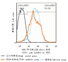
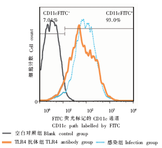
|

 )
)