
CHINESE JOURNAL OF PARASITOLOGY AND PARASITIC DISEASES ›› 2023, Vol. 41 ›› Issue (3): 286-293.doi: 10.12140/j.issn.1000-7423.2023.03.004
• ORIGINAL ARTICLES • Previous Articles Next Articles
YE Jingming( ), HE Wei, LIU Huiyuan, YU Xiao, LUO Bo, LIU Meichen, ZHOU Biying*()
Received:2022-09-06
Revised:2023-12-29
Online:2023-06-30
Published:2023-06-20
Contact:
*E-mail: Supported by:CLC Number:
YE Jingming, HE Wei, LIU Huiyuan, YU Xiao, LUO Bo, LIU Meichen, ZHOU Biying. Effect of excretory-secretory antigen TPx of Cysticercus cellulosae on activation of dendritic cells in piglets[J]. CHINESE JOURNAL OF PARASITOLOGY AND PARASITIC DISEASES, 2023, 41(3): 286-293.
Add to citation manager EndNote|Ris|BibTeX
URL: https://www.jsczz.cn/EN/10.12140/j.issn.1000-7423.2023.03.004
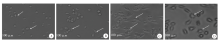
Fig. 1
Morphological changes of induced dendritic cell (DC) observed by light microscopy A: The DCs were ovoid on 1 d post-induction; B: The DCs showed pseudopods and spines on 4 d post-induction; C: The spines and pseudopods DCs further elongated on 7 d post-induction; D: More prominent irregular protrusions on the DCs surface on 9 d post-induction.
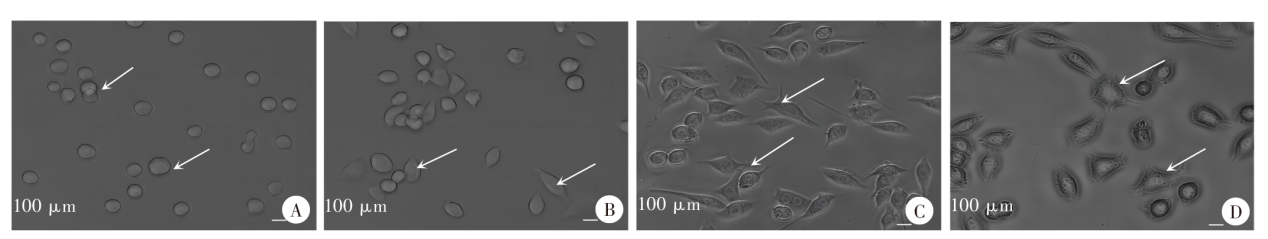

Fig. 2
Morphological changes of immature and mature DC observed by scanning electron microscope A: Immature DC cell population, a few pseudopods and spines are visible; B: Immature DC, surface irregular, with a few short protrusions from the cytosol; C: Mature DC cell population, numerous irregular protrusions are visible; D: Mature DC, long cloak-shaped, with a more irregular surface and cells radiating with protrusions of varying length.
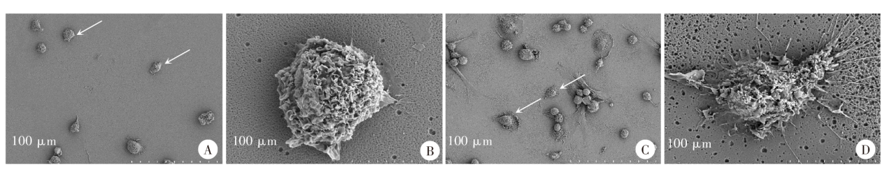
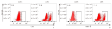
Fig. 3
The expression of immature and mature DC surface markers CD1 (A) and MHC-Ⅱ (B) detected by flow cytometry
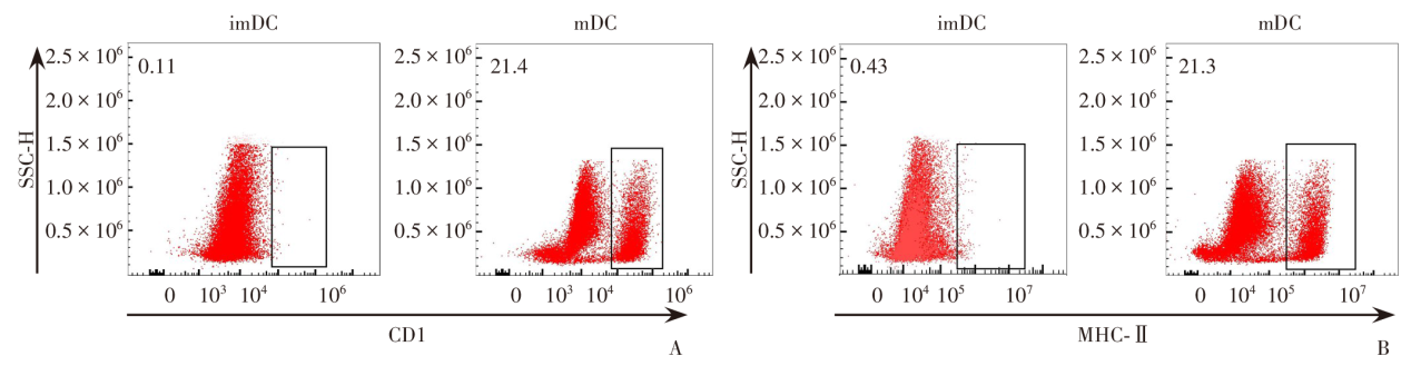
Fig. 4
Effect of TPx on the expression of DC surface markers MHC-Ⅱ (A), CD80 (B), and CD86 (C) detected by flow cytometry
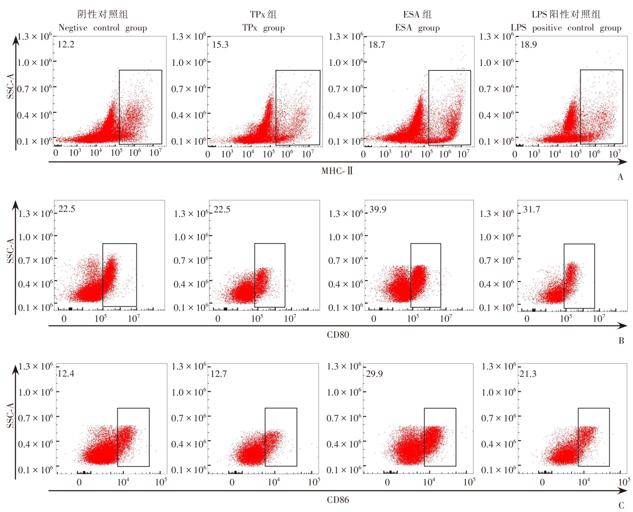
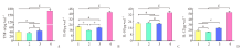
Fig. 5
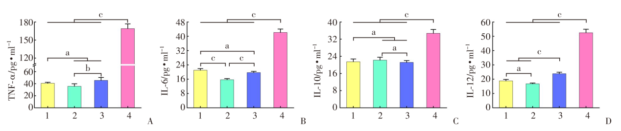
TPx protein induces the secretion of cytokines, including TNF-α (A), IL-6 (B), IL-10 (C)、IL-12 (D) in DC 1: Negative control group; 2: TPx group; 3: ESA group; 4: LPS positive control group. a: P > 0.05; b: P < 0.05; c: P < 0.01.
| [1] |
Gonzales I, Rivera JT, Garcia HH. Pathogenesis of Taenia solium taeniasis and cysticercosis[J]. Parasite Immunol, 2016, 38(3): 136-146.
doi: 10.1111/pim.12307 pmid: 26824681 |
| [2] |
Winkler AS. Neurocysticercosis in sub-Saharan Africa: a review of prevalence, clinical characteristics, diagnosis, and management[J]. Pathog Glob Health, 2012, 106(5): 261-274.
doi: 10.1179/2047773212Y.0000000047 pmid: 23265550 |
| [3] | Luo XN, Zheng YD, Cai XP. Epidemic situation and preventive strategies against taeniasis/cysticercosis[J]. J Pathog Biol, 2007, 2(3): 230-232, 238. (in Chinese) |
| (骆学农, 郑亚东, 才学鹏. 猪带绦虫/囊虫病的流行现状及防制策略[J]. 中国病原生物学杂志, 2007, 2(3): 230-232, 238.) | |
| [4] |
CystiTeam Group for Epidemiology and Modelling of Taenia solium Taeniasis/Cysticercosis. The World Health Organization 2030 goals for Taenia solium: insights and perspectives from transmission dynamics modelling[J]. Gates Open Res, 2019, 3: 1546.
doi: 10.12688/gatesopenres |
| [5] |
Braae UC, Hung NM, Satrija F, et al. Porcine cysticercosis (Taenia solium and Taenia asiatica): mapping occurrence and areas potentially at risk in East and Southeast Asia[J]. Parasit Vectors, 2018, 11(1): 613.
doi: 10.1186/s13071-018-3203-z |
| [6] | Li HZ, Zang XZ, Qian MB, et al. Current status and research progress of cysticercosis[J]. Chin J Schisto Control, 2018, 30(1): 99-103. (in Chinese) |
| (李焕璋, 臧新中, 钱门宝, 等. 囊尾蚴病流行现况及研究进展[J]. 中国血吸虫病防治杂志, 2018, 30(1): 99-103.) | |
| [7] | Chen T, Zhao GH, Ning CS, et al. Research progress on epidemiology of cysticercosis in China[J]. Shanghai J Animal Husb Vet Med, 2006(5): 18-20. (in Chinese) |
| (陈同, 赵光辉, 宁长申, 等. 我国囊虫病流行病学研究进展[J]. 上海畜牧兽医通讯, 2006(5): 18-20.) | |
| [8] |
Rosales-Mendoza S, Monreal-Escalante E, González-Ortega O, et al. Transplastomic plants yield a multicomponent vaccine against cysticercosis[J]. J Biotechnol, 2018, 266: 124-132.
doi: S0168-1656(17)31770-4 pmid: 29253519 |
| [9] | Fan XM, Zhou BY. Research advances on the relationship between cestode excretory/secretory products and host immune response[J]. Chin J Parasitol Parasit Dis, 2020, 38(1): 128-133. (in Chinese) |
| (范贤敏, 周必英. 绦虫排泄分泌物与宿主免疫效应的相关研究进展[J]. 中国寄生虫学与寄生虫病杂志, 2020, 38(1): 128-133.) | |
| [10] |
Fan X, Zhang Y, Ouyang R, et al. Cysticercus cellulosae regulates T-cell responses and interacts with the host immune system by excreting and secreting antigens[J]. Front Cell Infect Microbiol, 2021, 11: 728222.
doi: 10.3389/fcimb.2021.728222 |
| [11] | Zhang L, Fan XM, Ma Q, et al. Effects of an excretory secretory antigen of cysticercus cellulosae on the maturation and activation of DCs in piglets[J]. J Pathog Biol, 2021, 16(11): 1249-1253. (in Chinese) |
| (张雷, 范贤敏, 马琴, 等. 猪囊尾蚴排泄分泌抗原对仔猪DC成熟活化的影响[J]. 中国病原生物学杂志, 2021, 16(11): 1249-1253.) | |
| [12] | He W, Li LZ, Sun XQ, et al. Screening, validation, T-cell antigenic epitopes prediction and eukaryotic expression of Cysticercus cellulosae excretory-secretory antigen thioredoxin peroxidase protein[J]. J Pathog Biol, 2023, 18(2): 174-179, 184. (in Chinese) |
| (何威, 李丽竹, 孙晓晴, 等. 猪囊尾蚴排泄分泌抗原TPx蛋白的筛选验证、 T细胞抗原表位预测及真核表达[J]. 中国病原生物学杂志, 2023, 18(2): 174-179, 184.) | |
| [13] | He W, Luo B, Zhou BY. Research progress of recombinant thioredoxin peroxidase of important human parasites involved in immunoregulation, immunodiagnosis and immunoprophylaxis[J]. Chin J Endem, 2022, 41(10): 856-860. (in Chinese) |
| (何威, 罗波, 周必英. 人体重要寄生虫重组TPx参与免疫调控、免疫诊断及免疫预防的研究进展[J]. 中华地方病学杂志, 2022, 41(10): 856-860.) | |
| [14] |
Guo X, Zhang J, Li R, et al. Molecular cloning and functional characterization of a thioredoxin peroxidase gene in Echinococcus multilocularis[J]. Mol Biochem Parasitol, 2021, 245: 111408.
doi: 10.1016/j.molbiopara.2021.111408 |
| [15] | Li YG. Cloning, expression and antigenicity of thioredoxin peroxidase gene from Taenia polycephala[D]. Lanzhou: Gansu Agricultural University, 2009: 47-48. (in Chinese) |
| (李永光. 多头带绦虫硫氧还蛋白过氧化物酶基因的克隆、 表达及抗原性研究[D]. 兰州: 甘肃农业大学, 2009: 47-48.) | |
| [16] |
Hilligan KL, Ronchese F. Antigen presentation by dendritic cells and their instruction of CD4+ T helper cell responses[J]. Cell Mol Immunol, 2020, 17(6): 587-599.
doi: 10.1038/s41423-020-0465-0 pmid: 32433540 |
| [17] | Liu XX, Zhu M, Xu Y, et al. Parasitic infection to dendritic cell subsets[J]. Chin J Zoonoses, 2012, 28(10): 1020-1024. (in Chinese) |
| (刘晓霞, 朱明, 徐琦, 等. 寄生虫感染对树突状细胞亚群的影响[J]. 中国人兽共患病学报, 2012, 28(10): 1020-1024.) | |
| [18] |
Falcón C, Carranza F, Martínez FF, et al. Excretory-secretory products (ESP) from Fasciola hepatica induce tolerogenic properties in myeloid dendritic cells[J]. Vet Immunol Immunopathol, 2010, 137(1/2): 36-46.
doi: 10.1016/j.vetimm.2010.04.007 |
| [19] | Li LZ. Effect of proteomics-basted cysticercus cellulosae excretory secretory antigen LRRC15 protein on T-cell immune response in piglets[D]. Zunyi: Zunyi Medical University, 2022: 52-55. (in Chinese) |
| (李丽竹. 基于蛋白质组学研究猪囊尾蚴排泄分泌抗原LRRC15蛋白对仔猪T细胞免疫应答的影响[D]. 遵义: 遵义医科大学, 2022: 52-55.) | |
| [20] |
White RR, Artavanis-Tsakonas K. How helminths use excretory secretory fractions to modulate dendritic cells[J]. Virulence, 2012, 3(7): 668-677.
doi: 10.4161/viru.22832 pmid: 23221477 |
| [21] |
Wang Y, Zhou H, Shen Y, et al. Impairment of dendritic cell function and induction of CD4+CD25+Foxp3+ T cells by excretory-secretory products: a potential mechanism of immune evasion adopted by Echinococcus granulosus[J]. BMC Immunol, 2015, 16: 44.
doi: 10.1186/s12865-015-0110-3 |
| [22] | Li Y. Prokaryotic expression of GAPDH gene and Tpx gene of Baylisascaris schroederi and the evaluation of diagnostic value of recombinant antigens[D]. Yaan: Sichuan Agricultural University, 2017: 6-8. (in Chinese) |
| (李宇. 西氏贝蛔虫GAPDH基因和Tpx基因的原核表达与重组抗原诊断价值的评估[D]. 雅安: 四川农业大学, 2017: 6-8.) | |
| [23] | Yin C, Luo XN, Wang S, et al. Prokaryotic expression and biological properties of thioredoxin peroxidase from Taenia solium[J]. Acta Vet Zootechnica Sin, 2014, 45(9): 1512-1517. (in Chinese) |
| (尹才, 骆学农, 王帅, 等. 猪带绦虫硫氧还蛋白过氧化物酶的原核表达及其生物学特性分析[J]. 畜牧兽医学报, 2014, 45(9): 1512-1517.) | |
| [24] |
He W, Sun X, Luo B, et al. Regulation of piglet T-cell immune responses by thioredoxin peroxidase from Cysticercus cellulosaeexcretory-secretory antigens[J]. Front Microbiol, 2022, 13: 1019810.
doi: 10.3389/fmicb.2022.1019810 |
| [25] |
Moll H. Dendritic cells and host resistance to infection[J]. Cell Microbiol, 2003, 5(8): 493-500.
pmid: 12864809 |
| [26] |
Steinman RM, Hawiger D, Nussenzweig MC. Tolerogenic dendritic cells[J]. Annu Rev Immunol, 2003, 21: 685-711.
pmid: 12615891 |
| [27] | Sun LY, Ding Z, Chen P, et al. Research progress on tolerogenic dendritic cells[J]. Life Sci Res, 2020, 24(4): 314-320. (in Chinese) |
| (孙庐云, 丁喆, 陈鹏, 等. 耐受性树突状细胞的研究进展[J]. 生命科学研究, 2020, 24(4): 314-320.) | |
| [28] |
Maldonado RA, von Andrian UH. How tolerogenic dendritic cells induce regulatory T cells[J]. Adv Immunol, 2010, 108: 111-165.
doi: 10.1016/B978-0-12-380995-7.00004-5 pmid: 21056730 |
| [29] | Xu HY, He XZ. Research progress of tolerant dendritic cells[J]. Organ Transplant, 2014, 5(1): 49-53. (in Chinese) |
| (徐海燕, 何小舟. 耐受性树突状细胞的研究进展[J]. 器官移植, 2014, 5(1): 49-53.) | |
| [30] |
Na H, Cho M, Chung Y. Regulation of Th2 cell immunity by dendritic cells[J]. Immune Netw, 2016, 16(1): 1-12.
doi: 10.4110/in.2016.16.1.1 pmid: 26937227 |
| [31] | Dong LY. Various phenotype dendritic cells of rats mediated immune tolerance of liver transplantation in vitro[D]. Kungming: Kunming Medical University, 2012: 31-32. (in Chinese) |
| (董丽英. 大鼠不同表型树突状细胞介导肝移植免疫耐受的体外实验研究[D]. 昆明: 昆明医科大学, 2012: 31-32.) | |
| [32] |
Cvetkovic J, Ilic N, Gruden-Movsesijan A, et al. DC-SIGN signalling induced by Trichinella spiralis products contributes to the tolerogenic signatures of human dendritic cells[J]. Sci Rep, 2020, 10(1): 20283.
doi: 10.1038/s41598-020-77497-x pmid: 33219293 |
| [33] |
Lamendour L, Deluce-Kakwata-Nkor N, Mouline C, et al. Tethering innate surface receptors on dendritic cells: a new avenue for immune tolerance induction?[J]. Int J Mol Sci, 2020, 21(15): 5259.
doi: 10.3390/ijms21155259 |
| [34] |
Nam JH, Lee JH, Choi SY, et al. Functional ambivalence of dendritic cells: tolerogenicity and immunogenicity[J]. Int J Mol Sci, 2021, 22(9): 4430.
doi: 10.3390/ijms22094430 |
| [35] | Qiao YC, Shen J, He L, et al. Changes of regulatory T cells and of proinflammatory and immunosuppressive cytokines in patients with type 2 diabetes mellitus: a systematic review and meta-analysis[J]. J Diabetes Res, 2016, 2016: 3694957. |
| [36] |
Qiao YC, Pan YH, Ling W, et al. The Yin and Yang of regulatory T cell and therapy progress in autoimmune disease[J]. Autoimmun Rev, 2017, 16(10): 1058-1070.
doi: 10.1016/j.autrev.2017.08.001 |
| [37] |
Korn T, Hiltensperger M. Role of IL-6 in the commitment of T cell subsets[J]. Cytokine, 2021, 146: 155654.
doi: 10.1016/j.cyto.2021.155654 |
| [38] |
Yang XO, Nurieva R, Martinez GJ, et al. Molecular antagonism and plasticity of regulatory and inflammatory T cell programs[J]. Immunity, 2008, 29(1): 44-56.
doi: 10.1016/j.immuni.2008.05.007 pmid: 18585065 |
| [39] |
Samanta A, Li B, Song X, et al. TGF-beta and IL-6 signals modulate chromatin binding and promoter occupancy by acetylated FOXP3[J]. Proc Natl Acad Sci USA, 2008, 105(37): 14023-14027.
doi: 10.1073/pnas.0806726105 pmid: 18779564 |
| [40] |
Essig K, Hu D, Guimaraes JC, et al. Roquin suppresses the PI3K-mTOR signaling pathway to inhibit T helper cell differentiation and conversion of Treg to Tfr cells[J]. Immunity, 2017, 47(6): 1067-1082. e12.
doi: S1074-7613(17)30485-5 pmid: 29246441 |
| [41] |
Svensson MN, Doody KM, Schmiedel BJ, et al. Reduced expression of phosphatase PTPN2 promotes pathogenic conversion of Tregs in autoimmunity[J]. J Clin Invest, 2019, 129(3): 1193-1210.
doi: 10.1172/JCI123267 pmid: 30620725 |
| [42] |
Li D, Kong C, Tsun A, et al. MiR-125a-5p decreases the sensitivity of Treg cells toward IL-6-mediated conversion by inhibiting IL-6R and STAT3 expression[J]. Sci Rep, 2015, 5: 14615.
doi: 10.1038/srep14615 pmid: 26424054 |
| [43] |
Hsieh WC, Hsu TS, Chang YJ, et al. IL-6 receptor blockade corrects defects of XIAP-deficient regulatory T cells[J]. Nat Commun, 2018, 9(1): 463.
doi: 10.1038/s41467-018-02862-4 |
| [44] | Zhang CK. Study on immune tolerance induced by bone marrow mesenchymal stem cells combined with IL-6 monoclonal antibody in heart transplantation[D]. Suzhou: Soochow University, 2012: 41-43. (in Chinese) |
| (章传凯. 骨髓间充质干细胞联合IL-6单克隆抗体诱导心脏移植免疫耐受的研究[D]. 苏州: 苏州大学, 2012: 41-43.) |
| [1] | ZHANG Ling-hui, CHEN Gen, CHONG Shi-gui, SHEN Hui, MA Hui, ZHAO Yu-min. Research progress on the immune regulation mechanism in alveolar echinococcosis [J]. CHINESE JOURNAL OF PARASITOLOGY AND PARASITIC DISEASES, 2022, 40(1): 109-113. |
| [2] | CHEN Sui-lin, GAO Yuan-li, GUO Shuai, FAN Yong-ling, LIU Tai-ping, XU Wen-yue. Effect and mechanism of high-dose clodronate liposomes treatment on Plasmodium yoelii growth in mice [J]. CHINESE JOURNAL OF PARASITOLOGY AND PARASITIC DISEASES, 2022, 40(1): 28-35. |
| [3] | ZHOU Yan, CHEN Shao-hong, LI Hao, CHENG Na, HONG Jia-lin, XU Xue-nian. Cloning, expression of the thioredoxin peroxidase gene of Paragonimus westermani and its immunodiagnostic potential [J]. CHINESE JOURNAL OF PARASITOLOGY AND PARASITIC DISEASES, 2021, 39(1): 20-26. |
| [4] | YU Xiao-dong, YALI Ya-sen, WANG Jia-ling, LI Meng, YE Jian-rong. Establishment of BALB/c mouse model of Echinococcus granulosus-induced sensitization and changes of related immune cells [J]. CHINESE JOURNAL OF PARASITOLOGY AND PARASITIC DISEASES, 2020, 38(4): 412-416. |
| [5] | Bei JIANG, Xiao-jun XIAO, Chun-yan OUYANG, Xin-ping LUO, Bao-qing SUN, Jing LI, Zhi-gang LIU. Cloning and expression of must mite allergen Der f 32 and its activation on mouse dendritic cells [J]. CHINESE JOURNAL OF PARASITOLOGY AND PARASITIC DISEASES, 2019, 37(3): 279-285. |
| [6] | Jian-da PANG, Yi-ning SONG, Xin-rui WANG, Ming-yuan LIU, Shu-min SUN. Proteomic analysis of cyst fluid of Cysticercus cellulosae [J]. CHINESE JOURNAL OF PARASITOLOGY AND PARASITIC DISEASES, 2019, 37(2): 144-149. |
| [7] | Shang-hua WU, E-yan GENG, Jing ZHANG, Heng ZHANG, Zhi-qiang SHI, Shan WANG, Wei LU, Yi-zhen WU, Gui-jun WANG, Yong WANG, Qian-ming XU. Effects of recombinant autophagy related 5 protein on the maturation of dendritic cells stimulated by Toxoplasma gondii [J]. CHINESE JOURNAL OF PARASITOLOGY AND PARASITIC DISEASES, 2018, 36(5): 464-468. |
| [8] | Yong FU, Ru MENG, Hai-fang XUE, Hai-ning FAN, Hai-feng NIU, Zi-jia ZHOU, Hong-bin WANG. Morphological observation and phenotypic detection of dendritic cells from peripheral blood of patients with alveolar echinococcosis [J]. CHINESE JOURNAL OF PARASITOLOGY AND PARASITIC DISEASES, 2018, 36(5): 474-477. |
| [9] | Rui-xue YE, Yu-ju WU, Qing-zhi WANG, Min CAO, Tiao-ying LI, Xing-wang CHEN, Huan ZHOU. Status of Cysticercus cellulosae infection and its impact on mathematical learning ability among school-aged children in Tibetan agricultural areas of Sichuan Province [J]. CHINESE JOURNAL OF PARASITOLOGY AND PARASITIC DISEASES, 2017, 35(6): 580-585. |
| [10] | Jin LI, Yan WANG, Qing-kuan WEI, Feng-ju JIA, Ting XIAO, Hui SUN, Zhen-hua YU, Bing-cheng HUANG. Screening of antigens specific for the anti-Cysticercus cellulosae IgG4 antibody [J]. CHINESE JOURNAL OF PARASITOLOGY AND PARASITIC DISEASES, 2017, 35(5): 509-511. |
| [11] | Fen FANG, Zhe LIU, Gui-jun WANG, Qian-ming XU. In vivo characterization of mouse dendritic cells infected with Cryptosporidium parvum in the presence of Toll-like receptor 4 [J]. CHINESE JOURNAL OF PARASITOLOGY AND PARASITIC DISEASES, 2017, 35(3): 265-269. |
| [12] | Guang CHEN, Lei LIU, Fang-fang WANG, Sheng BI, Lan LUO, Ju-xiang SU, Hui-ming ZHANG, Lian-shun CAI, Zi-lin GONG. The regulatory effect of dendritic cells on Th17 cell differentiation and function in mouse infected with Plasmodium yoelii [J]. CHINESE JOURNAL OF PARASITOLOGY AND PARASITIC DISEASES, 2017, 35(1): 8-12. |
| [13] | LIU Bo-yu, WANG Cheng, XING Xin, CHEN Hong-liang, JIANG Jing, CAI Ya-nan,WANG Chun-feng, YANG Gui-lian*. Dynamic Changes of Dectin-2 Expression on Dendritic Cells in Mice Infected with Trichinella spiralis [J]. , 2016, 34(2): 3-105-108. |
| [14] | WANG Yue-qi1,ZHOU Yan1,CHENG Na1,CHEN Mu-xin1,AI Lin1,LIU Yu-hua2,. Cloning,Expression and Immunodiagnostic Evaluation of the Fasciola gigantica Thioredoxin Peroxidase [J]. , 2015, 33(2): 1-81-85. |
| [15] | JIANG Jing1,2,ZHAO Quan2,YANG Gui-lian1 *. The Role of Dendritic Cells in Host Immunity against Helminth Infections [J]. , 2015, 33(2): 16-147-150. |
| Viewed | ||||||
|
Full text |
|
|||||
|
Abstract |
|
|||||