
CHINESE JOURNAL OF PARASITOLOGY AND PARASITIC DISEASES ›› 2021, Vol. 39 ›› Issue (6): 779-783.doi: 10.12140/j.issn.1000-7423.2021.06.008
• ORIGINAL ARTICLES • Previous Articles Next Articles
MA Wen-mei1( ), SANG Wei1, AIMAITI Ya-sen2, ZUO Li-ke1, FU Li1, MIAO Na1,*()
Received:2021-06-28
Revised:2021-09-02
Online:2021-12-30
Published:2021-12-15
Contact:
MIAO Na
E-mail:marge10@126.com;namiao1980@163.com
Supported by:CLC Number:
MA Wen-mei, SANG Wei, AIMAITI Ya-sen, ZUO Li-ke, FU Li, MIAO Na. Roles of nuclear factor-κB/myeloid differentiation factor 88 in the liver fibrosis of cystic echinococcosis patients[J]. CHINESE JOURNAL OF PARASITOLOGY AND PARASITIC DISEASES, 2021, 39(6): 779-783.
Table 1
Primer sequences for qRT-PCR
| 基因名称Gene name | 引物序列(5′→3′) Primer sequence (5′→3′) |
|---|---|
| α-平滑肌肌动蛋白 ɑ-SMA | F: AGGCACCCCTGAACCCCAA |
| R: CAGCACCGCCTGGATAGCC | |
| 核因子-κB p65 NF-κB p65 | F: AACAGCAGATGGCCCATACCT |
| R: ACGCTGAGGTCCATCTCCTTG | |
| 髓样分化分子88 MyD88 | F: GGCTGCTCTCAACATGCGA |
| R: CTGTGTCCGCACGTTCAAGA | |
| 甘油醛-3-磷酸脱氢酶 GAPDH | F: CTGCTCCTCCTGTTCGACAGT |
| R: CCGTTGACTCCGACCTTCAC |
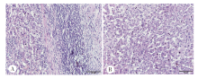
Fig. 1
Pathological changes of surgically resected liver tissues of hepatic CE patients(HE staining, × 200) A: CE proximal-lesion tissue, there was massive lymphocyte infiltration accompanied by granulocyte increase; B: Distant normal tissue, the morphology was normal.
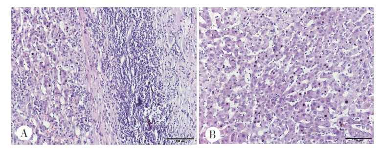
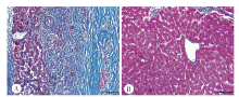
Fig. 2
Collagen deposition of surgically resected liver tissues of hepatic CE patients(masson staining, × 200) A: CE proximal-lesion tissue, massive collagen deposition was observed; B: Distant normal tissue, only less amount collagen was deposited.
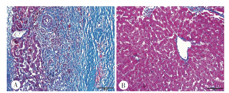
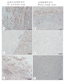
Fig. 3
Expression of α-SMA, NF-κB p65, MyD88 in the liver tissue of CE patients (immunohistochemical staining, × 200) A, C, E: Massive α-SMA, NF-κB p65, MyD88 positive cells were observed; B, D, F: Less α-SMA, NF-κB p65, MyD88 positive cells were observed.

| [1] | Hao W, Lucine V, Tuerhongjiang T, et al. Echinococcosis in the 21st century[J]. Clin Microbiol Rev, 2019, 32(2): e00075-93. |
| [2] |
Budke CM, Carabin H, Patrick CN, et al. A systematic review of the literature on cystic echinococcosis frequency worldwide and its associated clinical manifestations[J]. Am J Trop Med Hyg, 2013, 88(6): 1011-1027.
doi: 10.4269/ajtmh.12-0692 |
| [3] | Craig PS, Larrieu E. Control of cystic echinococcosis/hydatidosis: 1863—2002[J]. Adv Parasitol, 2006, 61: 443-508. |
| [4] |
Labsi M, Soufli I, Khelifi L, et al. In vivo treatment with IL-17A attenuates hydatid cyst growth and liver fibrogenesis in an experimental model of echinococcosis[J]. Acta Trop, 2018, 181: 6-10.
doi: 10.1016/j.actatropica.2018.01.014 |
| [5] |
Hong KK, Gwak MJ, Song J, et al. NF-κB pathway activation and PTEN downregulation in psoriasis[J]. Br J Dermatol, 2016, 174(2): 433-435.
doi: 10.1111/bjd.14106 pmid: 26302365 |
| [6] |
Tsuruta D. NF-kappaB links keratinocytes and lymphocytes in the pathogenesis of psoriasis[J]. Recent Pat Inflamm Allergy Drug Discov, 2009, 3(1): 40-48.
doi: 10.2174/187221309787158399 |
| [7] | Ji R, Liang RW, Guan ZY, et al. The role of TLR4/NF-κB signaling pathway in Cryptosporidim parvum infection[J]. Chin J Parasitol Parasit Dis, 2018, 36(4): 361-365. (in Chinese) |
| (汲蕊, 梁瑞文, 管志玉, 等. TLR4/NF-κB信号通路在微小隐孢子虫感染中的作用机制[J]. 中国寄生虫学与寄生虫病杂志, 2018, 36(4): 361-365.) | |
| [8] |
Wang S, Zhang Z, Wang Y, et al. Toxoplasma gondii excretory/secretory antigens (TgESAs) suppress pro-inflammatory cytokine secretion by inhibiting TLR-induced NF-κB activation in LPS-stimulated murine macrophages[J]. Oncotarget, 2017, 8(51): 88351-88359.
doi: 10.18632/oncotarget.v8i51 |
| [9] |
Zhang C, Shao Y, Yang S, et al. T-cell tolerance and exhaustion in the clearance of Echinococcus multilocularis: role of inoculum size in a quantitative hepatic experimental model[J]. Sci Rep, 2017, 7(1): 11153.
doi: 10.1038/s41598-017-11703-1 |
| [10] |
Lopez-Luis BA, Valdivia-Cayoja AR, Belaunzaran-Zamudio PF, et al. An immunocompromised patient & multiorgan cystic echinococcosis[J]. QJM-INT J MED, 2019, 112(3): 215-217.
doi: 10.1093/qjmed/hcy306 |
| [11] |
Joyce JA, Fearon DT. T cell exclusion, immune privilege, and the tumor microenvironment[J]. Science, 2015, 348(6230): 74-80.
doi: 10.1126/science.aaa6204 |
| [12] | Reynaert H, Thompson MG, Thomas T, et al. Hepatic stellate cells: roles in mivrovirulation and pathophysiology of portal hypertension[J]. Gat, 2002, 50: 571-581. |
| [13] | Dietrich GG, Gotze O, Geier A. Molecular changes in hepatic metabolism and transport in cirrhosis and their functional importance[J]. World J Gastroentrol, 2016, 22(1): 72-88. |
| [14] | Cai JY, Shao YX, Wang K, et al. Paeoniflorin inhibits activation of Raw 264.7 macrophages induced by high glucose via JAK2/STAT3 signaling pathway[J]. Chin Pharmacol Bull, 2019, 35(1): 56-62. (in Chinese) |
| (蔡建月, 邵云侠, 王坤, 等. 芍药苷通过JAK2/STAT3信号通路抑制高糖诱导的RAW264.7巨噬细胞激活[J]. 中国药理学通报, 2019, 35(1): 56-62.) | |
| [15] |
Zhong J, Wang H, Chen W, et al. Ubiquitylation of MFHAS1 by the ubiquitin ligase praja2 promotes M1 macrophage polarization by activating JNK and p38 pathways[J]. Cell Death Dis, 2017, 8(5): e2763.
doi: 10.1038/cddis.2017.102 |
| [16] |
Nakano T, Fukuda D, Koga J, et al. Delta-Like ligand 4-Notch signaling in macrophage activation[J]. Arterioscler Thromb Vasc Biol, 2016, 36(10): 2038-2047.
doi: 10.1161/ATVBAHA.116.306926 |
| [17] |
Czimmerer Z, Daniel B, Horvath A, et al. The transcription factor STAT6 mediates direct repression of inflammatory enhancers and limits activation of alternatively polarized macrophages[J]. Immunity, 2018, 48(1): 75-90.
doi: S1074-7613(17)30568-X pmid: 29343442 |
| [18] |
Sarah T, Dalila M, Zine-Charaf AT, et al. Potential role of NF-κB pathway in the immuno-inflammatory responses during human cystic echinococcosis[J]. Acta Trop, 2020, 203(8): 105306.
doi: 10.1016/j.actatropica.2019.105306 |
| [19] |
Caamano J, Hunter CA. NF-κB family of transcription factors: central regulators of innate and adaptive immune functions[J]. Clin Microbiol Rev, 2002, 15(3): 414-429.
doi: 10.1128/CMR.15.3.414-429.2002 |
| [1] | LI Tianxing, ZHANG Jiaming, XU Chenxi, WANG Zige, GUO Jingjie, LI Shan. Mechanism of a Chinese patent medicine in the treatment of liver fibrosis caused by infection of Clonorchis sinensis based on network pharmacology [J]. CHINESE JOURNAL OF PARASITOLOGY AND PARASITIC DISEASES, 2023, 41(4): 510-515. |
| [2] | KUI Yan, XUE Chuizhao, WANG Xu, LIU Baixue, WANG Ying, WANG Liying, YANG Shijie, HAN Shuai, WU Weiping, XIAO Ning. Progress of echinococcosis control in China, 2021 [J]. CHINESE JOURNAL OF PARASITOLOGY AND PARASITIC DISEASES, 2023, 41(2): 142-148. |
| [3] | LU Weimin, YANG Xiaotao, ZHU Ying, ZHANG Hong, LI Jiwei, WANG Yanchun. A child case of pulmonary cystic echinococcosis [J]. CHINESE JOURNAL OF PARASITOLOGY AND PARASITIC DISEASES, 2023, 41(2): 253-256. |
| [4] | GUO Lu, WU Xiao-xia, DUAN Lan-li, WANG Bing-jie, XU Ning, AREAI Ahatai, WU Yun-hua, ZHAO Li, BAN Wan-li, CHEN Yun-ying, YU Wan-rong, LIU Shuai, PAN Xing-yu, WULIJIANG Kamali, XU Jing, MUNILA Teliewuhan, ZHANG Zhuang-zhi. Prevalence and gene polymorphism analysis of Echinococcus granulosus in cattle and sheep in part areas of Xinjiang [J]. CHINESE JOURNAL OF PARASITOLOGY AND PARASITIC DISEASES, 2022, 40(5): 603-609. |
| [5] | HOU Jiao, WEN Hao, WANG Ming-kun, JIANG Tie-min, FANG Bin-bin, LI Jing, ZHANG Chuan-shan, WANG Hui. Analysis of the influencing factors of lesion activity in hepatic cystic echinococcosis patients [J]. CHINESE JOURNAL OF PARASITOLOGY AND PARASITIC DISEASES, 2022, 40(3): 309-314. |
| [6] | GAO Yuan, ZHANG Xiao-cheng, HU Yuan, CAO Jian-ping. Study on the inhibitory effect of natural killer cells on liver fibrosis of schistosomiasis [J]. CHINESE JOURNAL OF PARASITOLOGY AND PARASITIC DISEASES, 2022, 40(2): 168-174. |
| [7] | ZHANG Ya-lan, JIANG Tian-tian, HE Zhi-quan, DENG Yan, CHEN Wei-qi, ZHU Yan-kun, ZHANG Hong-wei, ZHAO Dong-yang. Differential expression of microRNA in the liver of mice infected by Capillaria hepatica [J]. CHINESE JOURNAL OF PARASITOLOGY AND PARASITIC DISEASES, 2022, 40(1): 56-60. |
| [8] | GAO Yuan, HU Yuan, CAO Jian-ping. Research progress on the role of immune cells in liver fibrosis due to schistosomiasis [J]. CHINESE JOURNAL OF PARASITOLOGY AND PARASITIC DISEASES, 2022, 40(1): 88-93. |
| [9] | LI Wen-ding, WEN Hao, HOU Jiao, WANG Ming-kun, LI liang, LI Jing, ZHANG Chuan-shan, SUN Bing, WANG Hui. Role of extracellular matrix protein 1 in the liver fibrosis induced by Echinococcus multilocularis infection in mice [J]. CHINESE JOURNAL OF PARASITOLOGY AND PARASITIC DISEASES, 2021, 39(3): 296-303. |
| [10] | SUN Lei, HU Yuan, SHEN Yu-juan, CAO Jian-ping. γδ T cells-secreted IL-17A aggravates liver fibrosis in mice infected with Schistosoma japonicum via activating hepatic stellate cells [J]. CHINESE JOURNAL OF PARASITOLOGY AND PARASITIC DISEASES, 2020, 38(3): 299-303. |
| [11] | SHEN Shuang, LUO Jun-tao, YE Jian-ping. Mitochondrial function regulated by Beclin1 in liver fibrosis in schistosomiasis [J]. CHINESE JOURNAL OF PARASITOLOGY AND PARASITIC DISEASES, 2020, 38(1): 41-46. |
| [12] | Yong-hua ZHOU, Chen XU, Ying-ying YANG, Chun-gang ZHOU, Cong-jin MEI, Xue SAI, Yong-liang XU, Jun-qi YANG, Li-juan SHEN. Effect of artesunate on expression of heat shock protein 47 in mice with liver fibrosis induced by Schistosoma japonicum [J]. CHINESE JOURNAL OF PARASITOLOGY AND PARASITIC DISEASES, 2019, 37(2): 115-121. |
| [13] | Zhi-hong GONG, Yun XU, Ke-xing LIU, Kun-jiao DAI, Xiao-jun ZENG, An NING, Hui-qun XIE, Ai-ming QI, Cheng-jian HUANG, Yong-hong TU, Jing XU, Zu-lu GAO, Wei-sheng JIANG. Analysis of a case report of pulmonary cystic echinococcosis in a child in Jiangxi Province [J]. CHINESE JOURNAL OF PARASITOLOGY AND PARASITIC DISEASES, 2019, 37(2): 228-231. |
| [14] | Rui JI, Rui-wen LIANG, Zhi-yu GUAN, Rui-fang LI, Yu-rong FU, Hong-yan WANG. The role of TLR4/NF-κB signaling pathway in Cryptosporidim parvum infection [J]. CHINESE JOURNAL OF PARASITOLOGY AND PARASITIC DISEASES, 2018, 36(4): 361-366. |
| [15] | SHAN Jiao-yu1,2, REBIYA·Nuli1, LI Rui1, LIN Ren-yong2, WEN Hao2,3, LI Hai-tao2,3*. Expression of the regulatory T cell transcription factor Foxp3 and interleukin-8 in liver tissues of patients with echinococcosis [J]. , 2018, 36(3): 4-218-223. |
| Viewed | ||||||||||||||||||||||||||||||||||||||||||||||||||
|
Full text 112
|
|
|||||||||||||||||||||||||||||||||||||||||||||||||
|
Abstract 400
|
|
|||||||||||||||||||||||||||||||||||||||||||||||||