
CHINESE JOURNAL OF PARASITOLOGY AND PARASITIC DISEASES ›› 2021, Vol. 39 ›› Issue (5): 659-665.doi: 10.12140/j.issn.1000-7423.2021.05.014
• ORIGINAL ARTICLES • Previous Articles Next Articles
YIN Meng( ), ZHANG Hao-bing*(), TAO Yi, JIANG Bin, LIU Hua
Received:2021-03-09
Revised:2021-06-17
Online:2021-10-30
Published:2021-11-10
Contact:
ZHANG Hao-bing
E-mail:yinmeng@nipd.chinacdc.cn;zhanghb@nipd.chinacdc.cn
Supported by:CLC Number:
YIN Meng, ZHANG Hao-bing, TAO Yi, JIANG Bin, LIU Hua. Evaluation on the in vivo efficacy of malarone and atovaquone-azithromycin combination against Babesia microti in mice under different immune status[J]. CHINESE JOURNAL OF PARASITOLOGY AND PARASITIC DISEASES, 2021, 39(5): 659-665.
Add to citation manager EndNote|Ris|BibTeX
URL: https://www.jsczz.cn/EN/10.12140/j.issn.1000-7423.2021.05.014
Fig. 1
Trends in EIR(A)and the relative value of the B. microti 18S rRNA gene (B)of BALB/c mice in the different groups after infection in drug-suppression test Start medication 4 hours after infection and continue medication for 10 days.
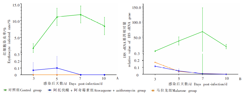
Fig. 2
EIR in BALB/c mice infected with B. microti in different groups after infection in a drug-therapy test Start medication 7 days after infection and continue medication for 10 days.
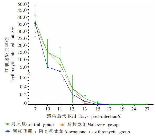
Table 1
EIR in BALB/c mice injected with an immunosuppressant/%
| 免疫抑制后天数/d Days post-immunosuppressant injection/d | 对照组 Control group | ATQ + AZM组 ATQ + AZM group | 马拉龙组 Malarone group |
|---|---|---|---|
| 3 | 0.06 ± 0.13 | 0 | 0 |
| 5 | 0.25 ± 0.07 | 0.10 ± 0.14 | 0.10 ± 0.14 |
| 7 | 1.56 ± 1.31 | 0.52 ± 0.46 | 3.20 ± 2.58 |
| 9a | 3.04 ± 5.15 | 0.20 ± 0.28 | 0.33 ± 0.38 |
Table 2
EIR in BALB/c mice receiving blood in the subpassage test/%
| 继代接种感染后天数/d Days post subpassage/d | 对照组 Control group | ATQ + AZM组 ATQ + AZM group | 马拉龙组 Malarone group |
|---|---|---|---|
| 7 | 3.34 ± 1.44 | 0.12 ± 0.11 | 0.42 ± 0.41 |
| 9 | 3.12 ± 2.72 | 0.04 ± 0.09 | 0.10 ± 0.12 |
| 12 | 32.60 ± 13.99 | 0 | 0 |

Fig. 3
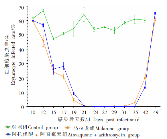
Trends in EIR in NOD/SCID mice infected with B.microti in different treatment groupsStart medication 10 days after infection and continue for 10 days.
| [1] |
Vannier E, Krause PJ. Human babesiosis[J]. N Engl J Med, 2012, 366(25):2397-2407.
doi: 10.1056/NEJMra1202018 |
| [2] |
Vannier EG, Diuk-Wasser MA, Ben Mamoun C, et al. Babesiosis[J]. Infect Dis Clin N Am, 2015, 29(2):357-370.
doi: 10.1016/j.idc.2015.02.008 |
| [3] |
Ruebush TK. Human babesiosis in north America[J]. Trans R Soc Trop Med Hyg, 1980, 74(2):149-152.
doi: 10.1016/0035-9203(80)90231-X |
| [4] |
Patel KM, Johnson JE, Reece R, et al. Babesiosis-associated splenic rupture: case series from a hyperendemic region[J]. Clin Infect Dis, 2019, 69(7):1212-1217.
doi: 10.1093/cid/ciy1060 |
| [5] | Gray EB, Herwaldt BL. Babesiosis surveillance--United States, 2011—2015[J]. MMWR Surveill Summ, 2019, 68(6):1-11. |
| [6] |
Goo YK, Terkawi MA, Jia H, et al. Artesunate, a potential drug for treatment of Babesia infection[J]. Parasitol Int, 2010, 59(3):481-486.
doi: 10.1016/j.parint.2010.06.004 |
| [7] |
Lawres LA, Garg A, Kumar V, et al. Radical cure of experimental babesiosis in immunodeficient mice using a combination of an endochin-like quinolone and atovaquone[J]. J Exp Med, 2016, 213(7):1307-1318.
doi: 10.1084/jem.20151519 |
| [8] |
El-Sayed SAES, Rizk MA, Ringo AE, et al. Impact of using pyronaridine tetraphosphate-based combination therapy in the treatment of babesiosis caused by Babesia bovis, B. caballi, and B. gibsoni in vitro and B. microti in mice[J]. Parasitol Int, 2021, 81:102260.
doi: 10.1016/j.parint.2020.102260 |
| [9] |
Marley SE, Eberhard ML, Steurer FJ, et al. Evaluation of selected antiprotozoal drugs in the Babesia microti-hamster model[J]. Antimicrob Agents Chemother, 1997, 41(1):91-94.
pmid: 8980761 |
| [10] |
Wormser GP, Dattwyler RJ, Shapiro ED, et al. The clinical assessment, treatment, and prevention of Lyme disease, human granulocytic anaplasmosis, and babesiosis: clinical practice guidelines by the Infectious Diseases Society of America[J]. Clin Infect Dis, 2006, 43(9):1089-1134.
pmid: 17029130 |
| [11] |
Vial HJ, Gorenflot A. Chemotherapy against babesiosis[J]. Vet Parasitol, 2006, 138(1/2):147-160.
doi: 10.1016/j.vetpar.2006.01.048 |
| [12] |
Sanchez E, Vannier E, Wormser GP, et al. Diagnosis, treatment, and prevention of Lyme disease, human granulocytic anaplasmosis, and babesiosis: a review[J]. JAMA, 2016, 315(16):1767-1777.
doi: 10.1001/jama.2016.2884 pmid: 27115378 |
| [13] |
Krause PJ, Gewurz BE, Hill D, et al. Persistent and relapsing babesiosis in immunocompromised patients[J]. Clin Infect Dis, 2008, 46(3):370-376.
doi: 10.1086/525852 pmid: 18181735 |
| [14] |
Wormser GP, Prasad A, Neuhaus E, et al. Emergence of resistance to azithromycin-atovaquone in immunocompromised patients with Babesia microti infection[J]. Clin Infect Dis, 2010, 50(3):381-386.
doi: 10.1086/649859 pmid: 20047477 |
| [15] |
Guirao-Arrabal E, González LM, García-Fogeda JL, et al. Imported babesiosis caused by Babesia microti--a case report[J]. Ticks Tick Borne Dis, 2020, 11(4):101435.
doi: 10.1016/j.ttbdis.2020.101435 |
| [16] |
Vyas JM, Telford SR, Robbins GK. Treatment of refractory Babesia microti infection with atovaquone-proguanil in an HIV-infected patient: case report[J]. Clin Infect Dis, 2007, 45(12):1588-1590.
doi: 10.1086/523731 |
| [17] | Blanshard A, Hine P. Atovaquone-proguanil for treating uncomplicated Plasmodium falciparum malaria[J]. Cochrane Database Syst Rev, 2021, 1: CD004529. |
| [18] | Xu SY, Bian RL, Chen X. Pharmacological experiment methodology[M]. 3 ed. Beijing: People’s Medical Publishing House, 2002: 1597-1600. (in Chinese) |
| ( 徐叔云, 卞如濂, 陈修. 药理实验方法学[M]. 3版. 北京: 人民卫生出版社, 2002: 1597-1600.) | |
| [19] | Zhao W, Sun GZ. Dose conversion between different kinds of experimental animals[J]. Chin J Animal Husb Vet Med, 2010(5):52-53. (in Chinese) |
| ( 赵伟, 孙国志. 不同种实验动物间用药量换算[J]. 畜牧兽医科技信息, 2010(5):52-53.) | |
| [20] |
Teal AE, Habura A, Ennis J, et al. A new real-time PCR assay for improved detection of the parasite Babesia microti[J]. J Clin Microbiol, 2012, 50(3):903-908.
doi: 10.1128/JCM.05848-11 |
| [21] |
Beshbishy AM, Batiha GE, Yokoyama N, et al. Ellagic acid microspheres restrict the growth of Babesia and Theileria in vitro and Babesia microti in vivo[J]. Parasit Vectors, 2019, 12(1):269.
doi: 10.1186/s13071-019-3520-x pmid: 31138282 |
| [22] |
Yao JM, Zhang HB, Liu CS, et al. Inhibitory effects of 19 antiprotozoal drugs and antibiotics on Babesia microti infection in BALB/c mice[J]. J Infect Dev Ctries, 2015, 9(9):1004-1010.
doi: 10.3855/jidc.5500 |
| [23] |
Mordue DG, Wormser GP. Could the drug tafenoquine revolutionize treatment of Babesia microti infection?[J]. J Infect Dis, 2019, 220(3):442-447.
doi: 10.1093/infdis/jiz119 pmid: 31099380 |
| [24] |
Tuvshintulga B, Vannier E, Tayebwa DS, et al. Clofazimine, a promising drug for the treatment of Babesia microti infection in severely immunocompromised hosts[J]. J Infect Dis, 2020, 222(6):1027-1036.
doi: 10.1093/infdis/jiaa195 pmid: 32310272 |
| [25] |
Nehrbass-Stuedli A, Boykin D, Tidwell RR, et al. Novel diamidines with activity against Babesia divergens in vitro and Babesia microti in vivo[J]. Antimicrob Agents Chemother, 2011, 55(7):3439-3445.
doi: 10.1128/AAC.01482-10 pmid: 21537025 |
| [26] |
Wang S, Li M, Luo X, et al. Inhibitory effects of fosmidomycin against Babesia microti in vitro[J]. Front Cell Dev Biol, 2020, 8:247.
doi: 10.3389/fcell.2020.00247 pmid: 32411701 |
| [27] |
Guo JY, Luo XY, Wang S, et al. Xanthohumol and gossypol are promising inhibitors against Babesia microti by in vitro culture via high-throughput screening of 133 natural products[J]. Vaccines, 2020, 8(4):613.
doi: 10.3390/vaccines8040613 |
| [28] |
AbouLaila M, Sivakumar T, Yokoyama N, et al. Inhibitory effect of terpene nerolidol on the growth of Babesia parasites[J]. Parasitol Int, 2010, 59(2):278-282.
doi: 10.1016/j.parint.2010.02.006 pmid: 20178862 |
| [1] | SUN Jiahui, SONG Peng, CHEN Muxin, ZHOU Yan, LIN Lin, CHEN Jiaxu, CAI Yuchun. Expression and functional analysis of recombinant peptidyl-prolyl cis-trans isomerase gene of Babesia microti [J]. CHINESE JOURNAL OF PARASITOLOGY AND PARASITIC DISEASES, 2023, 41(1): 29-35. |
| [2] | SONG Peng, CAI Yu-chun, LU Yan, AI Lin, CHEN Mu-xin, CHEN Shao-hong, CHEN Jia-xu. Establishment of mouse infection model of Babesia microti Lishui isolate and consequent pathological changes [J]. CHINESE JOURNAL OF PARASITOLOGY AND PARASITIC DISEASES, 2022, 40(4): 493-499. |
| [3] | ZHANG Yan, XU Ai-fang, ZHANG Jia-qi, YAO Li-nong, GU Kai-long, XUE Li-zhi, PAN Ke-nv. Differential diagnosis of a case of Babesia microti infection previously misdiagnosed as malaria [J]. CHINESE JOURNAL OF PARASITOLOGY AND PARASITIC DISEASES, 2020, 38(4): 445-448. |
| [4] | Zi-yue WANG, Yi-chao YANG, Zhi-pin CHEN, Yun-liang SHI. Infection of Plasmodium knowlesi and Babesia microti in farmed monkeys in Guangxi [J]. CHINESE JOURNAL OF PARASITOLOGY AND PARASITIC DISEASES, 2019, 37(4): 494-496. |
| [5] | Xiu-feng LIU, Jia-hui SUN, Bin XU, Jun-hu CHEN, Wei HU. Expression and evaluation of diagnostic candidate antigens from Babesia microti [J]. CHINESE JOURNAL OF PARASITOLOGY AND PARASITIC DISEASES, 2017, 35(6): 549-553. |
| [6] | Wei RUAN, Ling-ling ZHANG, Hua-liang CHEN, Qiao-yi LU, Xuan ZHANG, Yan FENG, Li-nong YAO. Investigation of the source regions of Babesia spp. infection in the central and south areas of Zhejiang Province [J]. CHINESE JOURNAL OF PARASITOLOGY AND PARASITIC DISEASES, 2017, 35(2): 125-130. |
| [7] | Fang-zhen XIAO, Xiu-qing Peng, Guo-ying XU, Yang CHEN, Dai-hua LIN, Yan-qin DENG. Investigation and genetic identification on Babesia infection in rodents in some areas of Fujian Province [J]. CHINESE JOURNAL OF PARASITOLOGY AND PARASITIC DISEASES, 2017, 35(1): 63-67. |
| [8] | WANG Dong, ZHANG Yuan-Yuan. Therapeutic Efficacy of Allitridin and Azithromycin on Cryptosporidium Infection in Mice [J]. , 2013, 31(6): 15-477-479. |
| [9] | YINWei-dong;GAOQuan-cheng;LIUXiang-dong*;TANGHong-wei. In vivo Effect of Dihydroartemisinin and Azithromycin on the Ultrastructure of Toxoplasma gondii Tachyzoites [J]. , 2009, 27(4): 19-327. |
| Viewed | ||||||
|
Full text |
|
|||||
|
Abstract |
|
|||||