中国寄生虫学与寄生虫病杂志 ›› 2018, Vol. 36 ›› Issue (5): 474-477.

• 论著 • 上一篇    下一篇

多房棘球蚴病患者血源树突状细胞形态观察和表型检测

付永1,2, 孟茹3, 薛海芳4, 樊海宁4, 牛海峰4, 周子佳4, 王宏宾4,*()   

  1. 1 青海大学畜牧兽医科学院,西宁 810016
    2 青海大学三江源生态与高原农牧业国家重点实验室,西宁 810016
    3 西宁市畜牧兽医站,西宁 810016
    4 青海大学附属医院,西宁 810001
  • 收稿日期:2018-04-27 出版日期:2018-10-30 发布日期:2018-11-13
  • 通讯作者: 王宏宾
  • 基金资助:
    青海省科学技术厅基础研究项目(No.2016-ZJ-760)

Morphological observation and phenotypic detection of dendritic cells from peripheral blood of patients with alveolar echinococcosis

Yong FU1,2, Ru MENG3, Hai-fang XUE4, Hai-ning FAN4, Hai-feng NIU4, Zi-jia ZHOU4, Hong-bin WANG4,*()   

  1. 1 Academy of Animal and Veterinary Medicine, Qinghai University, Xining 810016, China
    2 State Key Laboratory of Plateau Ecology and Agriculture, Qinghai University, Xining 810016, China
    3 Animal Husbandry and Veterinary Station of Xining, Xining 810016, China
    4 Affiliated Hospital of Qinghai University, Xining 810001, China
  • Received:2018-04-27 Online:2018-10-30 Published:2018-11-13
  • Contact: Hong-bin WANG
  • Supported by:
    Supported by the Science and Technology Project of Qinghai Province(No.2016-ZJ-760)

摘要:

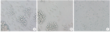
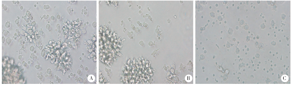
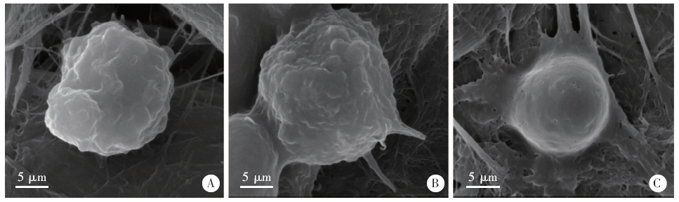
目的 了解多房棘球蚴病患者血源树突状细胞(DCs)形态和表型特点。方法 分别收集多房棘球蚴病病例(AE组)10例、汉族健康志愿者(HH组)10人、藏族健康志愿者(TH组)10人的外周血,分离单核细胞贴壁培养,应用重组人集落刺激因子和重组人白细胞介素-4诱导获得DCs。分别收集各组培养第1、3、5和7天的DCs,采用倒置显微镜和扫描电镜观察细胞形态。3组DCs诱导培养至第7天时,流式细胞术检测细胞表面标志分子的阳性表达率,采用SPSS 22.0统计学软件进行分析。结果 体外诱导培养第1天,AE组、HH组和TH组DCs大部分呈单个圆形、边界清晰、细胞质透亮、体积较小,浮于细胞液中。诱导培养第3天,3组DCs聚集呈半悬浮状,细胞变大呈不规则形态。诱导培养第5天,3组DCs形成集落,多数DCs边缘不光滑呈毛刺状。诱导培养至第7天,3组DCs呈半悬浮生长,边缘具有丝状刺突;AE组中诱导分化的DCs与HH组和TH组相比所形成的集落数量相对较少,细胞胞体所形成的不规则突起不明显且细胞表面树突状突起较少,呈非典型的DCs形态;AE组诱导分化的DCs表面协同共刺激分子CD1a、CD80和CD86阳性表达率分别为12.73% ± 1.73%、12.41% ± 2.83%和16.34% ± 3.59%,与HH组和TH组相比差异有统计学意义(P < 0.05),而HH组(18.40% ± 1.20%、20.77% ± 3.40%、31.78% ± 5.02%)和TH组(17.50% ± 1.44%、23.75% ± 5.33%、33.20% ± 2.47%)相比差异无统计学意义(P > 0.05)。结论 AE组诱导分化的DCs形态不典型、其重要细胞表面标志分子阳性表达率降低,表现为成熟障碍。

关键词: 多房棘球蚴病, 树突状细胞, 细胞形态, 免疫表型

Abstract:

Objective To understand the morphological and phenotypic characteristics of dendritic cells (DCs) from peripheral blood of patients with alveolar echinococcosis. Methods Peripheral blood was collected from 10 patients with alveolar echinococcosis (AE), 10 healthy Han participants (HH) (n = 10) and 10 healthy Tibetan participants (TH), respectively. Monocytes were isolated, and cultured by adherent culture. The monocytes were induced into DCs with recombinant human granulocyte macrophage colony stimulating factor and recombinant human interleukin 4. DCs were collected on days 1, 3, 5 and 7 of culture and cell morphology was observed by inverted microscopy and scanning electron microscopy. The positive rate of cell surface markers was detected by flow cytometry on day 7. Data were analyzed with SPSS 22.0 software. Results On day 1 of in vitro induction, the majority of DCs in the 3 groups had a single round shape with clear boundary, bright cytoplasm, and a small volume, floating in the culture medium. On day 3 of induction, the three groups of DCs aggregated into semi-suspension and the cells enlarged with an irregular shape. On day 5 of induction, the three groups of DCs formed colonies, and most DCs had a rough surface. On day 7 of induction, the three groups of DCs grew into semi-suspension with a filamentous cell edges. Compared to the HH and TH groups, the number of DC colonies in group AE was smaller, and the irregular protuberances of DC were not obvious, displaying an atypical DC morphology. The positive expression rates of CD1a, CD80 and CD86 on the surface of DCs induced differentiation in AE group (12.73% ± 1.73%, 12.41% ± 2.83% and 16.34% ± 3.59%, respectively) were significantly lower than those in the HH and TH groups (P < 0.05), while those of HH group (18.40% ± 1.20%, 20.77% ± 3.40%, 31.78% ± 5.02%) and TH group(17.50% ± 1.44%, 23.75% ± 5.33%, 33.20% ± 2.47%) were not significantly different (P > 0.05). Conclusion The induced DCs in the AE group have an atypical morphology and a decreased level of cell surface markers, indicating the occurrence of maturation disorder.

Key words: Alveolar echinococcosis, Dendritic cell, Cell morphology, Immunophenotype

中图分类号: