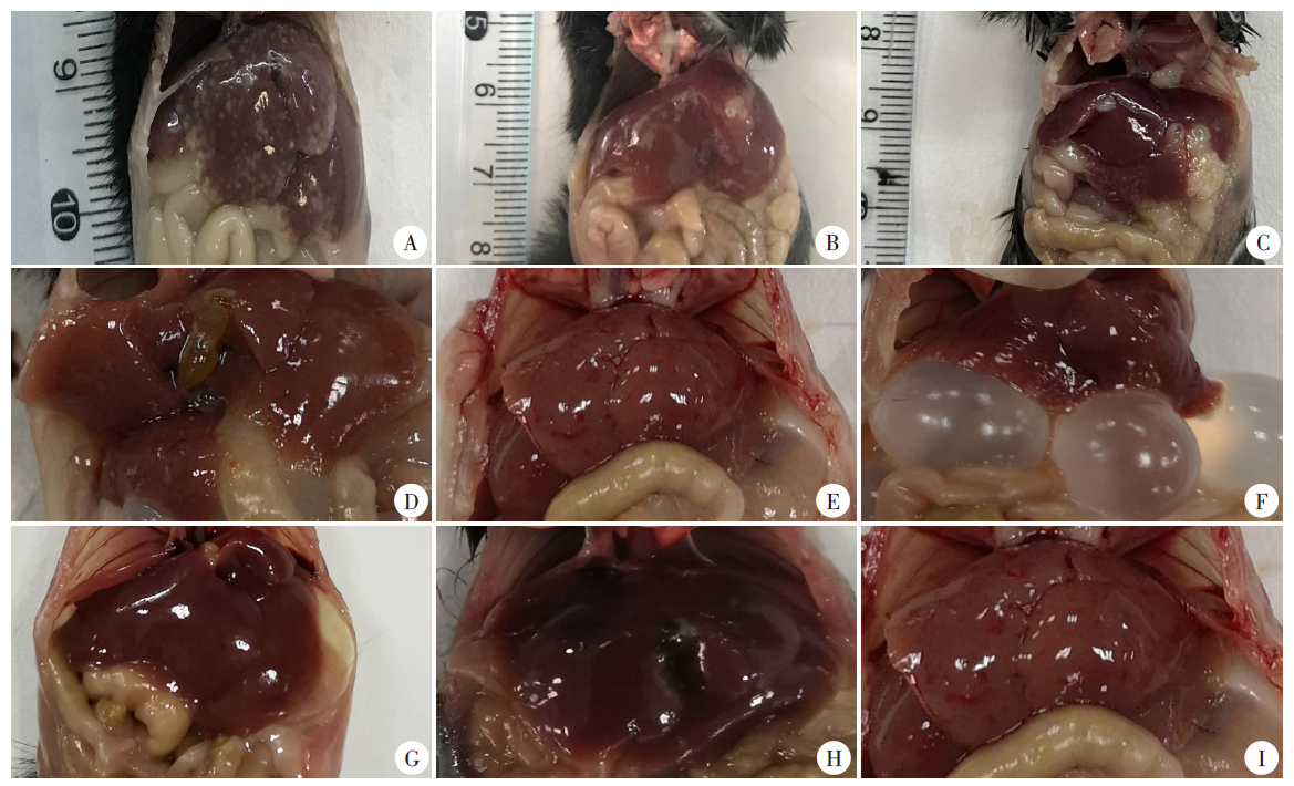
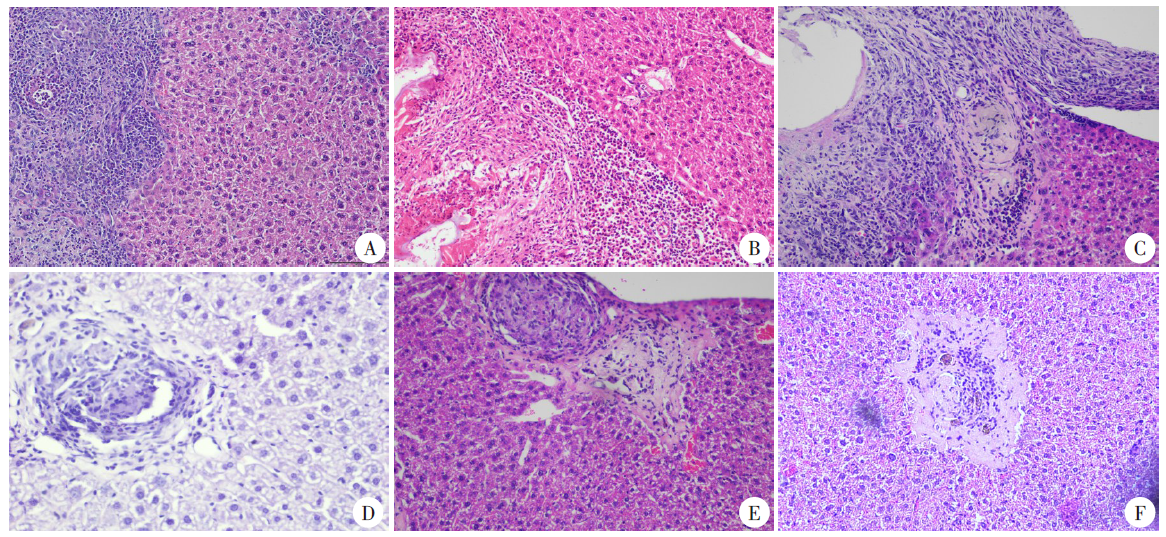
| [1] | Han S, Kui Y, Xue CZ, et al. The endemic status of echinococcosis in China from 2004 to 2020[J]. Chin J Parasitol Parasit Dis, 2022, 40(4): 475-480. (in Chinese) | | | (韩帅, 蒉嫣, 薛垂召, 等. 2004—2020年全国棘球蚴病疫情分析[J]. 中国寄生虫学与寄生虫病杂志, 2022, 40(4): 475-480.) | | [2] | Zheng CJ, Yang L, Zhang GJ, et al. Interpretation of technical scheme for echinococcosis control (edition 2019)[J]. J Trop Dis Parasitol, 2020, 18(4): 193-196, 201. (in Chinese) | | | (郑灿军, 杨柳, 张光葭, 等. 《包虫病防治技术方案(2019年版)》解读[J]. 热带病与寄生虫学, 2020, 18(4):193- 196, 201.) | | [3] | Pohnan R, Ryska M, Hytych V, et al. Echinococcosis mimicking liver malignancy: a case report[J]. Int J Surg Case Rep, 2017, 36: 55-58. | | [4] | Siles-Lucas M, Casulli A, Cirilli R, et al. Progress in the pharmacological treatment of human cystic and alveolar echinococcosis: compounds and therapeutic targets[J]. PLoS Negl Trop Dis, 2018, 12(4): e0006422. | | [5] | Yang C, He JY, Yang XW, et al. Surgical approaches for definitive treatment of hepatic alveolar echinococcosis: results of a survey in 178 patients[J]. Parasitology, 2019, 146(11): 1414-1420. | | [6] | Larrieu E, Herrero E, Mujica G, et al. Pilot field trial of the EG95 vaccine against ovine cystic echinococcosis in Rio Negro, Argentina: early impact and preliminary data[J]. Acta Trop, 2013, 127(2): 143-151. | | [7] | Zhang WB, Li J, You H, et al. Short report: Echinococcus granulosus from Xinjiang, PR China: cDNAS encoding the EG95 vaccine antigen are expressed in different life cycle stages and are conserved in the oncosphere[J]. Am J Trop Med Hyg, 2003, 68(1): 40-43. | | [8] | Bowles J, Blair D, McManus DP. Genetic variants within the genus Echinococcus identified by mitochondrial DNA sequencing[J]. Mol Biochem Parasitol, 1992, 54(2): 165-173. | | [9] | Nakao M, Xiao N, Okamoto M, et al. Geographic pattern of genetic variation in the fox tapeworm Echinococcus multilocularis[J]. Parasitol Int, 2009, 58(4): 384-389. | | [10] | Alvarez Rojas CA, Kronenberg PA, Aitbaev S, et al. Genetic diversity of Echinococcus multilocularis and Echinococcus granulosus sensu lato in Kyrgyzstan: the A2 haplotype of E. multilocularis is the predominant variant infecting humans[J]. PLoS Negl Trop Dis, 2020, 14(5): e0008242. | | [11] | Shang JY, Zhang GJ, Yu WJ, et al. Genetic polymorphism of Echinococcus multilocularis in northwestern China inferred from cox1 gene sequences[J]. J Pathog Biol, 2021, 16(2): 137-142. (in Chinese) | | | (尚婧晔, 张光葭, 喻文杰, 等. 中国西北部地区多房棘球绦虫cox1基因多态性研究[J]. 中国病原生物学杂志, 2021, 16(2): 137-142.) | | [12] | Konyaev SV, Yanagida T, Ingovatova GM, et al. Molecular identification of human echinococcosis in the Altai region of Russia[J]. Parasitol Int, 2012, 61(4): 711-714. | | [13] | Laurim?e T, Kronenberg PA, Alvarez Rojas CA, et al. Long-term (35 years) cryopreservation of Echinococcus multilocularis metacestodes[J]. Parasitology, 2020, 147(9): 1048-1054. | | [14] | Liu HD, Wang HB, Fan HN, et al. Alveolar echinococcosis and immune evasion[J]. Chin J Parasitol Parasit Dis, 2018, 36(6): 655-660. (in Chinese) | | | (刘寒冬, 王宏宾, 樊海宁, 等. 多房棘球蚴病的免疫逃避机制[J]. 中国寄生虫学与寄生虫病杂志, 2018, 36(6): 655-660.) | | [15] | Zhang LH, Chen G, Chong SG, et al. Research progress on the immune regulation mechanism in alveolar echinococcosis[J]. Chin J Parasitol Parasit Dis, 2022, 40(1): 109-113, 120. (in Chinese) | | | (张伶慧, 陈根, 种世桂, 等. 多房棘球蚴病中免疫细胞调控机制的研究进展[J]. 中国寄生虫学与寄生虫病杂志, 2022, 40(1): 109-113, 120.) | | [16] | Zheng HJ, Zhang WB, Zhang L, et al. The genome of the hydatid tapeworm Echinococcus granulosus[J]. Nat Genet, 2013, 45(10): 1168-1175. | | [17] | Tsai IJ, Zarowiecki M, Holroyd N, et al. The genomes of four tapeworm species reveal adaptations to parasitism[J]. Nature, 2013, 496(7443): 57-63. | | [18] | Guo BP. Study on correlation between pathogenic differences and mitochondrial genetic markers in Echinococcus multilocularis[D]. Shihezi: Shihezi University, 2019: 29-97. (in Chinese) | | | (郭宝平. 多房棘球绦虫致病差异与线粒体遗传标志相关性的研究[D]. 石河子: 石河子大学, 2019: 29-97.) | | [19] | Shang JY, Zhang GJ, Yu WJ, et al. Advances in researches on the genetic diversity of Echinococcus multilocularis[J]. Chin J Parasitol Parasit Dis, 2020, 38(5): 637-641, 646. (in Chinese) | | | (尚婧晔, 张光葭, 喻文杰, 等. 多房棘球绦虫遗传多态性研究进展[J]. 中国寄生虫学与寄生虫病杂志, 2020, 38(5): 637-641, 646.) | | [20] | Yin J. Clinical characteristics of patients with hepatic alveolar hydatid and cystic echinococcosis mixed infection[D]. Xining: Qinghai University, 2019: 36-44. (in Chinese) | | | (尹杰. 肝泡型合并囊型包虫混合感染患者的临床特点分析[D]. 西宁: 青海大学, 2019: 36-44.) |
|

 ), 任远1, 焦红杰3, 武娟4, 郭宝平1, 齐文静1, 李军1, 张文宝1,*(