中国寄生虫学与寄生虫病杂志 ›› 2025, Vol. 43 ›› Issue (1): 52-60.doi: 10.12140/j.issn.1000-7423.2025.01.009

• 论著 • 上一篇    下一篇

体外小鼠巨噬细胞与细粒棘球蚴原头节的相互作用

黎广1(), 姜慧娇1, 杜云峰1, 舒敏1, 罗雨盟1, 朱令懿2, 陈雪玲3, 吴向未1,2,*()   

  1. 1 石河子大学医学院第一附属医院肝胆外科,新疆 石河子 832000
    2 石河子大学医学院,国家卫生健康委中亚高发病防治重点实验室,新疆 石河子 832000
    3 石河子大学医学院免疫教研室,新疆 石河子 832000
  • 收稿日期:2024-02-06 修回日期:2024-04-22 出版日期:2025-02-28 发布日期:2025-03-26
  • 通讯作者: 吴向未,男,教授,博士生导师,从事肝胆外科良恶性疾病的临床与基础研究。E-mail:wxwshz@126.com
  • 作者简介:黎广,男,硕士研究生,从事普通外科疾病研究。E-mail:992586455@qq.com
  • 基金资助:
    新疆生产建设兵团促进科技成果转化引导计划(2021BB006);中国医学科学院中央级公益性科研院所基本科研业务费专项资金(2020-PT330-003)

Interaction between mouse macrophages and protoscolex of Echinococcus granulosus in vitro

LI Guang1(), JIANG Huijiao1, DU Yunfeng1, SHU Min1, LUO Yumeng1, ZHU Lingyi2, CHEN Xueling3, WU Xiangwei1,2,*()   

  1. 1 Department of Hepatobiliary Surgery, The First Affiliated Hospital of Medical College, Shihezi University, Shihezi 832000, Xinjiang, China
    2 School of Medicine, Shihezi University/NHC Key Laboratory of Prevention and Treatment of Central Asia High Incidence Diseases, Shihezi 832000, Xinjiang, China
    3 Department of Immunization, School of Medicine, Shihezi University, Shihezi 832000, Xinjiang, China
  • Received:2024-02-06 Revised:2024-04-22 Online:2025-02-28 Published:2025-03-26
  • Contact: E-mail: wxwshz@126.com
  • Supported by:
    Early immune escape mechanism and clinical exploration of hydatid infection(2021BB006);Non-profit Central Research Institute Fund of Chinese Academy of Medical Sciences(2020-PT330-003)

摘要:

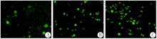
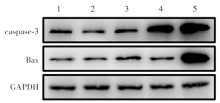
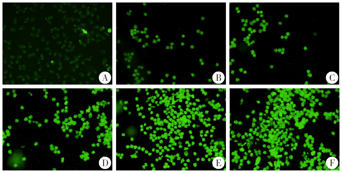
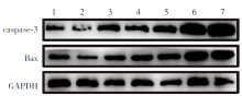
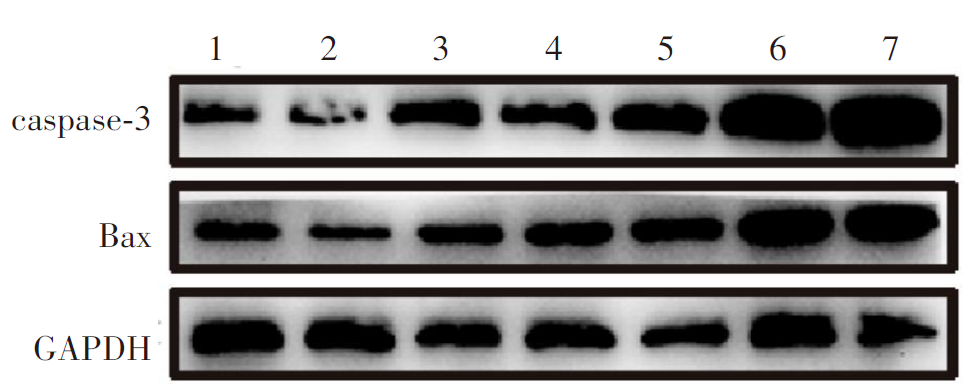
目的 探究小鼠免疫细胞对细粒棘球蚴原头节的杀伤作用,了解发挥功能的免疫细胞类型及其细胞因子的分泌变化。方法 分离健康C57BL/6小鼠的腹腔巨噬细胞和脾细胞。收集羊细粒棘球蚴包囊中的原头节并分组(3 000个/组)。巨噬细胞共培养组、脾细胞共培养组分别与6 × 106个巨噬细胞和脾细胞共培养,选择对原头节具有更强抑制作用的免疫细胞类型;共培养1~5组分别与1.2 × 10⁶、2.4 × 10⁶、4.8 × 10⁶、7.2 × 10⁶、9.6 × 10⁶个细胞共培养,选择适宜的细胞数量,建立共培养体系。共培养体系中分别加入细粒棘球蚴囊液和肿瘤坏死因子α(TNF-α)抑制剂,观察原头节活性和共培养上清中TNF-α、白细胞介素6(IL-6)、IL-10、转化生长因子β(TGF-β)浓度的变化。伊红染色检测原头节活性,二氯荧光素二乙酸酯(DCFH-DA)法检测活性氧水平,JC-1法检测线粒体膜电位,蛋白质免疫印迹(Western blotting)检测促凋亡蛋白Bcl-2相关X蛋白(Bax)和天冬氨酸蛋白水解酶3(caspase-3)表达水平,ELISA检测培养上清中细胞因子的浓度。两组间比较采用独立样本t检验,多组间比较采用单因素方差分析。结果 共培养第6天,巨噬细胞共培养组和脾细胞共培养组的原头节活性分别为(25.07 ± 0.40)%和(76.18 ± 0.31)%,巨噬细胞共培养组各时期的原头节活性低于脾细胞共培养组(F = 564.20,P < 0.05);共培养第4天,巨噬细胞共培养组的原头节活性氧相对荧光强度为32.20 ± 7.85,高于脾细胞共培养组的12.44 ± 2.93(t = 7.07,P < 0.05);共培养第6天,巨噬细胞共培养组和脾细胞共培养组的caspase-3蛋白相对表达水平分别为1.28 ± 0.02和1.16 ± 0.02,Bax蛋白相对表达水平分别为1.29 ± 0.01和0.46 ± 0.01,巨噬细胞共培养组各时期的caspase-3、Bax蛋白相对表达水平均高于脾细胞共培养组(F = 55.87、167.20,均P < 0.05),巨噬细胞对原头节的抑制作用强于脾细胞。共培养第6天,共培养1~5组原头节的线粒体膜电位相对荧光强度分别为20.15 ± 8.96、24.40 ± 9.71、48.41 ± 10.20、94.62 ± 8.72、112.85 ± 24.23,均高于原头节对照组的2.50 ± 1.02(F = 26.18,P < 0.01)。共培养第6天,共培养4、5组原头节的caspase-3蛋白相对表达水平分别为1.35 ± 0.03和1.49 ± 0.05,高于原头节对照组的0.28 ± 0.01(t = 17.03、10.60,均P < 0.05);共培养4、5组原头节的Bax蛋白相对表达水平分别为1.34 ± 0.01和1.38 ± 0.04,高于原头节对照组的0.78 ± 0.04(t = 6.68、6.46,均P < 0.05)。细胞共培养4组上清的TNF-α、IL-6和TGF-β浓度分别为(240.90 ± 17.29)、(435.90 ± 12.33)、(137.10 ± 6.62)pg/ml,均高于细胞对照组的(42.02 ± 0.52)、(65.72 ± 1.91)、(24.72 ± 1.78)pg/ml(t = 54.52、15.97、17.59,均P < 0.05);细胞共培养4组上清的IL-10浓度为(42.16 ± 1.45)pg/ml,与细胞对照组的(45.64 ± 1.03)pg/ml差异无统计学意义(t = 1.29,P > 0.05);共培养组各时期上清中TNF-α、IL-6、IL-10和TGF-β的浓度均高于细胞对照组(F = 294.66、450.50、687.72、660.15,均P < 0.05)。共培养第1、3、5、7天,囊液组的原头节线粒体膜电位相对荧光强度分别为4.46 ± 1.25、4.33 ± 0.39、4.89 ± 0.77、7.97 ± 0.62,均低于巨噬细胞组的5.67 ± 1.72、13.60 ± 0.50、35.28 ± 5.65、77.50 ± 9.60(F = 115.90,P < 0.01)。共培养第6天,囊液组上清中TNF-α、IL-6、IL-10和TGF-β的浓度分别为(64.12 ± 2.65)、(1 049.65 ± 25.70)、(230.30 ± 12.98)、(138.57 ± 13.71)pg/ml,巨噬细胞组的浓度分别为(41.61 ± 1.31)、(68.00 ± 0.42)、(56.15 ± 6.43)、(32.94 ± 4.90)pg/ml,囊液组各时期上清中TNF-α、IL-6、IL-10和TGF-β的浓度均高于巨噬细胞组(F = 289.80、366.50、145.40、32.94,均P < 0.05)。共培养第7天,巨噬细胞组和抑制剂组的原头节活性分别为(21.18 ± 1.61)%和(94.31 ± 2.58)%,抑制剂组各时期的原头节活性高于巨噬细胞组(F = 1 810.00,P < 0.05)。共培养第2、4、6天,抑制剂组的TNF-α浓度分别为(33.55 ± 7.48)、(13.78 ± 4.96)、(19.20 ± 0.69)pg/ml,均低于巨噬细胞组的(209.24 ± 9.90)、(209.47 ± 10.55)、(211.36 ± 13.66)pg/ml(t = 33.16、30.46、23.76,均P < 0.05)。结论 与细粒棘球蚴原头节在体外共培养的巨噬细胞能够表达TNF-α等细胞因子,抑制原头节的活性,促进原头节的凋亡。细粒棘球蚴囊液和TNF-α抑制剂能够降低巨噬细胞TNF-α的分泌,减轻巨噬细胞对原头节的杀伤作用。

关键词: 细粒棘球蚴, 巨噬细胞, 肿瘤坏死因子α, 天冬氨酸蛋白水解酶3, 共培养

Abstract:

Objective To explore the cytotoxic effect of mouse immune cells on the Echinococcus granulosus protoscolex and understand the types of immune cells involved and the cytokines secretion changes. Methods Healthy C57BL/6 mice were used to extract splenocytes and peritoneal macrophages. The protoscoleces from sheep E. granulosus cysts were collected and grouped (3 000 per group). To select the immune cell type exhibiting a stronger inhibitory effect on the protoscoleces, macrophage co-culture group and splenocyte co-culture group were co-cultured with 6 × 10⁶ macrophages or splenocytes, respectively. To choose an optimal cell count, co-culture group 1-5 were co-cultured with 1.2 × 10⁶, 2.4 × 10⁶, 4.8 × 10⁶, 7.2 × 10⁶ and 9.6 × 10⁶ immune cells, respectively. The co-culture system was established. The E. granulosus cyst fluid and tumor necrosis factor-α (TNF-α) inhibitor were added into the co-culture system respectively, and the activity of protoscoleces and the changes in concentration of TNF-α, interleukin 6 (IL-6), IL-10 and transforming growth factor-β (TGF-β) in the co-culture supernatant were observed. Eosin staining was used to detect the activity of protoscoleces, the dichlorodihydrofluorescein diacetate (DCFH-DA) method was used to measure the of reactive oxygen species levels, the JC-1 method was used to assess mitochondrial membrane potential, Western blotting was performed to detect the expression levels of the Bcl-2 associated X protein (Bax) and cysteinyl aspartate specific proteinase 3 (caspase-3), and ELISA was used to measure the concentration of cytokines in the culture supernatant. Independent samples t-test was used for comparisons between two groups and one-way ANOVA was used for multiple groups. Results On day 6 of co-culture, the protoscoleces activity in the macrophage co-culture group and the splenocyte co-culture group was (25.07 ± 0.40)% and (76.18 ± 0.31)%, respectively. The protoscoleces activity in the macrophage co-culture group was lower than that in the splenocyte co-culture group at all time points (F = 564.20, P < 0.05). On day 4 of co-culture, the relative fluorescent intensity of reactive oxygen species in the protoscoleces of the macrophage co-culture group was 32.20 ± 7.85, which was higher than that of the splenocyte co-culture group (12.44 ± 2.93) (t = 7.07, P < 0.05). On day 6 of co-culture, the relative expression levels of caspase-3 protein in the macrophage co-culture group and the splenocyte co-culture group were 1.28 ± 0.02 and 1.16 ± 0.02, respectively, and the relative expression levels of Bax protein were 1.29 ± 0.01 and 0.46 ± 0.01, respectively. The relative expression levels of caspase-3 and Bax protein in the macrophage co-culture group were higher than those in the splenocyte co-culture group at all time points (F = 55.87, 167.20; both P < 0.05). Macrophages exhibited a stronger inhibitory effect on protoscoleces than splenocytes. On day 6 of co-culture, the relative fluorescent intensities of mitochondrial membrane potential in protoscoleces from co-culture groups 1-5 were 20.15 ± 8.96, 24.40 ± 9.71, 48.41 ± 10.20, 94.62 ± 8.72 and 112.85 ± 24.23, respectively, all of which were higher than that of the protoscolex control group (2.50 ± 1.02) (F = 26.18, P < 0.01). On day 6 of co-culture, the relative expression levels of caspase-3 protein in protoscoleces from co-culture group 4 and 5 were 1.35 ± 0.03 and 1.49 ± 0.05, respectively, which were higher than that of the protoscolex control group (0.28 ± 0.01) (t = 17.03, 10.60; both P < 0.05). The relative expression levels of Bax protein in protoscoleces from co-culture groups 4 and 5 were 1.34 ± 0.01 and 1.38 ± 0.04, respectively, which were higher than that of the protoscolex control group (0.78 ± 0.04) (t = 6.68, 6.46; both P < 0.05). On day 4 of co-culture, the concentrations of TNF-α, IL-6 and TGF-β in the supernatants of co-culture group 4 were (240.90 ± 17.29), (435.90 ± 12.33) and (137.10 ± 6.62) pg/ml, respectively, all of which were higher than those in the cell control group [(42.02 ± 0.52), (65.72 ± 1.91), (24.72 ± 1.78) pg/ml] (t = 54.52, 15.97, 17.59; all P < 0.05). The concentration of IL-10 in the supernatant of co-culture group 4 was (42.16 ± 1.45) pg/ml, which was not significantly different from that of the cell control group [(45.64 ± 1.03) pg/ml] (t = 1.29, P > 0.05). The concentrations of TNF-α, IL-6, IL-10 and TGF-β in the supernatants of the co-culture groups at all time points were higher than those in the cell control group (F = 294.66, 450.50, 687.72, 660.15; all P < 0.05). On days 1, 3, 5 and 7 of co-culture, the relative fluorescent intensities of mitochondrial membrane potential in protoscoleces from the cyst fluid group were 4.46 ± 1.25, 4.33 ± 0.39, 4.89 ± 0.77 and 7.97 ± 0.62, respectively, all of which were lower than those in the macrophage group (5.67 ± 1.72, 13.60 ± 0.50, 35.28 ± 5.65, 77.50 ± 9.60) (F = 115.90, P < 0.01). On day 6 of co-culture, the concentrations of TNF-α, IL-6, IL-10 and TGF-β in the supernatant of the cyst fluid group were (64.12 ± 2.65), (1 049.65 ± 25.70), (230.30 ± 12.98) and (138.57 ± 13.71) pg/ml, respectively, and those in the macrophage group were (41.61 ± 1.31), (68.00 ± 0.42), (56.15 ± 6.43) and (32.94 ± 4.90) pg/ml, respectively. The concentrations of TNF-α, IL-6, IL-10 and TGF-β in the supernatants of the cyst fluid group at all time points were higher than those in the macrophage group (F = 289.80, 366.50, 145.40, 32.94; all P < 0.05). On day 7 of co-culture, the protoscoleces activity in the macrophage group and the inhibitor group was (21.18 ± 1.61)% and (94.31 ± 2.58)%, respectively. The protoscoleces activity in the inhibitor group was higher than that in the macrophage group at all time points (F = 1 810.00, P < 0.05). On days 2, 4 and 6 of co-culture, the concentrations of TNF-α in the inhibitor group were (33.55 ± 7.48), (13.78 ± 4.96) and (19.20 ± 0.69) pg/ml, respectively, all of which were lower than those in the macrophage group [(209.24 ± 9.90), (209.47 ± 10.55), (211.36 ± 13.66) pg/ml] (t = 33.16, 30.46, 23.76; all P < 0.05). Conclusion Macrophages co-cultured in vitro with the E. granulosus protoscoleces could express cytokines such as TNF-α, which inhibits the activity of the protoscoleces and promotes their apoptosis. Cyst fluid from E. granulosus and TNF-α inhibitors could reduce the secretion of TNF-α by macrophages, thereby alleviating the killing effect of macrophages on the protoscoleces.

Key words: Echinococcus granulosus, Macrophages, TNF-α, Caspase-3, Co-culture

中图分类号: