中国寄生虫学与寄生虫病杂志 ›› 2021, Vol. 39 ›› Issue (6): 771-778.doi: 10.12140/j.issn.1000-7423.2021.06.007

• 论著 • 上一篇    下一篇

多房棘球蚴感染小鼠脾脏巨噬细胞亚群及其极化表型的变化

侯娇1(), 温浩1, 王明坤2, 李文定1, 李亮1, 李静2, 张传山1,2, 王慧1,2,*()   

  1. 1 新疆医科大学第一附属医院,临床医学研究院,省部共建中亚高发病成因与防治国家重点实验室,新疆包虫病基础医学重点实验室,乌鲁木齐 830054
    2 新疆医科大学基础医学院,乌鲁木齐 830054
  • 收稿日期:2021-04-14 修回日期:2021-05-28 出版日期:2021-12-30 发布日期:2021-12-05
  • 通讯作者: 王慧
  • 作者简介:侯娇(1995-),女,硕士研究生,从事寄生虫感染与免疫。E-mail: houj526@163.com
  • 基金资助:
    新疆维吾尔自治区重点实验室开放课题(2019D04021);国家自然科学基金(81860359);国家自然科学基金(81660342);新疆维吾尔自治区天山青年计划“杰出青年科技人才”项目(2020Q007);省部共建中亚高发病成因与防治国家重点实验室开放课题项目(SKL-HIDCA-2020-5)

Changes of macrophage subsets and polarization in spleen of mice infected with Echinococcus multilocularis

HOU Jiao1(), WEN Hao1, WANG Ming-kun2, LI Wen-ding1, LI liang1, LI Jing2, ZHANG Chuan-shan1,2, WANG Hui1,2,*()   

  1. 1 State Key Laboratory of Pathogenesis, Prevention and Treatment of High Incidence Diseases in Central Asia, Clinical Medicine Institute, Xinjiang Key Laboratory of Echinococcosis, the First Affiliated Hospital of Xinjiang Medical University, Urumqi 830054, China
    2 Basic Medical College, Xinjiang Medical University, Urumqi 830054, China
  • Received:2021-04-14 Revised:2021-05-28 Online:2021-12-30 Published:2021-12-05
  • Contact: WANG Hui
  • Supported by:
    Open Project of State Key Laboratory of Xinjiang Uygur Autonomous Region(2019D04021);National Natural Science Foundation of China(81860359);National Natural Science Foundation of China(81660342);the“Outstanding Young Scientific and Technological Talents” Project of Tianshan Youth Program in Xinjiang Uygur Autonomous Region(2020Q007);Open Project of State Key Laboratory of Pathogenesis, Prevention and Treatment of High Incidence Diseases in Central Asia(SKL-HIDCA-2020-5)

摘要:

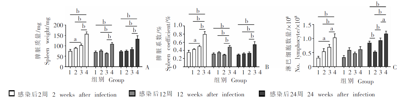
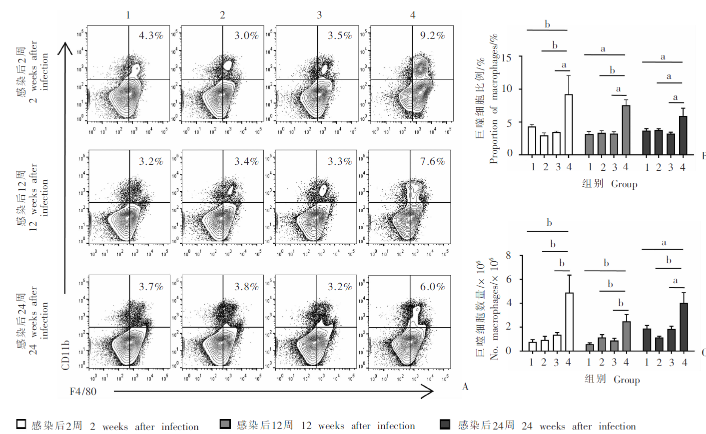
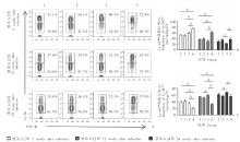
目的 研究不同数量多房棘球蚴原头节感染小鼠脾巨噬细胞亚群及其极化表型的变化。 方法 60只C57BL/6小鼠随机分为低、中、高感染组和对照组,每组15只。低、中、高感染组每只小鼠分别经肝门静脉穿刺接种50、500和2 000个多房棘球蚴原头节,对照组注射等量的生理盐水。感染后2、12和24周,各组随机选5只小鼠摘取脾脏,称量后计算脾脏系数;分离脾淋巴细胞,采用流式细胞术检测各组小鼠脾巨噬细胞不同亚群比例和数量;提取小鼠脾淋巴细胞总RNA,实时荧光定量PCR检测巨噬细胞M1和M2极化相关基因的mRNA相对转录水平,其中M1型极化相关基因为白细胞介素-1β(IL-1β)、γ干扰素(IFN-γ)、趋化因子(C-X-C基序)配体11(CXCL11)、趋化因子(C-C基序)受体7(CCR7)和CD86,M2型极化相关基因为甘露糖受体C-1型(MRC1)、抵抗素样分子α(Retnla)和精氨酸酶1(ARG1),以甘油醛-3-磷酸脱氢酶(GAPDH)为内参。采用SPSS 26.0软件进行统计学分析,不同时间点多组间的比较采用单因素方差分析,同一感染时间点的两两比较采用LSD法检验。 结果 感染后2周,高感染组小鼠脾脏质量、脾脏系数、脾淋巴细胞数量、脾巨噬细胞比例、脾巨噬细胞数量和Ly-6C高表达(Ly-6Chigh)巨噬细胞的比例分别为(157.2 ± 22.8)mg、(0.8 ± 0.1)%、(10.3 ± 2.9)× 107、(9.2 ± 6.4)%、(48.9 ± 32.7)× 105和(75.8 ± 4.6)%,均高于对照组[(75.0 ± 18.3)mg、(0.4 ± 0.1)%、(3.1 ± 1.3)× 107、(4.3 ± 0.7)%、(7.7 ± 4.1)× 105、(52.1 ± 8.4)%]、低感染组[(89.2 ± 7.4)mg、(0.4 ± 0.0)%、(5.4 ± 2.3)× 107、(3.0 ± 0.9)%、(9.3 ± 6.9)× 105、(50.1 ± 8.8)%]和中感染组[(102.6 ± 15.2)mg、(0.5 ± 0.1)%、(7.0 ± 2.1)× 107、(3.5 ± 0.3)%、(13.7 ± 3.9)× 105、(60.3 ± 8.7)%](F = 22.744、23.542、9.318、3.935、6.617、11.197,P < 0.05或P < 0.01);Ly-6C低表达(Ly-6Clow)巨噬细胞比例(20.3 ± 4.2)%低于对照组(39.0 ± 7.1)%、低感染组(41.2 ± 8.6)%和中感染组(34.2 ± 7.1)%(F = 9.157,P < 0.01)。感染后12周,高感染组小鼠脾脏质量、脾脏系数、脾巨噬细胞比例、巨噬细胞数量以及Ly-6Chigh巨噬细胞比例均高于对照组、低感染组和中感染组(F = 12.730、14.173、20.380、7.943和25.838,P < 0.01);Ly-6Clow巨噬细胞比例低于对照组、低感染组和中感染组(F = 27.668,P < 0.01)。感染后24周,高感染组小鼠脾脏质量、脾脏系数、脾淋巴细胞数量、脾巨噬细胞比例和数量均高于对照组、低感染组和中感染组(F = 8.664、7.318、13.047、3.315、6.007,P < 0.05或P < 0.01);Ly-6Chigh和Ly-6Clow巨噬细胞比例与对照组、低感染组和中感染组差异无统计学意义(F = 3.177、2.709,P > 0.05),且随着感染时间的延长,Ly-6Chigh巨噬细胞比例逐渐降低(F = 30.649,P < 0.01),Ly-6Clow巨噬细胞比例逐渐升高(F = 32.407,P < 0.01)。实时荧光定量PCR检测结果显示,感染后24周,高感染组M1型极化基因CXCL11的mRNA相对转录水平(28.2 ± 36.3)高于对照组(1.9 ± 2.7)(t = 2.243,P < 0.05),CD86(0.2 ± 0.1)低于对照组(0.5 ± 0.3)(t = -2.255,P < 0.05);M2型极化基因MRC1、Retnla和ARG1的mRNA相对转录水平(5.2 ± 2.9、201.8 ± 176.4、51.2 ± 69.6)均高于对照组(1.8 ± 1.5、0.8 ± 0.8、1.2 ± 0.8)(t = 2.313、3.470、2.185,P < 0.05)。 结论 感染后2周,多房棘球蚴原头节高感染组小鼠脾脏募集大量Ly-6C阳性单核来源的巨噬细胞,感染进程中Ly-6Chigh巨噬细胞逐渐向Ly-6Clow转变,且在感染后12周和24周脾巨噬细胞以M2型为主,诱导小鼠免疫耐受,利于多房棘球蚴的慢性寄生。

关键词: 多房棘球蚴病, 多房棘球蚴, 脾脏, 巨噬细胞, 极化

Abstract:

Objective To investigate the changes of splenic macrophages subsets and their polarization phenotype in mice infected with different numbers of Echinococcus multilocularis protoscoleces. Methods Sixty female C57BL/6 mice were randomly assigned to mild, moderate, and heavy infection groups and control group with 15 mice in each group. The mice in mild, moderate and heavy infection groups were inoculated with 50, 500, and 2 000 protoscoleces via hepatic portal vein puncture, respectively, while the control group was injected with the same volume of saline. At 2, 12, and 24 weeks after infection, the spleens from five mice randomly selected from each group were collected, followed by calculating the spleen coefficients after weighted, and isolating splenic lymphocytes. The proportions and numbers of different macrophages subsets were determined by flow cytometry. The total RNA of splenic lymphocytes was extracted, and the relative level of mRNA transcription of M1 and M2 polarization related genes in splenic macrophages were examined with real-time quantitative PCR. The M1 type polarization related genes were those involving interleukin-1β (IL-1β), interferon-γ (IFN-γ), chemokine (C-X-C motif) ligand 11 (CXCL11), chemokine (C-C motif) receptor 7 (CCR7) and CD86, and the M2 type polarization related genes were involving mannose receptor C-type 1 (MRC1), resistin like alpha (Retnla) and arginase 1 (ARG1). Glyceraldehyde-3-phosphate dehydrogenase (GAPDH) was used as an internal reference. SPSS 26.0 software was used for statistical analysis. The comparison between multiple groups at different time points was analyzed by one-way ANOVA, and the pairwise comparative analysis at the same time point was tested by LSD method. Results Two weeks after infection, the spleen weight, spleen coefficient, the number of splenic lymphocytes, the proportion of splenic macrophages, the number of splenic macrophages and Ly-6Chigh splenic macrophage subset proportion in heavy infection group were (157.2 ± 22.8) mg,(0.8 ± 0.1)%,(10.3 ± 2.9) × 107, (9.2 ± 6.4)%,(48.9 ± 32.7) × 105 and (75.8 ± 4.6)%, respectively, which were all significantly higher than that in the control group [(75.0 ± 18.3) mg, (0.4 ± 0.1)%, (3.1 ± 1.3) × 107, (4.3 ± 0.7)%, (7.7 ± 4.1) × 105, (52.1 ± 8.4)%], mild infection group [(89.2 ± 7.4) mg, (0.4 ± 0.0)%, (5.4 ± 2.3) × 107, (3.0 ± 0.9)%, (9.3 ± 6.9) × 105, (50.1 ± 8.8)%], and in moderate infection group [(102.6 ± 15.2) mg, (0.5 ± 0.1)%, (7.0 ± 2.1) × 107, (3.5 ± 0.3)%, (13.7 ± 3.9) × 105, (60.3 ± 8.7)%] (F = 22.744, 23.542, 9.318, 3.935, 6.617, 11.197, P < 0.05 or P < 0.01). Ly-6Clow macrophage subset proportion (20.3 ± 4.2)% was significantly milder than that in the control group (39.0 ± 7.1)%, mild infection group (41.2 ± 8.6)% and in moderate infection group (34.2 ± 7.1)%(F = 9.157, P < 0.01). At 12 weeks after infection, the spleen weight, spleen coefficient, the proportion and number of macrophages and the proportion of the Ly-6Chigh macrophage subset in the spleen were significantly higher in heavy infection group than that in the control group, mild and moderate infection groups (F = 12.730, 14.173, 20.380, 7.943, 25.838, P < 0.01), the proportion of Ly-6Clow macrophages was significantly lower than that in the control group, mild and moderate infection groups (F = 27.668, P < 0.01). At 24 weeks after infection, the spleen weight, spleen coefficient, the number of lymphocytes, the proportion and number of macrophages in the spleen were also significantly higher in heavy infection group (F = 8.664, 7.318, 13.047, 3.315, 6.007, P < 0.05 or P < 0.01). The proportions of Ly-6Chigh and Ly-6Clow macrophages were not significantly different from those of the control group, mild and moderate infection groups (F = 3.177 and 2.709, P > 0.05). With longer time of infection, the proportion of Ly-6Chigh macrophages was gradually decreased (F = 30.649, P < 0.01), and the proportion of Ly-6Clow macrophages was gradually increased (F = 32.407, P < 0.01). Real-time quantitative PCR showed that at 24 weeks after infection, the relative level of mRNA transcription of CXCL11 in heavy infection group (28.2 ± 36.3) was higher than that in control group (1.9 ± 2.7) (t = 2.243, P < 0.05), and the relative level of mRNA transcription of CD86 (0.2 ± 0.1) was significantly lower than that in the control group (0.5 ± 0.3) (t = -2.255, P < 0.05). The relative transcription level of mRNA of MRC1, Retnla, and ARG1 (5.2 ± 2.9, 201.8 ± 176.4, 51.2 ± 69.6) were significantly higher than those in control group (1.8 ± 1.5, 0.8 ± 0.8, 1.2 ± 0.8, respectively) (t = 2.313, 3.470 and 2.185, P < 0.05) Conclusion Two weeks after infection, large numbers of Ly-6C positive monocyte derived macrophages were recruited into spleen in the heavy infection group and the Ly-6Chigh macrophage subset gradually transformed shifted to the Ly-6Clow subset during infection. At 12 weeks and 24 weeks after infection, the macrophages in the spleen were dominated by the M2 type, inducing immune tolerance, favorable to chronic infection of Echinococcus multilocularis metacestode.

Key words: Alveolar echinococcoisis, Echinococcus multilocularis, Spleen, Macrophage, Polarization

中图分类号: