中国寄生虫学与寄生虫病杂志 ›› 2023, Vol. 41 ›› Issue (1): 23-28.doi: 10.12140/j.issn.1000-7423.2023.01.004

• 论著 • 上一篇    下一篇

细粒棘球蚴抗原B对小鼠巨噬细胞RAW264.7的极化作用

焦红杰1(), 齐文静2, 郭刚1, 包建玲1, 吴川川2, 宋传龙1, 李军1, 张文宝1,2, 严媚1,*()   

  1. 1.新疆医科大学第一附属医院,乌鲁木齐 830054
    2.新疆医科大学基础医学院,乌鲁木齐 830054
  • 收稿日期:2022-06-13 修回日期:2022-07-10 出版日期:2023-02-28 发布日期:2023-02-24
  • 通讯作者: * 严媚(1967-),女,博士,教授,主任医师,从事儿科血液及肿瘤研究。E-mail:yan10mei25@163.com
  • 作者简介:焦红杰(1987-),女,博士研究生,从事儿科血液及肿瘤研究。E-mail:374064774@qq.com
  • 基金资助:
    国家自然科学基金(82160031);国家自然科学基金(81830066);省部共建中亚高发病成因与防治国家重点实验室立项项目(SKL-HIDCA-2020-BC);新疆维吾尔自治区科技厅天山创新团队项目(2020D14027)

Polarization effect of Echinococcus granulosus antigen B on the mouse macrophage RAW264.7

JIAO Hongjie1(), QI Wenjing2, GUO Gang1, BAO Jianling1, WU Chuanchuan2, SONG Chuanlong1, LI Jun1, ZHANG Wenbao1,2, YAN Mei1,*()   

  1. 1. First Affiliated Hospital of Xinjiang Medical University, Urumqi 830054, China
    2. Basic Medical College of Xinjiang Medical University, Urumqi 830054, China
  • Received:2022-06-13 Revised:2022-07-10 Online:2023-02-28 Published:2023-02-24
  • Contact: * E-mail: yan10mei25@163.com
  • Supported by:
    National Natural Science Foundation of China(82160031);National Natural Science Foundation of China(81830066);project of State Key Laboratory of Pathogenesis and Prevention of Middle Asian High Disease(SKL-HIDCA-2020-BC);Tianshan Innovation Team Project of Science and Technology Department of Xinjiang Uygur Autonomous Region(2020D14027)

摘要:

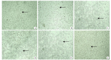
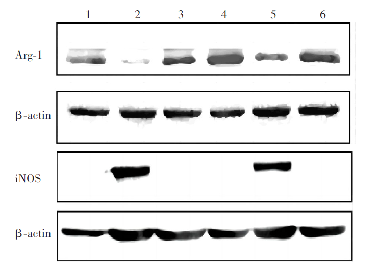
目的 探讨细粒棘球蚴抗原B(AgB)对巨噬细胞极化的调控作用。 方法 将RAW264.7巨噬细胞培养24 h后,分为M1、M2、AgB、AgB+M1、AgB+M2和空白对照组(M0组),每组3孔。待所有巨噬细胞贴壁3 h后,AgB、AgB+M1、AgB+M2组均加入羊源细粒棘球蚴囊液提取的天然AgB(终浓度为1 000 ng/ml),刺激1 h后,M1组和AgB+M1组加入脂多糖(LPS,终浓度为100 ng/ml)和γ干扰素(IFN-γ,终浓度为20 ng/ml)刺激分化20 h;M2组和AgB+M2组加入白细胞介素4(IL-4)、IL-13(终浓度均为20 ng/ml)刺激分化20 h;空白对照组不更换培养液,同步培养20 h。显微镜下观察巨噬细胞形态。提取各组巨噬细胞总RNA,RT-PCR检测刺激后巨噬细胞表面标志物精氨酸酶1(Arg-1)、肿瘤坏死因子α(TNF-α)的mRNA相对转录水平;蛋白质免疫印迹(Western blotting)分析巨噬细胞蛋白Arg-1、诱导型一氧化氮合酶(iNOS)的相对表达量;ELISA检测刺激后巨噬细胞培养上清中IL-10、TNF-α的表达变化。 结果 经刺激分化后,镜下可见M1组和AgB+M1组巨噬细胞大部分呈不规则形,有触角;M2组和AgB+M2组巨噬细胞大部分呈圆形或椭圆形,极少呈不规则形;M0组和AgB组巨噬细胞部分呈圆形、椭圆形,部分呈不规则形。RT-PCR结果显示,M2组和AgB+M2组巨噬细胞的Arg-1 mRNA相对转录水平分别为189.49 ± 68.43、435.83 ± 123.57(t = 246.30,P < 0.01),二者均高于M0组(1.00 ± 0.00)、M1组(1.87 ± 1.29)、AgB组(2.37 ± 2.06)、AgB+M1组(3.96 ± 1.92)(t = 188.50、187.60、187.10、185.50,均P < 0.01;t = 434.80、434.00、433.50、431.90,均P < 0.01);M1和AgB+M1组巨噬细胞的TNF-α mRNA相对转录水平分别为8.34 ± 2.92、8.10 ± 1.54(t = 0.24,P > 0.05),二者均高于M0组(1.00 ± 0.00)、M2组(1.37 ± 0.64)、AgB组(2.86 ± 0.44)、AgB+M2组(1.62 ± 0.27)(t = 7.34、6.97、5.48、6.71,均P < 0.01;t = 7.10、6.74、5.24、6.48,均P < 0.01)。Western blotting检测结果显示,M2组巨噬细胞的Arg-1蛋白相对表达量为1.18 ± 0.35,高于M1组(0.33 ± 0.18)、AgB+M1组(0.58 ± 0.10)(t = 0.67、0.61,均P < 0.01),与AgB组(1.05 ± 0.17)、AgB+M2组(0.97 ± 0.27)比较差异无统计学意义(t = 0.20、0.13,均P > 0.05);AgB+M2与M1组、AgB+M1组比较差异均有统计学意义(t = 0.52、0.48,均P < 0.05)。M1组和AgB+M1组iNOS蛋白相对表达量分别为0.95 ± 0.21、0.88 ± 0.02(t = 0.07,P > 0.05),二者均高于M0组(0.03 ± 0.00)、M2组(0)、AgB组(0)和AgB+M2组(0)(t = 0.92、0.95、0.95、0.95,均P < 0.01;t = 0.85、0.88、0.88、0.88,均P < 0.01)。ELISA检测结果显示,巨噬细胞培养上清液中,AgB+M1组和AgB+M2组IL-10细胞因子表达量分别为166.67 ± 56.67、213.33 ± 16.67,均高于M0组(0.00 ± 0.00)、M1组(43.33 ± 36.67)、M2组(50.00 ± 43.00)、AgB组(47.50 ± 25.00)(t = 166.70、123.30、116.70、119.20,均P < 0.05;t = 213.30、170.00、163.30、165.80,均P < 0.01)。M1、AgB+M1组TNF-α细胞因子表达量分别为833.13 ± 3.09、745.63 ± 118.00(t = 87.50,P > 0.05),均高于M0组(217.50 ± 32.26)、M2组(224.69 ± 17.68)、AgB组(308.44 ± 4.42)、AgB+M2组(251.25 ± 1.33)(t = 615.60、608.40、524.70、581.90,均P < 0.01;t = 528.10、520.90、437.20、494.40,均P < 0.01)。 结论 AgB可上调巨噬细胞Arg-1的表达,使其向M2型方向极化,可能是宿主和寄生虫免疫的重要调控分子,参与巨噬细胞的免疫调节。

关键词: 细粒棘球绦虫, 分泌抗原, 抗原B, 巨噬细胞极化

Abstract:

Objective To investigate the regulatory effect of Echinococcus granulosus antigen B (AgB) on macrophage polarization. Methods After cultivated for 24 h, the RAW264.7 macrophages cells were designated to 6 groups: M1, M2, AgB, AgB+M1, AgB+M2 and blank control (M0), 3 wells each group. After all the cells attached to the well wall for 3 h, the AgB、AgB+M1、AgB+M2 group was respectively added with natural AgB extracted from sheep hydatid cyst fluid (1 000 ng/ml, final concentration); 1 h post-stimulation, the M1 and AgB+M1 group was respectively added with lipopolysaccharide (LPS, final concentration 100 ng/ml), and IFN-γ (20 ng/ml, final concentration) to stimulate differentiation for 20 h; M2 and AgB+M2 group was added with interleukin 4 (IL-4) and IL-13 (final concentration 20 ng/ml) to stimulate differentiation for 20 h; the control group was cultured in parallel without changing medium. The morphology of macrophage cells were observed microscopically. Total RNA of the macrophages in all groups was extracted for performing RT-PCR to detect the relative transcription levels of the surface markers on stimulated macrophages, including arginase 1 (Arg-1) and tumor necrosis factor α (TNF-α). The relative expression levels of Arg-1 and inducible nitric oxide synthase (iNOS) were analyzed by Western blotting. The change of IL-10 and TNF-α expression in the culture supernatant of stimulated macrophages were detected by ELISA. Results After stimulation and differentiation, most cells in the M1 group and AgB+M1 group were irregularly shaped and had antennae. The cells of M2 group and AgB+M2 group were mostly round or oval, and very few were irregular. The cells of M0 group and AgB group were partly round and oval, and partly irregular. RT-PCR showed that the relative transcription levels of Arg-1 mRNA in the M2 group and the AgB+M2 group were 189.49 ± 68.43 and 435.83 ± 123.57, respectively (t = 246.30, P < 0.01). They were higher than those in the M0 group (1.00 ± 0.00), M1 group (1.87 ± 1.29), AgB group (2.37 ± 2.06), AgB+M1 group (3.96 ± 1.92) (t = 188.50, 187.60, 187.10, 185.50, P < 0.01; t = 434.80, 434.00, 433.50, 431.90, all P < 0.01). The relative transcription levels of TNF-α mRNA in the M1 group and the AgB+M1 group were 8.34 ± 2.92 and 8.10 ± 1.54, respectively (t = 0.24, P > 0.05). They were higher than that of the M0 group (1.00 ± 0.00), M2 group (1.37 ± 0.64), AgB group (2.86 ± 0.44) and AgB+M2 group (1.62 ± 0.27) (t = 7.34, 6.97, 5.48, 6.71, P < 0.01; t = 7.10, 6.74, 5.24, 6.48, P < 0.01). Western blotting showed that the relative expression level of Arg-1 protein in the M2 group was 1.18 ± 0.35, which was higher than that in the M1 group (0.33 ± 0.18) and the AgB+M1 group (0.58 ± 0.10) (t = 0.67,0.61, P < 0.01). There was no significant difference on the Arg-1 protein relative expression level between the AgB group (1.05 ± 0.17) and the AgB+M2 group (0.97 ± 0.27) (t =0.20, 0.13, P > 0.05). There was statistical significance in AgB+M2 group compared with M1 group and AgB+M1 group (t = 0.52, 0.48, P < 0.05). The iNOS relative expression levels of M1 group and AgB+M1 group were 0.95 ± 0.21 and 0.88 ± 0.02 (t = 0.07, P > 0.05), respectively. They were higher than those in M0 group (0.03 ± 0.00), M2 group (0), AgB group (0) and AgB+M2 group (0) (t = 0.92, 0.95, 0.95, 0.95, P < 0.01; t = 0.85, 0.88, 0.88, 0.88, P < 0.01). The ELISA results showed that the expression level of IL-10 cytokine in the cell supernatant was 166.67 ± 56.67 in the AgB+M1 group and 213.33 ± 16.67 in the AgB+M2 group, respectively. They were higher than those in the M0 group (0.00 ± 0.00), M1 group (43.33 ± 36.67), M2 group (50.00 ± 43.00) and AgB group (47.50 ± 25.00) (t = 166.70, 123.30, 116.70, 119.20, all P < 0.05. t = 213.30, 170.00, 163.30, 165.80, P < 0.01). The expression levels of TNF-α cytokines in M1 group and AgB+M1 group were 833.13 ± 3.09 and 745.63 ± 118.00, respectively (t = 87.50, P > 0.05). They were higher than those in M0 group (217.50 ± 32.26), M2 group (224.69 ± 17.68), AgB group (308.44 ± 4.42), AgB+M2 group (251.25 ± 1.33) (t = 615.60, 608.40, 524.70, 581.90, P < 0.01; t = 528.10, 520.90, 437.20, 494.40, P < 0.01). Conclusion AgB can up-regulate the expression of Arg-1 in macrophages and polarize it towards M2 type, which may be an important regulatory molecule in the host-parasite immune responses, involving in the immune regulation of macrophages.

Key words: Echinococcus granulosus, Secreted antigen, Antigen B, Polarization of macrophages

中图分类号: