| [1] |
World Health Organization . World Malaria Report 2019[R]. Geneva: WHO, 2019.
|
| [2] |
Idro R, Kakooza-Mwesige A, Balyejjussa S , et al. Severe neurological sequelae and behaviour problems after cerebral malaria in Ugandan children[J]. BMC Res Notes, 2010,3:104.
|
| [3] |
Kessler A, Dankwa S, Bernabeu M , et al. Linking EPCR-binding PfEMP1 to brain swelling in pediatric cerebral malaria[J]. Cell Host Microbe, 2017, 22(5): 601-614.e5.
|
| [4] |
MacCormick IJ, Beare NA, Taylor TE , et al. Cerebral malaria in children: using the Retina to study the brain[J]. Brain, 2014,137(Pt 8):2119-2142.
|
| [5] |
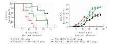
Besnard AG, Guabiraba R, Niedbala W , et al. IL-33-mediated protection against experimental cerebral malaria is linked to induction of type 2 innate lymphoid cells, M2 macrophages and regulatory T cells[J]. PLoS Pathog, 2015,11(2):e1004607.
|
| [6] |
El-Assaad F, Combes V, Grau GE , et al. Potential efficacy of citicoline as adjunct therapy in treatment of cerebral malaria[J]. Antimicrob Agents Chemother, 2014,58(1):602-605.
|
| [7] |
Waknine-Grinberg JH, Even-Chen S, Avichzer J , et al. Glucocorticosteroids in nano-sterically stabilized liposomes are efficacious for elimination of the acute symptoms of experimental cerebral malaria[J]. PLoS One, 2013,8(8):e72722.
|
| [8] |
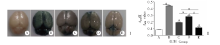
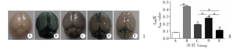
Tunon-Ortiz A, Lamb TJ . Blood brain barrier disruption in cerebral malaria: beyond endothelial cell activation[J]. PLoS Pathog, 2019,15(6):e1007786.
|
| [9] |
Shen J, Tian YP, Song GH , Mechanisms of cytoadherence in severe cerebral falciparum malaria[J]. Chin J Parasitol Parasit Dis, 2003,21(1):46-49. (in Chinese)
|
|
( 沈娟, 田野苹, 宋关鸿 . 重症恶性脑型疟疾的细胞粘附机制[J]. 中国寄生虫学与寄生虫病杂志, 2003,21(1):46-49.)
|
| [10] |
Zheng L, Sun XD, Pan YY , et al. Characteristic comparison of CD4 + Th immune responses and apoptosis in BALB/c mice infected with different plasmodia [J]. J Microbiol, 2011,31(1):19-23. (in Chinese)
|
|
( 郑丽, 孙晓丹, 潘艳艳 , 等. BALB/c小鼠感染不同疟原虫CD4 + Th应答和凋亡特点的比较研究 [J]. 微生物学杂志, 2011,31(1):19-23.)
|
| [11] |
Liew FY, Pitman NI , McInnes IB. Disease-associated functions of IL-33: the new kid in the IL-1 family[J]. Nat Rev Immunol, 2010,10(2):103-110.
|
| [12] |
Mirchandani AS, Salmond RJ, Liew FY . Interleukin-33 and the function of innate lymphoid cells[J]. Trends Immunol, 2012,33(8):389-396.
|
| [13] |
Chen G, Liu L . Functional change of dendritic cells/regulatory T cells/t-helper 17 cells and the mechanism of its cross-regulation in malaria[J]. Chin J Parasitol Parasit Dis, 2014,32(4):304-307. (in Chinese)
|
|
( 陈光, 刘蕾 . 疟疾发病中树突状细胞/调节性T细胞/Th17细胞的功能变化及其交叉调控机制[J]. 中国寄生虫学与寄生虫病杂志, 2014,32(4):304-307.)
|
| [14] |
Gogtay N, Kannan S, Thatte UM , , et al. Artemisinin-based combination therapy for treating uncomplicated Plasmodium vivax malaria[J]. Cochrane Database Syst Rev, 2013(10): CD008492.
|
| [15] |
Ayimba E, Hegewald J, Ségbéna AY , et al. Proinflammatory and regulatory cytokines and chemokines in infants with uncomplicated and severe Plasmodium falciparum malaria[J]. Clin Exp Immunol, 2011,166(2):218-226.
|
| [16] |
Nie CQ, Bernard NJ, Schofield L , et al. CD4 +CD25 + regulatory T cells suppress CD4 + T-cell function and inhibit the development of Plasmodium berghei-specific TH1 responses involved in cerebral malaria pathogenesis [J]. Infect Immun, 2007,75(5):2275-2282.
|
| [17] |
Gorman S, Kuritzky LA, Judge MA , et al. Topically applied 1,25-dihydroxyvitamin D3 enhances the suppressive activity of CD4 +CD25 + cells in the draining lymph nodes [J]. J Immunol, 2007,179(9):6273-6283.
|
| [18] |
Ait-Belgnaoui A, Durand H, Cartier C , et al. Prevention of gut leakiness by a probiotic treatment leads to attenuated HPA response to an acute psychological stress in rats[J]. Psychoneuroendocrinology, 2012,37(11):1885-1895.
|
| [19] |
McCusker RH, Kelley KW . Immune-neural connections: how the immune system’s response to infectious agents influences behavior[J]. J Exp Biol, 2013,216(Pt 1):84-98.
|
| [20] |
Coban C, Ishii KJ, Uematsu S ,et al. Pathological role of Toll-like receptor signaling in cerebral malaria[J]. Int Immunol, 2007,19(1):67-79.
|
| [21] |
Liu TP, Fu Y, Xu WY . Immunopathological mechanism of cerebral malaria[J]. Chin J Parasitol Parasit Dis, 2011,29(1):64-67. (in Chinese)
|
|
( 刘太平, 付雍, 徐文岳 . 脑型疟发生的免疫病理机制[J]. 中国寄生虫学与寄生虫病杂志, 2011,29(1):64-67.)
|
| [22] |
Yang GH, Yang L, Wang WD , et al. Discovery and validation of extracellular/circulating microRNAs during idiopathic pulmonary fibrosis disease progression[J]. Gene, 2015,562(1):138-144.
|
| [23] |
Chen L, Sha ML, Li D , et al. Relaxin abrogates renal interstitial fibrosis by regulating macrophage polarization via inhibition of Toll-like receptor 4 signaling[J]. Oncotarget, 2017,8(13):21044-21053.
|

 ), Wei ZHAO2, Ya-ming CAO3, Lan XU1