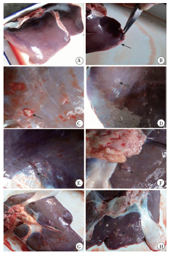
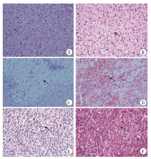
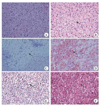
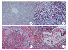
| [1] | Urquhart GM.Veterinary parasitology[M]. 2nd ed. Blackwell Science. 1996. | | [2] | 刘倩, 程娜, 周岩, 等. 片形吸虫病研究进展[J]. 中国寄生虫学与寄生虫病杂志, 2013, 31(3): 229-234. | | [3] | 向才碧, 张青松, 张登斌, 等. 我国人群感染片形吸虫的调查分析[J]. 职业与健康, 2003, 19(11): 90-90. | | [4] | 张克琴, 陈宝杰, 张克志, 等. 宾川县片形吸虫感染疫情的调查报告[J]. 卫生软科学, 2013, 27(3): 164-166. | | [5] | 顾伟, 苏慧勇, 邹静, 等. 云南省首次暴发巨片形吸虫感染的临床诊治分析[J]. 中国寄生虫学与寄生虫病杂志, 2012, 30(6): 455-459. | | [6] | 陈凤, 杨慧, 刘榆华, 等. 云南省宾川县片形吸虫病流行病学调查及慢性病例分析[J]. 中国寄生虫学与寄生虫病杂志, 2014, 32(6): 422-424. | | [7] | Zafra R, Buffoni L, Martínez-Moreno A, et al. A study of the liver of goats immunized with a synthetic peptide of the Sm14 antigen and challenged with Fasciola hepatica[J]. J Comp Pathol, 2008, 139(4): 169-176. | | [8] | Alvarez Rojas CA, Ansell BR, Hall RS, et al. Transcriptional analysis identifies key genes involved in metabolism, fibrosis/tissue repair and the immune response against Fasciola hepatica in sheep liver[J]. Parasit Vectors, 2015, 8: 124. | | [9] | Zafra R, Pérez-écija RA, Buffoni L, et al. Early and late peritoneal and hepatic changes in goats immunized with recombinant cathepsin L1 and infected with Fasciola hepatica[J]. J Comp Pathol, 2013, 148(4): 373-384. | | [10] | 梅雪芳, 施维, 张瑶瑶, 等. 大片吸虫分泌排泄抗原对小鼠肝损伤的研究[J]. 动物医学进展, 2016, 37(12): 34-38. | | [11] | Van Milligen F, Cornelissen J, Gaasenbeek C, et al. A novel exvivo rat infection model to study protective immunity against Fasciola hepatica at the gut level[J]. J Immunol Methods, 1998, 213(2): 183-190. | | [12] | Kawano J, Yamamoto T, Koga M, et al. Penetration in vitro of newly excysted juvenile flukes of Japanese Fasciola sp. through ligated intestines of rabbits, mice, rats and chickens[J]. J Vet Med Sci, 1992, 54(1): 69-73. | | [13] | Rajasekariah G, Howell M.The fate of Fasciola hepatica metacercariae following challenge infection of immune rats[J]. J Helminthol, 1977, 51(4): 289-294. | | [14] | Burden D, Bland A, Hammet N, et al. Fasciola hepatica: migration of newly excysted juveniles in resistant rats[J]. Exp Parasitol, 1983, 56(2): 277-288. | | [15] | Kendall S, Peirce M.Synergism in the chemotherapy of fascioliasis[J]. Br Vet J, 1969, 125(2): 82-86. | | [16] | Hayes T.Further evidence for the early expression of immunity to Fasciola hepatica in rats[J]. J Parasitol, 1978, 64(2): 374-376. | | [17] | 全琛宇. 小鼠模型感染大片形吸虫研究平台初探[D]. 南宁:广西大学, 2014. | | [18] | 范东, 李鹏, 孙华, 等. 肝片吸虫感染所致肝脓肿的CT表现[J]. 中华放射学杂志, 2006, 40(1): 191-194. | | [19] | Bottari N, Mendes R, Lucca N, et al. Oxidative stress associated with pathological lesions in the liver of rats experimentally infected by Fasciola hepatica[J]. Exp Parasitol, 2015, 159: 24-28. |
|

 )
)