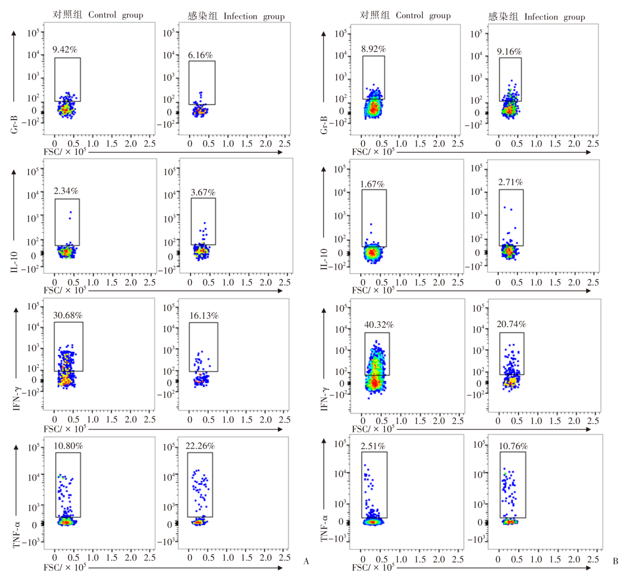
| [1] | Carmena D, Benito A, Eraso E. The immunodiagnosis of Echinococcus multilocularis infection[J]. Clin Microbiol Infect, 2007, 13(5): 460-475. | | [2] | Davidson RK, Romig T, Jenkins E, et al. The impact of globalisation on the distribution of Echinococcus multilocularis[J]. Trends Parasitol, 2012, 28(6): 239-247. | | [3] | Zhang CS, Lin RY, Li ZD, et al. Immune exhaustion of T cells in alveolar echinococcosis patients and its reversal by blocking checkpoint receptor TIGIT in a murine model[J]. Hepatology, 2020, 71(4): 1297-1315. | | [4] | 施阳, 李德伟, 阿比旦?艾尼瓦尔, 等. 多房棘球蚴感染对小鼠脾脏自然杀伤细胞Tim3表达的影响[J]. 中国血吸虫病防治杂志, 2023, 35(4): 366-373. | | | Shi Y, Abidan ANWE, Li DW, et al. Effect of Echinococcus multilocularis infection on Tim3 expression in spleen natural killer cells of mice[J]. Chin J Schisto Control, 2023, 35(4): 366-373. (in Chinese) | | [5] | 侯昕伶, 李亮, 李玲慧, 等. 泡球蚴感染对小鼠脾脏CD8+T细胞免疫功能耗竭的影响[J]. 中国血吸虫病防治杂志, 2020, 32(6): 591-597, 604. | | | Hou XL, Li L, Li LH, et al. Exhaustion of CD8+T cell immune functions in spleen of mice with different doses of Echinococcus multilocularis infections[J]. Chin J Schisto Control, 2020, 32(6): 591-597, 604. (in Chinese) | | [6] | 侯娇, 温浩, 王明坤, 等. 多房棘球蚴感染小鼠脾脏巨噬细胞亚群及其极化表型的变化[J]. 中国寄生虫学与寄生虫病杂志, 2021, 39(6): 771-778. | | | Hou J, Wen H, Wang MK, et al. Changes of macrophage subsets and polarization in spleen of mice infected with Echinococcus multilocularis[J]. Chin J Parasitol Parasit Dis, 2021, 39(6): 771-778. (in Chinese) | | [7] | 王宝辉. 脾脏CD49b-NK细胞的发育分化研究[D]. 合肥: 中国科学技术大学, 2019: 46-51. | | | Wang BH. The development and differentiation of spleen CD49b-NK cells[D]. Hefei: University of Science and Technology of China, 2019: 46-51. (in Chinese) | | [8] | Ding Y, Lavaert M, Grassmann S, et al. Distinct developmental pathways generate functionally distinct populations of natural killer cells[J]. Nat Immunol, 2024, 25(7): 1183-1192. | | [9] | Shannon MJ, Mace EM. Natural killer cell integrins and their functions in tissue residency[J]. Front Immunol, 2021, 12: 647358. | | [10] | Chen SM, Zhu HT, Jounaidi Y. Comprehensive snapshots of natural killer cells functions, signaling, molecular mechanisms and clinical utilization[J]. Signal Transduct Target Ther, 2024, 9: 302. | | [11] | Gasteiger G, Fan XY, Dikiy S, et al. Tissue residency of innate lymphoid cells in lymphoid and nonlymphoid organs[J]. Science, 2015, 350(6263): 981-985. | | [12] | Sojka DK, Plougastel-Douglas B, Yang LP, et al. Tissue-resident natural killer (NK) cells are cell lineages distinct from thymic and conventional splenic NK cells[J]. eLife, 2014, 3: e01659. | | [13] | Sivori S, Pende D, Quatrini L, et al. NK cells and ILCs in tumor immunotherapy[J]. Mol Aspects Med, 2021, 80: 100870. | | [14] | Michel T, Poli A, Domingues O, et al. Mouse lung and spleen natural killer cells have phenotypic and functional differences, in part influenced by macrophages[J]. PLoS One, 2012, 7(12): e51230. | | [15] | Taggenbrock RLRE, ILC1: development, maturation, and transcriptional regulation[J]. Eur J Immunol, 2023, 53(2): 2149435. | | [16] | Zhang CS, Shao YM, Yang ST, et al. T-cell tolerance and exhaustion in the clearance of Echinococcus multilocularis: role of inoculum size in a quantitative hepatic experimental model[J]. Sci Rep, 2017, 7: 11153. | | [17] | Zhang CS, Wang H, Li J, et al. Involvement of TIGIT in natural killer cell exhaustion and immune escape in patients and mouse model with liver Echinococcus multilocularis infection[J]. Hepatology, 2021, 74(6): 3376-3393. | | [18] | Crane GM, LiuYC, Chadburn A. Spleen: development, anatomy and reactive lymphoid proliferations[J]. Semin Diagn Pathol, 2021, 38(2): 112-124. | | [19] | Cenariu D, Iluta S, Zimta AA, et al. Extramedullary hematopoiesis of the liver and spleen[J]. J Clin Med, 2021, 10(24): 5831. | | [20] | Gutiérrez-Hoya A, Soto-Cruz I. NK cell regulation in cervical cancer and strategies for immunotherapy[J]. Cells, 2021, 10(11): 3104. | | [21] | Stojanovic A, Cerwenka A. ILC1-like NK cells as matchmakers for DC-T cell interactions[J]. Immunity, 2021, 54(10): 2185-2187. | | [22] | Van Acker N, Frenois FX, Gravelle P, et al. Spatial mapping of innate lymphoid cells in human lymphoid tissues and lymphoma at single-cell resolution[J]. Nat Commun, 2025, 16: 4545. | | [23] | He JM, Chen DL, Xiong W, et al. Eomesodermin spatiotemporally orchestrates the early and late stages of NK cell development by targeting KLF2 and T-bet, respectively[J]. Cell Mol Immunol, 2024, 21(7): 662-673. | | [24] | Samper N, Hardardottir L, Depierreux DM, et al. Kir6.1, a component of an ATP-sensitive potassium channel, regulates natural killer cell development[J]. Front Immunol, 2024, 15: 1490250. | | [25] | Flommersfeld S, B?ttcher JP, Ersching J, et al. Fate mapping of single NK cells identifies a type 1 innate lymphoid-like lineage that bridges innate and adaptive recognition of viral infection[J]. Immunity, 2021, 54(10): 2288-2304. e7. | | [26] | Lu JS, Hu ZQ, Jiang HJ, et al. Dual nature of type I interferon responses and feedback regulations by SOCS1 dictate malaria mortality[J]. J Adv Res, 2025, 73: 295-310. | | [27] | Montes de Oca M, Kumar R, de Labastida Rivera F, et al. Type I interferons regulate immune responses in humans with blood-stage Plasmodium falciparum infection[J]. Cell Rep, 2016, 17(2): 399-412. | | [28] | Hu Y, Wang XL, Wei YH, et al. Functional inhibition of natural killer cells in a BALB/c mouse model of liver fibrosis induced by Schistosoma japonicum infection[J]. Front Cell Infect Microbiol, 2020, 10: 598987. | | [29] | Zhou J, Peng H, Li K, et al. Liver-resident NK cells control antiviral activity of hepatic T cells via the PD-1-PD-L1 axis[J]. Immunity, 2019, 50(2): 403-417. e4. | | [30] | Abulizi A, Shao YM, Aji T, et al. Echinococcus multilocularis inoculation induces NK cell functional decrease through high expression of NKG2A in C57BL/6 mice[J]. BMC Infect Dis, 2019, 19(1): 792. |
|

 ), 阿比旦·艾尼瓦尔1, 葛聪蕙1, 唐娜1, 孙胜1, 王梦颖1, 高毅1, 阿依娜尔·吉恩斯1, 胡秋1, 李静1,2, 王慧1,3, 张传山1,3,*(
 )(
)(