中国寄生虫学与寄生虫病杂志 ›› 2025, Vol. 43 ›› Issue (6): 827-834.doi: 10.12140/j.issn.1000-7423.2025.06.012

• 论著 • 上一篇    下一篇

粉尘螨过敏原Der f 1引发人气道上皮细胞炎症反应的作用机制

姜尚德1()(), 臧潇1, 梅伟1, 马连政2, 饶莉娜1, 洪善超3,*()(), 汪伟1,4,*()()   

  1. 1 南京医科大学公共卫生学院,江苏 南京 211166
    2 江南大学无锡医学院,江苏 无锡 214122
    3 江南大学附属中心医院医学检验科,江苏 无锡 214062
    4 国家卫生健康委员会寄生虫病预防与控制技术重点实验室,江苏省寄生虫与媒介控制技术重点实验室,江苏省血吸虫病防治研究所,江苏 无锡 214064
  • 收稿日期:2025-08-09 修回日期:2025-09-04 出版日期:2025-12-30 发布日期:2025-12-25
  • 通讯作者: *汪伟(ORCID:0000-0002-5709-981X),男,硕士,研究员,从事热带病流行病学研究。E-mail:wangwei@jipd.com;洪善超(ORCID:0000-0003-1587-3253),女,博士,主任医师,从事尘螨致敏机制的研究。E-mail:hongshanchao1@163.com
  • 作者简介:姜尚德(ORCID:0009-0007-0921-7205),男,硕士研究生,从事尘螨诱导过敏性哮喘发病机制的研究。E-mail:2023121186@stu.njmu.edu.cn
  • 基金资助:
    江苏省无锡市“双百”中青年医疗卫生后备拔尖人才项目(HB2023024);无锡市卫生健康委重大科研项目(Z202520)

Mechanism underlying human airway epithelial cell inflammation triggered by dust mite allergen Der f 1

JIANG Shangde1()(), ZANG Xiao1, MEI Wei1, MA Lianzheng2, RAO Lina1, HONG Shanchao3,*()(), WANG Wei1,4,*()()   

  1. 1 School of Public Health, Nanjing Medical University, Nanjing 211166, Jiangsu, China
    2 Wuxi School of Medicine, Jiangnan University, Wuxi 214122, Jiangsu, China
    3 Department of Laboratory Medicine, Jiangnan University Medical Center, Wuxi 214062, Jiangsu, China
    4 National Health Committee Key Laboratory of Parasitic Diseases Prevention and Control, Jiangsu Provincial Key Laboratory on Parasites and Vector Control Technology, Jiangsu Institute of Parasitic Diseases, Wuxi 214064, Jiangsu, China
  • Received:2025-08-09 Revised:2025-09-04 Online:2025-12-30 Published:2025-12-25
  • Contact: *E-mail:wangwei@jipd.com;hongshanchao1@163.com
  • Supported by:
    Wuxi City“Double Hundred”Middle-aged and Young Medical and Health Reserve Talents Project of Jiangsu Province(HB2023024);Major Scientific Research Project of Wuxi Municipal Health Commission(Z202520)

摘要:

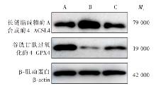
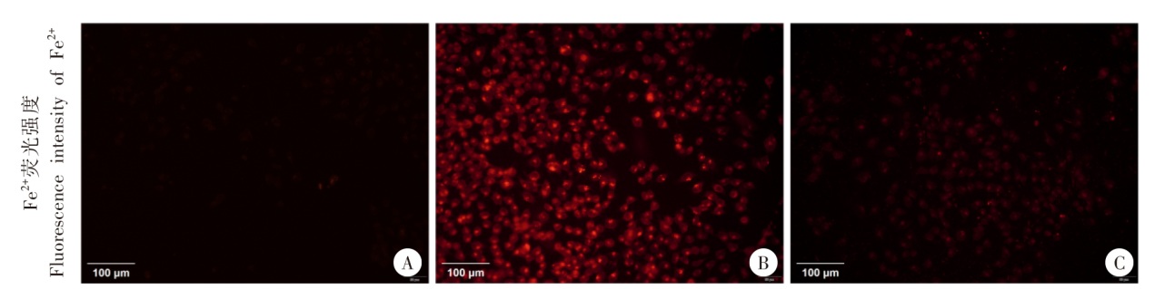
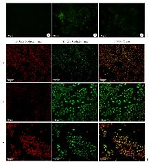
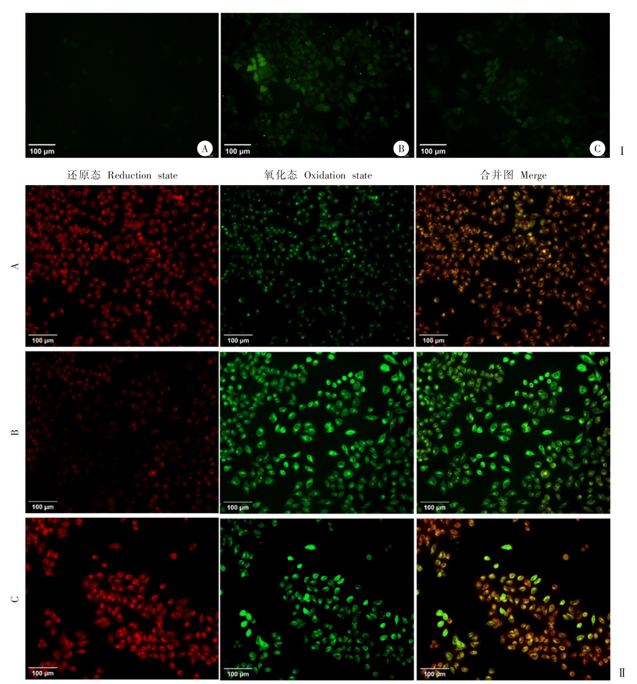
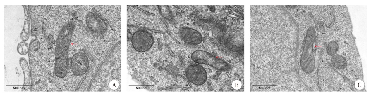
目的 探讨粉尘螨过敏原1(Der f 1)引发人气道上皮细胞炎症反应的作用机制。方法 将人支气管上皮细胞系BEAS-2B接种至96孔板中(5 × 10³个/孔),分为Der f 1组、Der f 1 + 铁抑素-1(Fer-1)组和阴性对照组,Der f 1组加40 μl Der f 1(100 μg/ml),Der f 1 + Fer-1组在加Der f 1前2 h加4 μl Fer-1(1 000 μmol/ml),阴性对照组加等量无酶水,培养24 h后,采用细胞计数试剂盒-8(CCK-8)进行细胞计数,ELISA测定3组细胞培养上清中的细胞因子白细胞介素-6(IL-6)、人胸腺基质淋巴细胞生成素(TSLP)、IL-33含量。将BEAS-2B细胞接种至6孔板中(5 × 10⁵个/孔),分为Der f 1组、Der f 1 + Fer-1组和阴性对照组,提取各组细胞总蛋白,十二烷基硫酸钠-聚丙烯酰胺凝胶电泳(SDS-PAGE)检测蛋白表达情况,以谷胱甘肽过氧化酶4(GPX4)抗体(1∶2 000)、长链脂酰辅酶A合成酶4(ACSL4)抗体(1∶2 000)为一抗,辣根过氧化物酶(HRP)标记的山羊抗兔IgG抗体为二抗(1∶10 000),蛋白质免疫印迹(Western blotting)检测细胞中GPX4和ACSL4蛋白表达水平。将BEAS-2B细胞接种至24孔板中(5 × 10⁴个/孔),分为Der f 1组、Der f 1 + Fer-1组和阴性对照组,荧光显微镜下观察各组细胞中亚铁离子(Fe2+)、活性氧(ROS)荧光强度,以及脂质过氧化物(LPO)染色后红色荧光与绿色荧光强度,并计算比值。将BEAS-2B细胞接种至6孔板中(5 × 10⁵个/孔),分为Der f 1组、Der f 1 + Fer-1组和阴性对照组,比色法检测各组细胞丙二醛(MDA)含量。将BEAS-2B细胞接种至6孔板中(5 × 10⁶个/孔),分为Der f 1组、Der f 1 + Fer-1组和阴性对照组,透射电子显微镜下观察各组细胞线粒体形态。采用GraphPad Prism 8.0.1软件进行统计学分析,组间比较采用单因素方差分析。结果 细胞活力检测结果显示,Der f 1组细胞活力为0.79 ± 0.03,低于阴性对照组(1.07 ± 0.08)(t = 6.663,P < 0.01);Der f 1 + Fer-1组细胞活力为0.94 ± 0.03,高于Der f 1组(t = 6.694,P < 0.01)。ELISA结果显示,Der f 1组上清中IL-6含量为(117.30 ± 21.32)pg/ml,高于阴性对照组(50.07 ± 5.82)pg/ml(t = 5.279,P < 0.01),Der f 1 + Fer-1组上清中IL-6含量为(50.31 ± 12.28)pg/ml,低于Der f 1组(t = 4.721,P < 0.01);Der f 1组上清中TSLP含量为(10.00 ± 2.37)pg/ml,高于阴性对照组(3.81 ± 0.92)pg/ml(t = 4.223,P < 0.05),Der f 1 + Fer-1组上清中TSLP含量为(4.41 ± 1.59)pg/ml,低于Der f 1组(t = 3.399,P < 0.05);Der f 1组上清中IL-33含量为(24.18 ± 2.53)pg/ml,高于阴性对照组(12.09 ± 2.08)pg/ml(t = 6.39,P < 0.01),Der f 1 + Fer-1组上清中IL-33含量为(15.76 ± 1.39)pg/ml,低于Der f 1组(t = 5.045,P < 0.01)。Western blotting结果显示,Der f 1组GPX4蛋白相对表达量为0.38 ± 0.08,低于阴性对照组的1.00 ± 0.00(t = 13.21,P < 0.01),Der f 1 + Fer-1组相对表达量为0.72 ± 0.08,高于Der f 1组(t = 5.122,P < 0.01);Der f 1组ACSL4蛋白相对表达量为1.74 ± 0.12,高于阴性对照组的1.00 ± 0.00(t = 10.65,P < 0.01);Der f 1 + Fer-1组相对表达量为1.33 ± 0.13,低于Der f 1组(t = 4.094,P < 0.05)。荧光显微镜观察结果显示,Der f 1组细胞内Fe2+荧光强度为37.19 ± 5.42,高于阴性对照组的11.93 ± 0.54(t = 8.035,P < 0.01);Der f 1 + Fer-1组为13.16 ± 1.89,低于Der f 1组(t = 7.253,P < 0.01)。Der f 1组细胞内ROS荧光强度为13.48 ± 3.36,高于阴性对照组的6.80 ± 0.60(t = 3.386,P < 0.05);Der f 1 + Fer-1组ROS荧光强度为7.35 ± 0.42,低于Der f 1组(t = 3.134,P < 0.05)。Der f 1组细胞内LPO程度为3.91 ± 1.65,高于阴性对照组的0.31 ± 0.13(t = 3.775,P < 0.05);Der f 1 + Fer-1组LPO程度为0.80 ± 0.15,低于Der f 1组(t = 3.262,P < 0.05)。比色法结果显示,Der f 1组细胞内MDA含量为(5.57 ± 1.66)nmol/mg,高于阴性对照组的(2.18 ± 0.51)nmol/mg(t = 3.393,P < 0.05),Der f 1 + Fer-1组MDA含量为(2.24 ± 0.38)nmol/mg,低于Der f 1组(t = 3.4,P < 0.05)。透射电子显微镜结果显示,阴性对照组细胞线粒体状态良好,线粒体边界清晰且线粒体嵴较多;Der f 1组与阴性对照组相比细胞线粒体体积减小、膜增厚并且线粒体嵴减少甚至消失;Der f 1 + Fer-1组与Der f 1组相比,细胞线粒体形态改变受到抑制,线粒体体积减小、膜增厚和线粒体嵴减少等形态变化减轻。结论 Der f 1可通过扰乱铁代谢、抑制抗氧化防御及促进脂质过氧化,诱导人气道上皮细胞发生铁死亡,进而增强IL-6、TSLP和IL-33等炎症因子的释放,促进气道炎症反应。

关键词: 尘螨, 粉尘螨主要过敏原1, 气道上皮细胞, 铁死亡, 炎症

Abstract:

Objective To decipher the mechanisms underlying human airway epithelial cell inflammation induced by the major allergen Der f 1 of dust mite. Methdos BEAS-2B cells were seeded onto 96-well plates at a density of 5 × 10³ cells per well and divided into three groups, including the Der f 1 group, Der f 1 + ferrostatin-1 (Fer-1) group, and negative control group. Cells in the Der f 1 group were treated with 40 μl of Der f 1 (100 μg/ml), and cells in the Der f 1 + Fer-1 group was pretreated with 4 μl Fer-1 (1 000 μmol/ml) 2 hours prior to Der f 1 treatment, while cells in the negative control group were given an equivalent volume of nuclease-free water. After 24 hours of incubation, cell viability was assessed in the Der f 1 group, Der f 1 + Fer-1 group and negative control group using the CCK-8 assay, and concentrations of interleukin-6 (IL-6), thymic stromal lymphopoietin (TSLP), and IL-33 were measured in the cell culture supernatants with enzyme-linked immunosorbent assay (ELISA). For Western blotting assay, BEAS-2B cells were seeded onto 6-well plates at a density of 5 × 10⁵ cells per well, and assigned into three groups, including the Der f 1 group, Der f 1 + Fer-1 group, and negative control group. Total protein was extracted from BEAS-2B cells, separated by sodium dodecyl sulfate-polyacrylamide gel electrophoresis (SDS-PAGE), and probed with primary antibodies against glutathione peroxidase 4 (GPX4, 1︰2 000 dilution) and acyl-CoA synthetase long-chain family member 4 (ACSL4, 1︰2 000 dilution), followed by incubation into horseradish peroxidase (HRP)-conjugated goat anti-rabbit IgG secondary antibody (1︰10 000 dilution). The GPX4 and ACSL4 protein expression was determined in cells using Western blotting assay. To assess ferroptosis-related indicators, BEAS-2B cells were seeded onto 24-well plates at a density of 5 × 10⁴ cells per well and assigned into three groups, including the Der f 1 group, Der f 1 + Fer-1 group, and negative control group. Intracellular fluorescence intensity of Fe²⁺ and reactive oxygen species (ROS) were measured under a fluorescence microscope, and the ratio of red to green fluorescence intensity was estimated following lipid peroxidation (LPO) staining. For colorimetric assay, BEAS-2B cells were seeded onto 6-well plates at a density of 5 × 10⁵ cells per well and assigned into three groups, including the Der f 1 group, Der f 1 + Fer-1 group, and negative control group, and malondialdehyde (MDA) content was measured using a colorimetric assay. In addition, BEAS-2B cells were seeded onto 6-well plates at a density of 5 × 10⁶ cells per well and assigned into three groups, including the Der f 1 group, Der f 1 + Fer-1 group, and negative control group, and mitochondrial morphology was examined under a transmission electron microscope. All statistical analyses were performed using the software GraphPad Prism 8.0.1, and differences of means among three groups were tested for statistical significance with one-way analysis of variance (ANOVA). Results The cell viability was significantly lower in the Der f 1 group (0.79 ± 0.03) than in the negative control (1.07 ± 0.08) (t = 6.663, P < 0.01), and co-treatment with Fer-1 restored cell viability to 0.94 ± 0.03 (t = 6.694, P < 0.01). ELISA showed that the IL-6 concentration in the supernatant of the Der f 1 group was (117.30 ± 21.32) pg/ml, which was higher than that of the negative control group (50.07 ± 5.82) pg/ml (t = 5.279, P < 0.01). The IL-6 concentration in the supernatant of the Der f 1 + Fer-1 group was (50.31 ± 12.28) pg/ml, which was lower than that of the Der f 1 group (t = 4.721, P < 0.01). The TSLP concentration in the supernatant of the Der f 1 group was (10.00 ± 2.37) pg/ml, which was higher than that of the negative control group (3.81 ± 0.92) pg/ml (t = 4.223, P < 0.05). The TSLP concentration in the supernatant of the Der f 1 + Fer-1 group was (4.41 ± 1.59) pg/ml, which was lower than that of the Der f 1 group (t = 3.399, P < 0.05). The IL-33 concentration in the supernatant of the Der f 1 group was (24.18 ± 2.53) pg/ml, which was higher than that of the negative control group (12.09 ± 2.08) pg/ml (t = 6.39, P < 0.01). The IL-33 concentration in the supernatant of the Der f 1 + Fer-1 group was (15.76 ± 1.39) pg/ml, which was lower than that of the Der f 1 group (t = 5.045, P < 0.01). Western blotting determined lower relative GPX4 protein expression in the Der f 1 group (0.38 ± 0.08) than in the negative control group (1.00 ± 0.00) (t = 13.21, P < 0.01), and higher GPX4 protein expression in the Der f 1 + Fer-1 group (0.72 ± 0.08) than in the Der f 1 group (t = 5.122, P < 0.01). Conversely, ACSL4 protein expression was upregulated in the Der f 1 group (1.74 ± 0.12 vs. 1.00 ± 0.00; t = 10.65, P < 0.01) and reduced by Fer-1 treatment (1.33 ± 0.13, t = 4.094, P < 0.05). Fluorescence microscopy showed increased intracellular fluorescence intensity of Fe²⁺ in the Der f 1 group (37.19 ± 5.42 vs. 11.93 ± 0.54; t = 8.035, P < 0.01), which was significantly lowered by Fer-1 treatment (13.16 ± 1.89, t = 7.253, P < 0.01). The intracellular ROS fluorescence intensity in the Der f 1 group was 13.48 ± 3.36, which was higher than that in the negative control group (6.80 ± 0.60, t = 3.386, P < 0.05). The fluorescence intensity in the Der f 1 + Fer-1 group was 7.35 ± 0.42, which was lower than that in the Der f 1 group (t = 3.134, P < 0.05). The intracellular LPO fluorescence intensity in the Der f 1 group was 3.91 ± 1.65, which was higher than that in the negative control group (0.31 ± 0.13, t = 3.775, P < 0.05). The LPO fluorescence intensity in the Der f 1 + Fer-1 group was 0.80 ± 0.15, which was lower than that in the Der f 1 group (t = 3.262, P < 0.05). The colorimetric assay results showed that the intracellular MDA content in the Der f 1 group was (5.57 ± 1.66) nmol/mg, which was higher than that in the negative control group (2.18 ± 0.51) nmol/mg (t = 3.393, P < 0.05). The MDA content in the Der f 1 + Fer-1 group was (2.24 ± 0.38) nmol/mg, which was lower than that in the Der f 1 group (t = 3.4, P < 0.05). Transmission electron microscopy displayed well mitochondrial status, clear mitochondrial boundary and many mitochondrial cristae in the negative control group, and Der f 1 exposure led to mitochondrial shrinkage, membrane thickening, and reduction or loss of cristae. These structural alterations were markedly attenuated in the Der f 1 + Fer-1 group, including weakening of mitochondrial shrinkage, membrane thickening and reduced numbers of mitochondrial cristae. Conclusion Der f 1 induces ferroptosis in human airway epithelial cells through disrupting iron homeostasis, suppressing antioxidant defense, and promoting lipid peroxidation, thereby potentiating the release of IL-6, TSLP, and IL-33 to exacerbate airway inflammation.

Key words: Dust mite, Dermatophagoides farinae allergen 1, Airway epithelial cell, Ferroptosis, Inflammation

中图分类号: