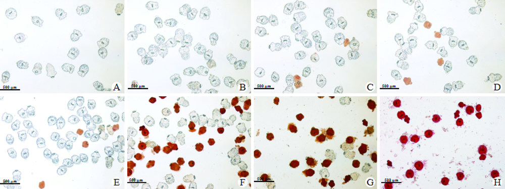
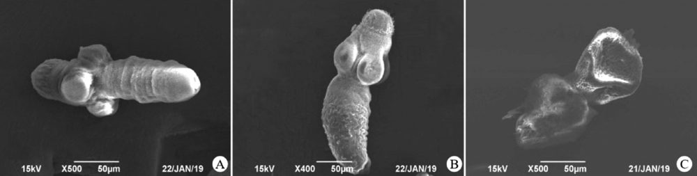
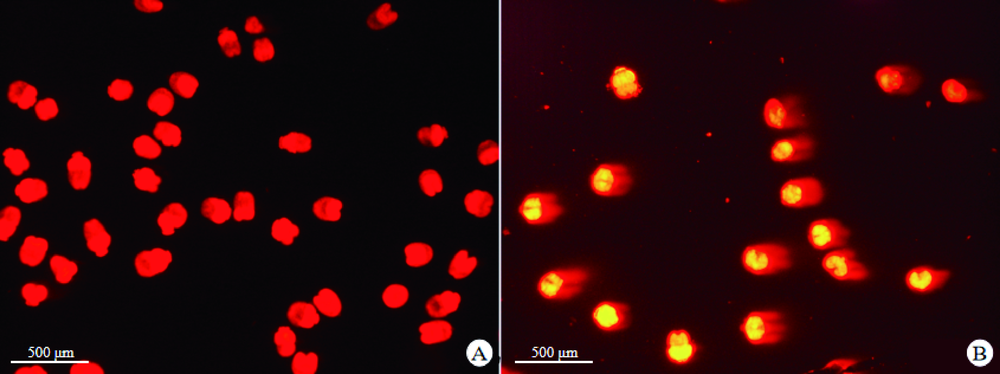
| [1] | Tanki H, Singh H, Raswan US, et al. Pediatric intracranial hydatid cyst: a series with literature review[J]. Pediatr Neurosurg, 2018, 53(5): 299-304. | | [2] | 庞楠楠, 安梦婷, 张峰波, 等. 细粒棘球蚴感染中TGF-β/Smad信号通路对IL-9调控作用的研究[J]. 中国寄生虫学与寄生虫病杂志, 2018, 36(5): 460-463, 468. | | [3] | 刘平, 李金花, 李印, 等. 包虫病病原在我国的流行现状及成因分析[J]. 中国动物检疫, 2016, 33(1): 48-51. | | [4] | Fadel SA, Asmar K, Faraj W, et al. Clinical review of liver hydatid disease and its unusual presentations in developing countries[J]. Abdom Radiol (NY), 2019, 44(4): 1331-1339. | | [5] | 张梦媛, 伍卫平, 官亚宜, 等. 我国棘球蚴病疾病负担分析[J]. 中国寄生虫学与寄生虫病杂志, 2018, 36(1): 15-19, 25. | | [6] | 伍卫平, 王虎, 王谦, 等. 2012-2016年中国棘球蚴病抽样调查分析[J]. 中国寄生虫学与寄生虫病杂志, 2018, 36(1): 1-14. | | [7] | 王永珍, 韩秀敏, 郭亚民. 肝包虫病的诊断与治疗研究进展[J]. 寄生虫病与感染性疾病, 2018, 16(1): 47-51. | | [8] | Goja S, Saha SK, Yadav SK, et al. Surgical approaches to hepatic hydatidosis ranging from partial cystectomy to liver transplantation[J]. Ann Hepatobiliary Pancreat Surg, 2018, 22(3): 208-215. | | [9] | Das AK.Anticancer effect of antimalarial artemisinin compounds[J]. Ann Med Health Sci Res, 2015, 5(2): 93-102. | | [10] | Ashley EA, Dhorda M, Fairhurst RM, et al. Spread of artemisinin resistance in Plasmodium falciparum malaria[J]. N Engl J Med, 2014, 371(5): 411-423. | | [11] | 魏晗. 新剂型阿苯达唑乳剂对肝囊型包虫病患者的疗效观察[J]. 临床医药文献电子杂志, 2016, 3(10): 1977-1977, 1980. | | [12] | 高惠静, 陈蓓, 张海波, 等. 阿苯达唑纳米脂质体冻干粉在大鼠体内药动学及肝靶向研究[J]. 中国医院药学杂志, 2017, 37(8): 702-706. | | [13] | 阿卜来海提·麦提色依提. 阿苯达唑壳聚糖微球抗小鼠泡球蚴药效实验研究[D]. 石河子: 石河子大学, 2015. | | [14] | 李亚芬, 赵军, 吕国栋, 等. Veliparib联合青蒿琥酯体外抗细粒棘球蚴作用机制的研究[J]. 中国病原生物学杂志, 2018, 13(7): 733-739. | | [15] | 史红娟, 吕海龙, 雷颖, 等. 布洛芬抑制体外细粒棘球蚴原头节生长的实验研究[J]. 中国病原生物学杂志, 2016, 11(3): 220-224. | | [16] | 郑璇, 卢帅, 赵军, 等. siRNA特异性干扰EgRad9基因表达对细粒棘球蚴原头节DNA氧化损伤机制的影响[J]. 中国寄生虫学与寄生虫病杂志, 2018, 36(4): 343-349. | | [17] | 郑海亚, 文丽梅, 吕国栋, 等. 青蒿琥酯体外抗细粒棘球蚴活性氧对DNA作用机制影响的研究[J]. 中国病原生物学杂志, 2017, 12(8): 751-761. | | [18] | 刘晓, 邵方元, 陈宏远. 阿霉素抗肿瘤分子机制的研究进展[J]. 中国医药生物技术, 2012, 7(5): 373-375. | | [19] | Deterding A, Dungey FA, Thompson KA, et al. Anti-trypanosomal activities of DNA topoisomerase inhibitors[J]. Acta Tropica, 2005, 93(3): 311-316. | | [20] | 相琳琳. EZH2在电离辐射诱导Hep3B细胞DNA损伤修复中对ATM/p53通路的调控作用研究[D]. 长春: 吉林大学, 2018. | | [21] | Harper JW, Elledge SJ.The DNA damage response: ten years after[J]. Mol Cell, 2007, 28(5): 739-745. | | [22] | Yeo CQX, Alexander I, Lin ZR, et al. P53 maintains genomic stability by preventing interference between transcription and replication[J]. Cell Rep, 2016, 15(1): 132-146. | | [23] | 赵芳, 徐斌, 蒋敬庭, 等. P53在肿瘤微环境、细胞代谢中的调控机制及检测技术的研究进展[J]. 临床检验杂志, 2017, 35(8): 621-623. | | [24] | 江阔. 辐射诱导DNA损伤修复中EZH2与ATM调控作用的研究[D]. 长春: 吉林大学, 2018. | | [25] | 李晶, 汪竹, 袁昕, 等. Ki67和TOPOⅡα在胃癌中的表达及临床意义[J]. 现代肿瘤医学, 2015, 23(5): 666-668. |
|

 ), Bei CHEN2,3, Shuai LU2,3, Li-mei WEN2,3, Jun ZHAO2,3, Xuan ZHENG1, Kuerbannisha·AMAHONG1, Yi GAO1, Jian-hua WANG2,3,*(