中国寄生虫学与寄生虫病杂志 ›› 2024, Vol. 42 ›› Issue (2): 160-168.doi: 10.12140/j.issn.1000-7423.2024.02.005

• 论著 • 上一篇    下一篇

脑型疟炎症微环境诱导星形胶质细胞活化及对神经元损伤的研究

仝国栋1,2(), 朱卿昊2, 王军2, 刘晓冉3, 沈燕2, 梁姣2, 李英辉2, 黄豫晓2, 王一2, 赵亚2,*()   

  1. 1 西北大学生命科学学院,陕西西安 710127
    2 空军军医大学基础医学院微生物与病原生物学教研室,陕西西安 710032
    3 空军军医大学基础医学院2021级临床医学(五年制),陕西西安 710032
  • 收稿日期:2023-08-02 修回日期:2023-09-27 出版日期:2024-04-30 发布日期:2023-12-22
  • 通讯作者: * 赵亚(1972—),男,博士,教授,从事疟原虫感染与免疫相关研究。E-mail:zhaoya@fmmu.edu.cn
  • 作者简介:仝国栋(1999—),男,硕士研究生,从事脑型疟发病机制及免疫辅助治疗研究。E-mail:870460728@qq.com
  • 基金资助:
    国家自然科学基金(82002158);国家自然科学基金(82072298);陕西省自然科学基础研究计划-重点项目(2022JZ-14)

Activation of astrocytes and neuronal damage induced by the inflammatory microenvironment of cerebral malaria

TONG Guodong1,2(), ZHU Qinghao2, WANG Jun2, LIU Xiaoran3, SHEN Yan2, LIANG Jiao2, LI Yinghui2, HUANG Yuxiao2, WANG Yi2, ZHAO Ya2,*()   

  1. 1 The College of Life Sciences, Northwest University, Xi’an 710127, Shaanxi, China
    2 Department of Medical Microbiology and Parasitology, Basic Medical College, Air Force Medical University, Xi’an 710032, Shaanxi, China
    3 Grade 2019 Clinical Medicine (Five-Year Program), Basic Medical College, Air Force Medical University, Xi’an 710032, Shaanxi, China
  • Received:2023-08-02 Revised:2023-09-27 Online:2024-04-30 Published:2023-12-22
  • Contact: * E-mail: zhaoya@fmmu.edu.cn
  • Supported by:
    National Natural Science Foundation of China(82002158);National Natural Science Foundation of China(82072298);Natural Science Basic Research Program of Shaanxi(2022JZ-14)

摘要:

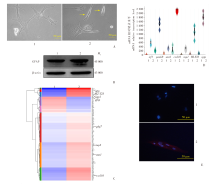
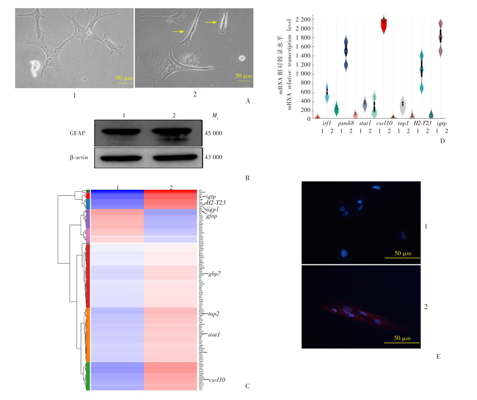
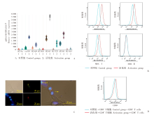
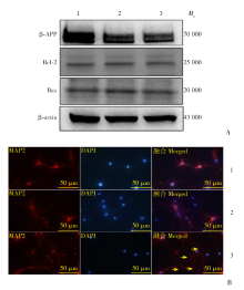
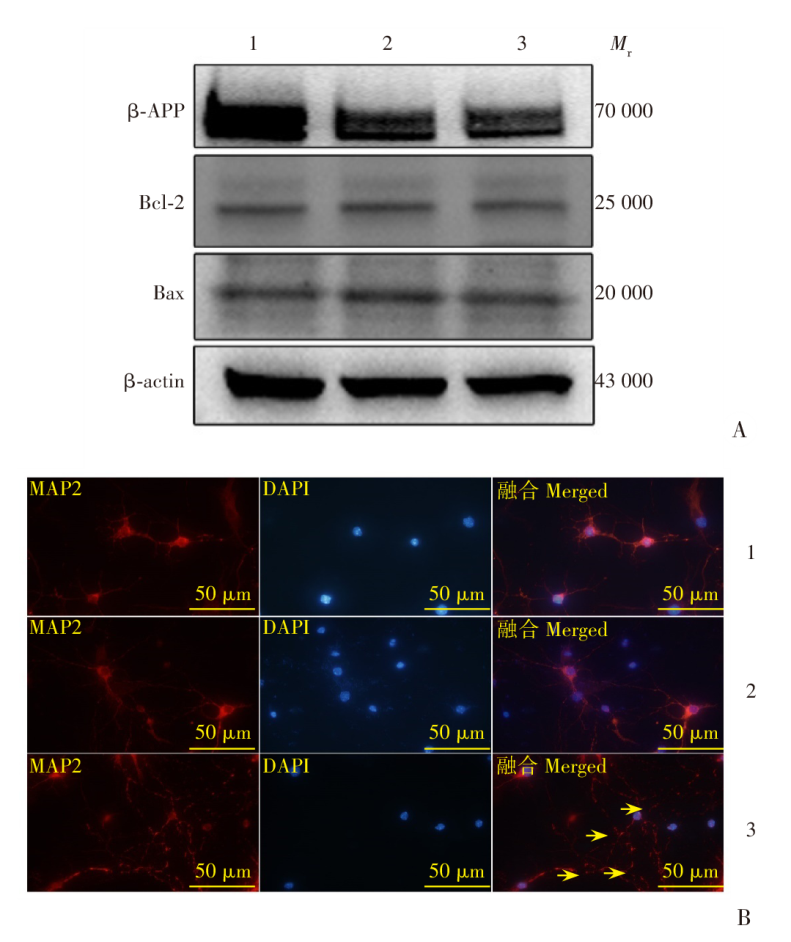
目的 探索脑型疟发病过程中,星形胶质细胞被脑血管周炎性微环境诱导活化及其机制,及其对神经元损伤的影响。方法 4~5周C57BL/6雄性小鼠经腹腔接种5 × 106个感染伯氏疟原虫ANKA株的红细胞,7~10 d后死亡,作为实验型脑型疟模型。新生3~5 d的乳鼠,安乐死后分离脑皮层原代星形胶质细胞;24 h内的乳鼠,安乐死后分离脑皮层原代神经元。以20 ng/ml γ干扰素(IFN-γ)、1 ng/ml肿瘤坏死因子α(TNF-α)和感染疟原虫的小鼠红细胞(pRBC)诱导星形胶质细胞活化,作为活化组,同时设置未处理静息态细胞作为对照组。24 h后用细胞裂解提取液提取总RNA测序,聚类分析生成热图,选取代表性的数个基因绘制小提琴图。ELISA测定细胞培养上清中CXCL10表达水平。分离脑型疟小鼠脾脏CD8+ T细胞,与活化的星形胶质细胞共孵育,荧光显微镜下观察免疫突触及与CXCL10抗体共孵育后的CXCL10水平,流式细胞仪检测CD80等分子表达水平。原代神经元加入两组星形胶质细胞培养上清,在MAP2抗体中孵育,设置空白培养基作为空白组,乳酸脱氢酶(LDH)检测试剂盒检测神经元损伤程度,CCK-8试剂盒检测神经元活力。Western blotting检测STAT1、STAT3及其磷酸化分子水平,活化组加入STAT1通路抑制剂氟达拉滨,免疫荧光染色观察神经元形态。采用PRISM Graph Pad 8.0软件进行统计学分析,两两比较采用t检验。结果 活化组星形胶质细胞形态由扁平星状变为长梭形,胶质纤维酸性蛋白(GFAP)相对含量为(1.36 ± 0.03),高于对照组的(1.00 ± 0.00)(t = 13.33,P < 0.01);活化组细胞培养上清中的CXCL10含量为(7.07 ± 0.81)ng/ml,高于对照组的(2.57 ± 0.28)ng/ml(t = 9.05,P < 0.01)。转录组分析显示,活化组抗原加工、提呈与T细胞趋化因子转录水平上调(均P < 0.01),CD80、CD86、MHC Ⅰ表达水平上调。活化组和CD8+ T细胞共孵育形成“免疫突触”,CD8+ T细胞CD69表达量提高。LDH结果显示,活化组上清刺激后神经元相对死亡率为(50.2 ± 2.4)%,高于空白组(0%)和对照组(0%)(t = 20.62、20.62,均P < 0.01);CCK-8检测结果显示,活化组上清刺激后神经元相对活力为0.52 ± 0.03,低于空白组(1.00 ± 0.00)和对照组(1.42 ± 0.06)(t = 18.92、16.65,均P < 0.01);Western blotting结果显示,神经元经活化星形胶质细胞上清刺激后β-淀粉样前体蛋白(β-APP)相对表达量为0.44 ± 0.02,低于空白组(1.00 ± 0.00)和对照组(0.55 ± 0.02)(t = 37.28、4.93,均P < 0.01);促凋亡蛋白Bax与抑凋亡蛋白Bcl-2的比例为1.01 ± 0.07,与空白组(1.00 ± 0.00)、对照组(1.00 ± 0.06)比较差异无统计学意义(t = 0.31、0.13,均P > 0.05)。Western blotting结果显示,活化组胞浆中STAT1、p-STAT1蛋白相对表达量分别为3.40 ± 1.08、4.00 ± 0.82,均高于对照组(1)(t = 3.13、5.13,均P < 0.05);活化组胞浆中STAT3、p-STAT3蛋白相对表达量分别为1.00 ± 0.03、1.01 ± 0.05,与对照组(1)差异无统计学意义(t = 0.27、0.52,均P > 0.05)。在活化组胞核中p-STAT1蛋白相对表达量为1.78 ± 0.21,高于对照组(1)(t = 5.081,P < 0.01)、p-STAT3蛋白相对表达量为1.02 ± 0.02,与对照组(1)差异无统计学意义(t = 1.38,均P > 0.05)。对MAP2免疫荧光染色结果显示,活化组上清可造成明显的神经元损伤,经STAT1抑制剂处理后损伤得到缓解。结论 脑型疟发病过程中血管周炎性微环境可通过STAT1分子诱导神经毒性星形胶质细胞产生,可导致神经元死亡。活化星形胶质细胞与活化的CD8+ T细胞相互作用,进一步加重中枢神经系统损伤。

关键词: 脑型疟, 星形胶质细胞, CD8+ T细胞, 神经元, 炎性微环境

Abstract:

Objective To explore the activation of astrocytes induced by the perivascular inflammatory microenvironment during the pathogenesis of the brain during the pathogenesis process of cerebral malaria and its mechanism, as well as its impact on neuronal damage. Methods C57BL/6 male mice aged 4-5 weeks were intraperitoneally inoculated with 5 × 10⁶ red blood cells infected with the ANKA strain of Plasmodium berghei, which would be died 7-10 d later and served as an experimental cerebral malaria model. Newborn suckling mice aged 3-5 d were euthanized to isolate primary astrocytes from the cerebral cortex; while the other suckling mince were euthanized within 24 hours for isolation of primary cortical neurons. The cell culture containing 20 ng/ml γ interferon (IFN-γ), 1 ng/ml tumor necrosis factor α (TNF-α) and mice red blood cells (pRBC) infected with malaria parasites were used to induce activation of astrocytes, and assigned as the activation group, while untreated resting-state cells were set as the control group. After cultivation for 24 h, total RNA was extracted by cell lysis and sequenced. Cluster analysis was performed to generate heatmaps, and representative genes were selected to draw violin diagram. ELISA was used to determine the expression level of CXCL10 in cell culture supernatant. The spleen CD8+ T cells of mice with cerebral malaria were separated and co-cultured with activated astrocytes to observe immune synapses and CXCL10 levels after co-incubation with CXCL10 antibodies by fluorescence microscopy, and CD80 and other molecular expression levels were detected using flow cytometry. Primary neurons were added into two sets of astrocyte culture supernatants and incubated in MAP2 antibody. Blank culture medium was set as the blank group, and lactate dehydrogenase (LDH) and CCK-8 assay kit were used to detect the degree of neuronal damage. Western blotting was used to detect the levels of STAT1, STAT3, and their phosphorylated molecules. The activation group was treated with STAT1 pathway inhibitor fludarabine, and the morphology of neurons was observed by immunofluorescence staining. Statistical analysis was conducted using PRISM Graph Pad 8.0 software, and pairwise comparisons were conducted using t-tests. Results The morphology of activated astrocytes changed from flat star-shaped to long spindle-shaped, and the relative content of glial fibrillary acidic protein (GFAP) was (1.36 ± 0.03), higher than that of the control group (1.00 ± 0.00) (t = 13.33, P < 0.01). The CXCL10 content in the supernatant of the activation group cell culture was (7.07 ± 0.81) ng/ml, which was higher than that of the control group (2.57 ± 0.28) ng/ml (t = 9.05, P < 0.01). Transcriptome analysis showed that antigen processing, presentation, and transcription levels of T cell chemokines were upregulated in the activation group (all P < 0.01), and the expression levels of CD80, CD86, and MHC Ⅰ were upregulated. The activation group co-incubated with CD8+ T cells to form "immune synapses", and the CD69 expression level of CD8+ T cells increased. The LDH results showed that the relative mortality rate of neurons in the activation group after supernatant stimulation was (50.2 ± 2.4)%, which was higher than that in the blank group (0% ) and the control group (0%) (t = 20.62, 20.62, both P < 0.01). The CCK-8 detection results showed that the relative activity of neurons in the activation group after supernatant stimulation was 0.52 ± 0.03, lower than that in the blank group (1.00 ± 0.00) and the control group (1.42 ± 0.06) (t = 18.92, 16.65, both P < 0.01). Western blotting results showed that relative expression level of neurons stimulated with activated astrocytes supernatant β-amyloid precursor protein (β-APP) was 0.44 ± 0.02, which was lower than that of the blank group (1.00 ± 0.00) and the control group (0.55 ± 0.02) (t = 37.28, 4.93, both P < 0.01). The ratio of pro apoptotic protein Bax to anti apoptotic protein Bcl-2 was 1.01 ± 0.07, and there was no statistically significant difference compared with the blank group (1.00 ± 0.00) and the control group (1.00 ± 0.06) (t = 0.31, 0.13, both P > 0.05). Western blotting results showed that the relative expression level of STAT1 and p-STAT1 protein in the cytoplasm of activation group were 3.40 ± 1.08 and 4.00 ± 0.82, both of which were higher than the control group (1) (t = 3.13, 5.13, both P < 0.05). The relative expression level of STAT3 and p-STAT3 protein in the cytoplasm of activation group were 1.00 ± 0.03 and 1.01 ± 0.05. There was no statistically significant difference compared to the control group (1) (t = 0.27, 0.52, both P > 0.05). The relative expression level of p-STAT1 protein in the nucleus of the activation group was 1.78 ± 0.21, higher than that of the control group (1) (t = 5.081, P < 0.01), and the relative expression level of p-STAT3 protein was 1.02 ± 0.02, with no statistically significant difference compared to the control group (1) (t = 1.38, all P > 0.05). The results of MAP2 immunofluorescence staining showed that the activation group supernatant could cause significant neuronal damage, which was alleviated after treatment with STAT1 inhibitors. Conclusion During the pathogenic process of cerebral malaria, the brain perivascular inflammatory microenvironment may induce production of neurotoxic astrocytes via the STAT1, and may cause death of neurons. This activated astrocytes could interact with activated CD8+ T cells, and further aggravate the damage of central nervous system.

Key words: Cerebral malaria, Astrocytes, CD8+ T cells, Neuron, Inflammatory microenvironment

中图分类号: