中国寄生虫学与寄生虫病杂志 ›› 2018, Vol. 36 ›› Issue (4): 352-356.

• 论著 • 上一篇    下一篇

氯舒隆和奥硝唑体外抗细粒棘球蚴原头节和多房棘球蚴的效果评价

辛奇, 高海军, 宋晓霞, 孙旭东, 吕薇, NabilPERVAIZ, 鲁俊, 景涛*()   

  1. 兰州大学基础医学院病原生物学研究所,兰州 730000
  • 收稿日期:2018-01-15 出版日期:2018-08-30 发布日期:2018-09-06
  • 通讯作者: 景涛
  • 基金资助:
    中央高校基本科研业务费专项资金(No. lzujbky-2018-90);国家自然科学基金(No. 81171632)

In vitro effects of clorsulon and ornidazole against Echinococcus granulosus protoscoleces and E. multilocularis metacestodes

Qi XIN, Hai-jun GAO, Xiao-xia SONG, Xu-dong SUN, Wei LV, PERVAIZ Nabil, Jun LU, Tao JING*()   

  1. Institute of Pathogen Biology, School of Basic Medical Sciences, Lanzhou University, Lanzhou 730000, China
  • Received:2018-01-15 Online:2018-08-30 Published:2018-09-06
  • Contact: Tao JING
  • Supported by:
    Supported by the Fundamental Research Funds for the Central Universities (No. lzujbky-2018-90) and the National Natural Science Foundation of China (No. 81171632)

摘要:

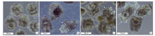
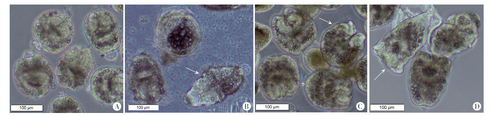
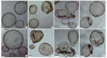
目的 研究氯舒隆和奥硝唑体外抗细粒棘球蚴原头节和多房棘球蚴的效果。方法 从感染细粒棘球蚴绵羊肝脏中收集原头节;从感染长爪沙鼠腹腔中分离多房棘球蚴,加入预先接种人肝癌细胞的DMEM培养基中培养2个月后,收集直径为1~5 mm囊泡。实验分氯舒隆组(实验组)、奥硝唑组(实验组)、阿苯达唑组(阳性对照)和0.2%二甲基亚砜(DMSO)组(溶剂对照组)。每种药物设2个平行孔,终浓度均为40 μmol/L,重复2次。每孔加细粒棘球蚴原头节约100个或多房棘球蚴囊泡25~35个。药物处理细粒棘球蚴原头节24、48、72、96、120、144和168 h后,显微镜下观察原头节形态,用台盼蓝染色,计算原头节存活率,组间存活率的比较采用方差分析。药物处理多房棘球蚴囊泡36 h和120 h后,显微镜下观察囊泡的形态学改变,测定培养上清液中碱性磷酸酶的活性,组间酶活性的比较采用卡方分析。结果 氯舒隆、奥硝唑和阿苯达唑作用于原头节后,原头节颜色变深、钙颗粒减少、头钩脱落、头节外翻并伸长,0.2% DMSO对原头节形态无影响。氯舒隆、臭硝唑和阿苯达唑作用24、48、72、96、120、144 和168 h后,原头节存活率分别为79%、70%、56%、42%、33%、16%、15%,86%、67%、63%、48%、32%、28%、21%和85%、71%、45%、36%、21%、15%、8%;0.2% DMSO组原头节存活率为100%。氯舒隆、奥硝唑和阿苯达唑组原头节存活率与0.2% DMSO组相比差异均有统计学意义(χ2 = 147.83、130.58、170.37,P < 0.05)。氯舒隆、奥硝唑和阿苯达唑作用后,多房棘球蚴囊泡均塌陷、皱缩,0.2% DMSO对多房棘球蚴囊泡形态无影响。作用36 h后氯舒隆、奥硝唑、阿苯达唑和0.2% DMSO组培养上清液碱性磷酸酶的吸光度(A405)值分别为0.196 ± 0.030、0.186 ± 0.004、0.244 ± 0.049和0.131 ± 0.020,作用120 h后分别为0.431 ± 0.006、0.271 ± 0.004、0.423 ± 0.007和0.116 ± 0.004。氯舒隆、奥硝唑和阿苯达唑组与0.2% DMSO组相比差异均有统计学意义(t = 0.006、0.004、0.007,P < 0.05)。结论 氯舒隆和奥硝唑对体外培养的细粒棘球蚴原头节和多房棘球蚴均具有较强的作用,是潜在的抗棘球蚴药物。

关键词: 细粒棘球蚴原头节, 多房棘球蚴, 氯舒隆, 奥硝唑

Abstract:

Objective To evaluate the effects of clorsulon and ornidazole against Echinococcus granulosus protoscoleces and E. multilocularis metacestodes in vitro. Methods E. granulosus protoscoleces were isolated from livers of sheep infected with E. granulosus, and E. multilocularis metacestodes were isolated from the peritoneal cavity of infected Meriones unguiculatus. They were cultured in Dulbecco’s modified eagle medium (DMEM) seeded with human hepatoma cells for 2 months, then vesicles of 1-5 mm in diameter were collected. Four groups were set in the experiment: the clorsulon treatment group (experimental group), ornidazole group (experimental group), albendazole group (positive control), and 0.2% DMSO group (vehicle control group). The experiment was performed in duplicate, and repeated twice. All drugs were used at a final concentration of 40 μmol/L. Each well contained about 100 E. granulosus protoscoleces, or 25-35 E. multilocularis vesicles. After being treated for 24, 48, 72, 96, 120, 144 and 168 h, the protoscoleces were stained by trypan blue and observed by microscopy. The viability of protoscoleces was calculated and analyzed with analysis of chi-square test. The E. multilocularis vesicles were treated for 36 and 120 h, followed by morphological observation by microscopy and measurement of alkaline phosphatase activity in the culture supernatant. The results were analyzed with ANOVA. Result After treatment with clorsulon, ornidazole and albendazole, the protoscoleces showed a darkened color, decreased calcium precipitates, hook shedding, and scolex evagination and elongation, while 0.2% DMSO did not show such effects. After treatment with clorsulon, ornidazole or albendazole for 24, 48, 72, 96, 120, 144 and 168 h, the viability of protoscoleces was 79%, 70%, 56%, 42%, 33%, 16%, 15%; 86%, 67%, 63%, 48%, 32%, 28%, 21%; and 85%, 71%, 45%, 36%, 21%, 15%, 8%, respectively. In the 0.2% DMSO group, 100% of protoscoleces survived. The viability after clorsulon, ornidazole and albendazole treatment was significantly lower than that in the 0.2% DMSO group (χ2 = 147.83, 130.58, 170.37; P < 0.05). Compared to the intact vesicles after 0.2% DMSO treatment, clorsulon, ornidazole and albendazole all caused collapse and shrinkage of E. multilocularis vesicles. At 36 h of treatment with clorsulon, ornidazole, albendazole and 0.2% DMSO treatment, the A405 values of alkaline phosphatase were 0.196 ± 0.030, 0.186 ± 0.004, 0.244 ± 0.049 and 0.131 ± 0.020, respectively, and at 120 h, they were 0.431 ± 0.006, 0.271 ± 0.004, 0.423 ± 0.007 and 0.116 ± 0.004, respectively. There were significant differences in the alkaline phosphatase activity in the three drug treatment groups versus the 0.2% DMSO group (t = 0.006, 0.004, 0.007; P < 0.05). Conclusion Both clorsulon and ornidazole have strong effects against E. granulosus protoscoleces and E. multilocularis metacestodes in vitro, and are potential drugs for echinococcosis.

Key words: Echinnococcus granulosus, E. multilocularis, Clorsulon, Ornidazole

中图分类号: