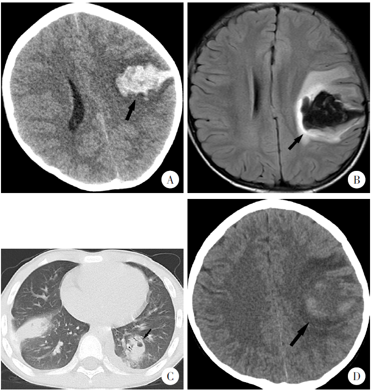
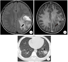
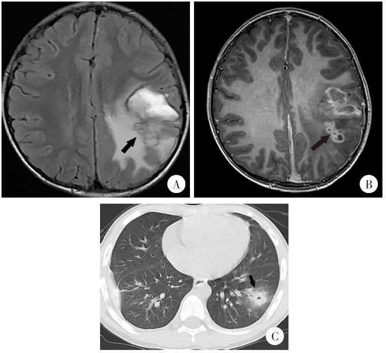
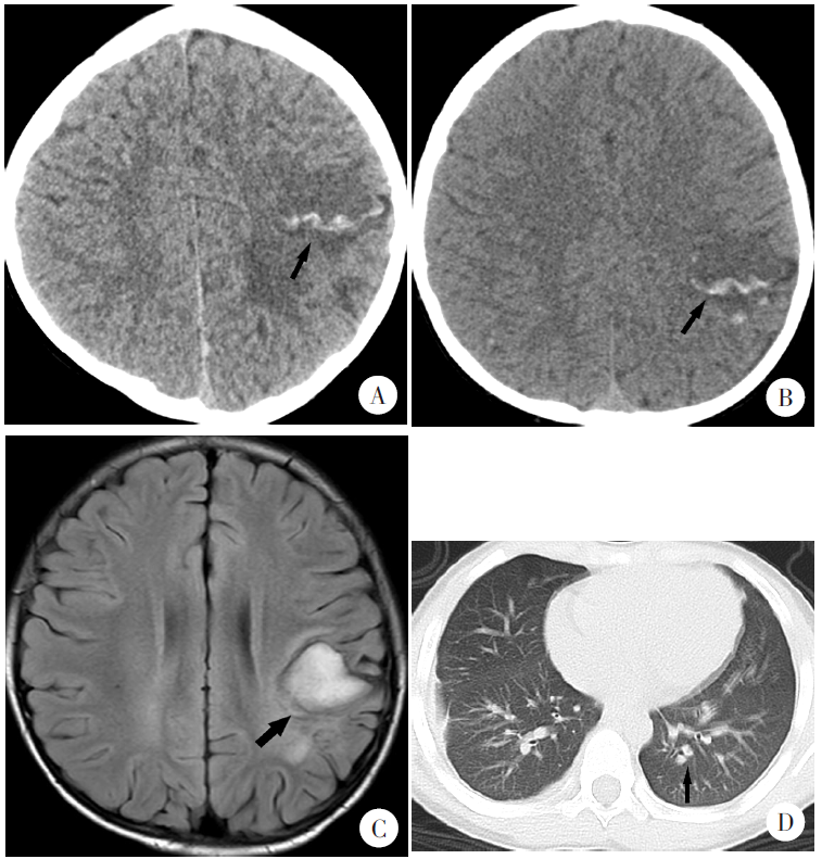
| [1] | 李呜皋, 陈兆义, 胡清锡, 等. 贵州省斯氏狸殖吸虫病的分布[J]. 实用寄生虫病杂志, 1993(1): 36-38. | | | Li WG, Chen ZY, Hu QX, et al. Distribution of pagumogonimiasis skrjabini in Guizhou Province[J]. Parasitoses Infect Dis, 1993(1): 36-38. (in Chinese) | | [2] | 龚志红, 龚红卡, 徐芸, 等. 江西省2011—2020年并殖吸虫病病例回顾性分析[J]. 中国寄生虫学与寄生虫病杂志, 2022, 40(2): 247-251. | | | Gong ZH, Gong HK, Xu Y, et al. Retrospective analysis of paragonimiasis cases in Jiangxi Province from 2011 to 2020[J]. Chin J Parasitol Parasit Dis, 2022, 40(2): 247-251. (in Chinese) | | [3] | 韦捷. 儿童中枢神经系统肺吸虫病的影像特点[D]. 重庆: 重庆医科大学, 2022: 12-19. | | | Wei J. Imaging features of central nervous system paragonimiasis in children[D]. Chongqing: Chongqing Medical University, 2022: 12-19. (in Chinese) | | [4] | Coogle B, Sosland S, Bahr NC. A clinical review of human disease due to Paragonimus kellicotti in North America[J]. Parasitology, 2022, 149(10): 1327-1333. | | [5] | Kim JG, Ahn CS, Kang I, et al. Cerebral paragonimiasis: Clinicoradiological features and serodiagnosis using recombinant yolk ferritin[J]. PLoS Negl Trop Dis, 2022, 16(3): e0010240. | | [6] | Li LS, Zhang YT, Zhu J, et al. Intracranial pseudoaneurysm caused by cerebral paragonimiasis in pediatric patients[J]. Pediatr Neurol, 2020, 109: 47-51. | | [7] | Qian MN, Li F, Zhang YH, et al. A retrospective clinical analysis of pediatric paragonimiasis in a Chinese children’s hospital from 2011 to 2019[J]. Sci Rep, 2021, 11(1): 2005. | | [8] | Blair D. Lung flukes of the genus Paragonimus: Ancient and re-emerging pathogens[J]. Parasitology, 2022, 149(10): 1286-1295. | | [9] | 王铂. 肺吸虫脑病1例[C]. 杭州: 浙江省神经外科学学术大会, 2020: 123. | | | Wang B. One case of Paragonimus encephalopathy[C]. Hangzhou: Zhejiang Medical Association Society of Neurosurgery, 2020: 123. (in Chinese) | | [10] | Villanueva-Villegas R, Diaz-Mendoza J, Salas-Lopez J, et al. Paragonimiasis misdiagnosed as pulmonary tuberculosis: A case report[J]. Cureus, 2023, 15(3): e36169. | | [11] | Blair D. Paragonimiasis[J]. Adv Exp Med Biol, 2024, 1454:203-238. | | [12] | 王忠诚. 神经外科手术学[M]. 北京: 科学出版社, 2000: 454-455. | | | Wang ZC. Operative neurosurgery[M]. Beijing: Science Press, 2000: 454-455. (in Chinese) | | [13] | 蔡泽政, 杨萍, 廖倩倩. 儿童并殖吸虫复杂性感染1例[J]. 中国寄生虫学与寄生虫病杂志, 2024, 42(6): 813-816. | | | Cai ZZ, Yang P, Liao QQ. A child case of complicated Paragonimus infection[J]. Chin J Parasitol Parasit Dis, 2024, 42(6): 813-816. (in Chinese) | | [14] | Wang QS, Hou LM, Liu L. Diagnosis and treatment of hemorrhagic cerebral paragonimiasis: Three case reports and literature review[J]. Turk Neurosurg, 2020, 30(4): 624-628. |
|

 ), 王军浩1, 曾茜2, 徐卡娅2,*(