中国寄生虫学与寄生虫病杂志 ›› 2021, Vol. 39 ›› Issue (1): 35-42.doi: 10.12140/j.issn.1000-7423.2021.01.005

• 论著 • 上一篇    下一篇

丰宫并殖吸虫感染大鼠后诱导肺组织细胞自噬的初探

马志强1,4(), 王霖1, 李生浩1, 徐晶晶1, 李才信1, 刘媛1, 张燕玲1, 束秋红2, 庄杉杉3, 何姝美琪4, 王文林4, 王卫群4,*()   

  1. 1 昆明市第三人民医院,昆明 650000
    2 昆明医科大学第二附属医院,昆明 650000
    3 昆明市延安医院,昆明 650000
    4 昆明医科大学基础医学院,昆明 650000
  • 收稿日期:2020-06-16 修回日期:2020-08-20 出版日期:2021-02-28 发布日期:2021-03-10
  • 通讯作者: 王卫群
  • 作者简介:马志强(1990-),男,硕士,检验医师,主要从事病原微生物研究。E-mail: 18468279606@163.com
  • 基金资助:
    云南省科技厅-昆明医科大学应用基础研究联合专项(2019FE001-161);云南省地方本科高校基础研究联合专项基金(2018FH001-087);云南省教育厅科学研究基金(2018JS157);昆明市卫生科技人才培养项目“千”工程后备人选[2020-SW(后备)-72,2020-SW(后备)-60]([2020?SW(reserve)?72, (2020?SW(reserve)?60])

Preliminary study on autophagy of lung tissue cells in rats infected with Paragonimus proliferus

MA Zhi-qiang1,4(), WANG Lin1, LI Sheng-hao1, XU Jing-jing1, LI Cai-xin1, LIU Yuan1, ZHANG Yan-ling1, SHU Qiu-hong2, ZHUANG Shan-shan3, HE Shu Mei-qi4, WANG Wen-lin4, WANG Wei-qun4,*()   

  1. 1 The Third People’s Hospital of Kunming, Kunming 650500, China
    2 The Second Affiliated Hospital of Kunming Medical University, Kunming 650500, China
    3 Yan’an Hospital of Kunming City, Kunming 650500, China
    4 College of Basic Medicine, Kunming Medical University, Kunming 650500, China
  • Received:2020-06-16 Revised:2020-08-20 Online:2021-02-28 Published:2021-03-10
  • Contact: WANG Wei-qun
  • Supported by:
    Yunnan Provincial Department of Science and Technology?Kunming Medical University Applied Basic Research Joint Special Project(2019FE001-161);Joint Special Fund for Basic Research of Local Undergraduate Universities in Yunnan Province(2018FH001-087);Scientific Research Fund Project of Yunnan Provincial Department of Education(2018JS157);Scientific Research Fund Project of Yunnan Provincial Department of Education([2020?SW(reserve)?72, (2020?SW(reserve)?60])

摘要:

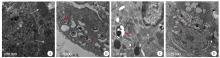
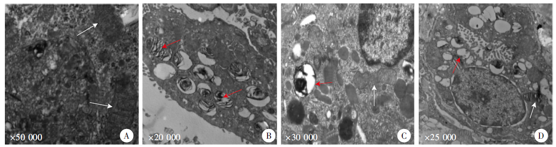
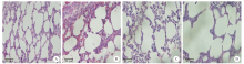
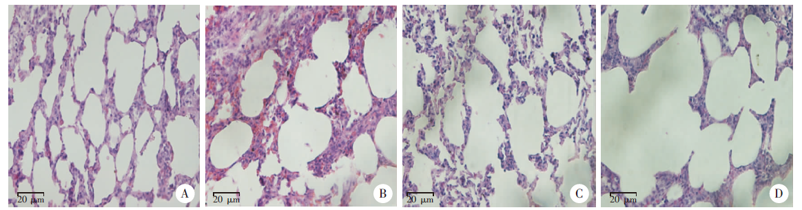
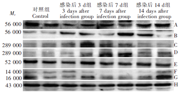
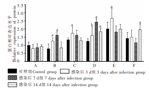
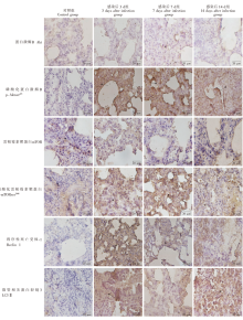
目的 通过检测蛋白激酶B(Akt)/哺乳动物雷帕霉素靶蛋白(mTOR)信号通路相关因子的表达,探讨丰宫并殖吸虫感染大鼠是否可导致肺组织细胞发生自噬。 方法 40只SD大鼠随机分为健康对照组,感染后3、7、14 d组,每组10只,感染组每鼠腹壁皮下注射6条丰宫并殖吸虫后尾蚴,分别在感染后3、7、14 d取各组大鼠血清和肺组织,ELISA检测血清中IL-1、IL-6水平。取肺组织用于透射电镜观察自噬体,HE染色观察肺组织病理学改变,蛋白质免疫印迹(Western blotting)和免疫组化检测Akt、雷帕霉素靶蛋白(mTOR)、程序性死亡受体-1(Beclin 1)及微管相关蛋白轻链3(LC3Ⅱ)相关因子的蛋白表达。采用SPSS 19.0软件对数据进行统计学分析。 结果 ELISA检测结果显示,感染后3、7、14 d组血清中IL-1水平分别为(1 558.0 ± 123.6)、(1 511.0 ± 213.1)和(1 448.0 ± 176.8)pg/ml,均高于对照组的(1 222.0 ± 112.8)pg/ml(P < 0.05);感染后3、7 d组IL-6水平分别为(1 481.0 ± 197.9)、(1 423.0 ± 210.0)pg/ml,均高于对照组的(1 221.0 ± 138.9)pg/ml(P < 0.05)。透射电镜观察在不同的感染阶段,肺组织中线粒体均出现自噬现象。大鼠组织肺病理学检测结果显示,各感染组细胞排列紊乱,肺泡结构遭到不同程度的破坏。各感染组Akt蛋白表达水平与对照组比较,差异无统计学意义(P > 0.05);感染后3、7 d组磷酸化蛋白激酶B(p-Aktser 473)蛋白表达水平为(1.288 ± 0.109)、(1.619 ± 0.132),均高于对照组(0.733 ± 0.135)(P < 0.01);感染后3、7、14 d组磷酸化雷帕霉素靶蛋白(p-mTORser 2448)蛋白表达水平分别为(1.574 ± 0.278)、(2.384 ± 0.125)和(1.808 ± 0.121),均高于对照组(1.260 ± 0.087)(P < 0.05);感染后3 d组mTOR、Beclin 1蛋白表达水平分别为(1.714 ± 0.217)和(2.736 ± 0.333),均高于对照组(1.345 ± 0.067)和(1.974 ± 0.225)(P < 0.01);感染后14 d组LC3Ⅱ蛋白表达(1.938 ± 0.191)高于对照组(1.401 ± 0.200)(P < 0.01)。免疫组化分析结果显示,对照组肺组织细胞呈蓝色,阳性呈棕黄色,各因子阳性定位于细胞膜和细胞浆。各感染组Akt、mTOR与对照组相比,肺组织细胞中棕黄色显色不明显,A450值与对照组比较差异无统计学意义(P > 0.05);感染后3、7、14 d组p-Aktser 473、p-mTORser 2448及Beclin 1与对照组相比,棕黄色显色加深明显,A450值分别为(0.104 ± 0.010)、(0.143 ± 0.022)、(0.088 ± 0.013),(0.100 ± 0.007)、(0.151 ± 0.006)、(0.120 ± 0.012)和(0.129 ± 0.005)、(0.047 ± 0.004)、(0.050 ± 0.005),均高于相应对照组(0.032 ± 0.001)、(0.065 ± 0.002)和(0.031 ± 0.001)(P < 0.05);感染后3、14 d组LC3Ⅱ与对照组比较棕黄色明显加深,A450值分别为(0.056 ± 0.006)、(0.120 ± 0.007),均高于对照组(0.042 ± 0.004)(P < 0.05)。 结论 丰宫并殖吸虫感染大鼠所致的肺损伤,炎症反应可诱导肺组织细胞发生自噬,该自噬作用可通过检测Akt/mTOR信号通路相关因子的表达来初步实现。

关键词: 丰宫并殖吸虫, 感染, 大鼠, Akt/mTOR信号通路, 肺组织细胞, 自噬

Abstract:

Objective To explore whether Paragonimus proliferus infection in rats can cause autophagy in lung tissue cells, by detecting the expression of factors related to the protein kinase B (Akt)/mammalian target of rapamycin (mTOR) signaling pathway. Methods Forty SD rats were randomly divided into 4 groups (n = 10 in each group). The rats in all groups (except the control group) were intraperitoneally injected with 6 P. proliferus metacercaria, and were sacrificed on days 3, 7 and 14 after injection, respectively. Serum samples collected from the rats were used to detect the levels of IL-1 and IL-6 by ELISA. The lung tissues were examined for autophagosome formation by transmission electron microscopy (TEM), pathological changes by HE staining, and protein expression of Akt, mTOR, Beclin 1 and LC3Ⅱ by Western blotting and immunohistochemistry. Statistical analysis was performed with the SPSS 19.0 software. Results The ELISA results showed that the expression levels of IL-1 on days 3, 7 and 14 after infection was (1 558.0 ± 123.6), (1 511.0 ± 213.1) and (1 448.0 ± 176.8) pg/ml, respectively, all significantly higher than the normal group (1 222.0 ± 112.8) pg/ml (P < 0.05); the expression levels of IL-6 on days 3 and 7 after infection was (1 481.0 ± 197.9) and (1 423.0 ± 210.0) pg/ml, respectively, both higher than the control group (1 221.0 ± 138.9) pg/ml (P < 0.05). TEM revealed autophagy in mitochondria at different stages of infection. HE staining results showed that the cells in each infection group were arranged disorderly and the alveolar structure showed varied degrees of impairment. There was no significant difference in the protein level of Akt in the infection groups, compared to the control group (P > 0.05); the protein levels of p-Aktser 473 on days 3 and 7 after infection were (1.288 ± 0.109) and (1.619 ± 0.132), respectively, both higher than that of the normal group (0.733 ± 0.135) (P < 0.01). The protein levels of p-mTORser 2448 in the infection groups were (1.574 ± 0.278), (2.384 ± 0.125) and (1.808 ± 0.121), all higher than the normal group (1.260 ± 0.087) (P < 0.05). The protein levels of mTOR and Beclin 1 on day 3 after infection were (1.714 ± 0.217) and (2.736 ± 0.333), respectively, both higher than (1.345 ± 0.067) and (1.974 ± 0.225) in the normal group (P < 0.01). The LC3Ⅱ protein level on day 14 after infection was (1.938 ± 0.191), higher than that of the normal group (1.401 ± 0.200) (P < 0.01). The results of IHC showed that the lung tissue cells of the negative control group were blue, while the positive staining was in brown yellow; the proteins stained positive were all located in the cell membrane and cytoplasm. Compared with the control group, Akt and mTOR in each infection group showed no obvious brown color in the lung tissue cells, and there was no significant difference in the optical density of Akt and mTOR between the infection groups and the control group (P > 0.05). The staining for p-Aktser 473, p-mTORser 2448 and Beclin 1 in the groups 3 d, 7 d, and 14 d after infection showed denser brown yellow signals compared with the control group, and the optical densities were (0.104 ± 0.010), (0.143 ± 0.022), (0.088 ± 0.013); (0.100 ± 0.007), (0.151 ± 0.006), (0.120 ± 0.012); and (0.129 ± 0.005), (0.047 ± 0.004), (0.050 ± 0.005), which were higher than those in the normal group brown yellow signals[(0.032 ± 0.001), (0.065 ± 0.002) and (0.031 ± 0.001)] (P < 0.05). The LC3Ⅱ showed significantly darker brown yellow signals than the control group on days 3 and 14 after infection, and the optical densities were (0.056 ± 0.006) and (0.120 ± 0.007), which were higher than that of the normal group (0.042 ± 0.004) (P < 0.05). Conclusion P. proliferus infection in rats causes lung injury and inflammatory response, which may induce autophagy of liver cells. The autophagy event could be detected by assessing the expression of factors related to the Akt/mTOR signaling pathway.

Key words: Paragonimus proliferus, Infection, Rat, Akt/mTOR signaling pathway, Lung tissue cells, Autophagy

中图分类号: