中国寄生虫学与寄生虫病杂志 ›› 2026, Vol. 44 ›› Issue (1): 35-41.doi: 10.12140/j.issn.1000-7423.2026.01.006

• 论著 • 上一篇    下一篇

中性粒细胞在肝多房棘球蚴病患者中的临床意义初探

樊羿辰1()(), 张传山1, 侯娇2,3, 唐钊元1, 鲁雪梅1, 何荣东1, 加依达尔·胡玛尔汗1, 宋志浩1, 阚明轩1, 王明娟1, 孙立1, 温浩1,*()()   

  1. 1 省都共建中亚高发病成因与防治国家重点实验室新疆医科大学第一附属医院,肝胆包虫病外科新疆乌鲁木齐 830054
    2 青海大学附属医院肝胆胰外科青海西宁 810001
    3 青海省棘球蚴病研究重点实验室青海西宁 810001
  • 收稿日期:2025-09-16 修回日期:2026-02-18 出版日期:2026-02-28 发布日期:2026-02-24
  • 通讯作者: 温浩(ORCID:0000-0001-7144-220X),男,博士,主任医师,从事棘球蚴病防治与研究。E-mail:dr.wenhao@163.com
  • 作者简介:樊羿辰(ORCID:0009-0006-0143-5219),男,硕士研究生,从事寄生虫感染与免疫研究。E-mail:fanyichen1203@163.com
  • 基金资助:
    新疆维吾尔自治区青年科学基金(2022D01C743);省部共建中亚高发病成因与防治国家重点实验室开放课题青年基金(SKL-HIDCA-2023-28)

Preliminary investigation of the clinical significance of neutrophils in patients with hepatic alveolar echinococcosis

FAN Yichen1()(), ZHANG Chuanshan1, HOU Jiao2,3, TANG Zhaoyuan1, LU Xuemei1, HE Rongdong1, JIAYIDAER Humaerhan1, SONG Zhihao1, KAN Mingxuan1, WANG Mingjuan1, SUN Li1, WEN Hao1,*()()   

  1. 1 State Key Laboratory of Pathogenesis and Prevention of High-Incidence Diseases in Central Asia, Department of Hepatobiliary Echinococcosis Surgery, First Affiliated Hospital of Xinjiang Medical University, Urumqi 830054, Xinjiang, China
    2 Department of Hepatobiliary and Pancreatic Surgery, Affiliated Hospital of Qinghai University, Xining 810001, Qinghai, China
    3 Qinghai Provincial Key Laboratory for Echinococcosis, Xining 810001, Qinghai, China
  • Received:2025-09-16 Revised:2026-02-18 Online:2026-02-28 Published:2026-02-24
  • Contact: E-mail: dr.wenhao@163.com
  • Supported by:
    Xinjiang Uygur Autonomous Region Youth Science Foundation(2022D01C743);Open Project Youth Fund of the Ministry-Coordinated Central Asia High Incidence Disease Causes and Prevention State Key Laboratory(SKL-HIDCA-2023-28)

摘要:

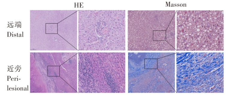
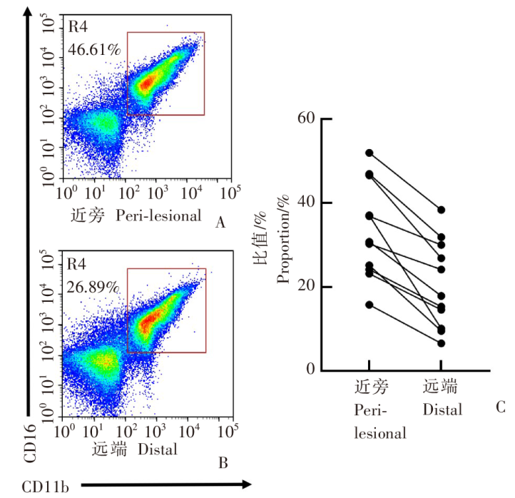
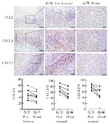
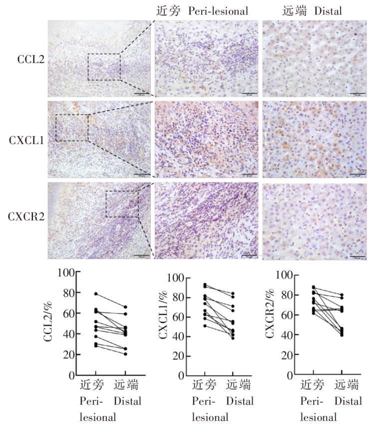
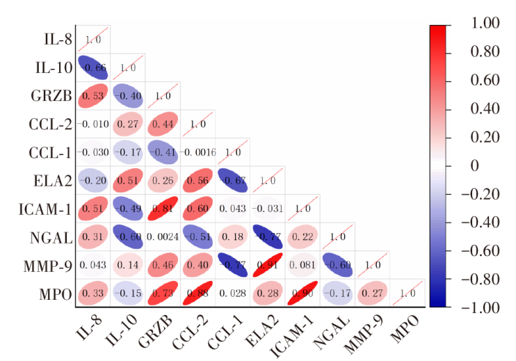
目的 探讨肝多房棘球蚴病(AE)患者外周血及肝脏中中性粒细胞水平、免疫微环境特征及其与疾病严重程度的关系。 方法 纳入2024年7月至2025年9月在青海大学附属医院接受手术治疗的24例AE患者(AE组)和24位健康者(HC组)为研究对象。AE组患者均经影像学与病理学确诊。收集研究对象性别、年龄、中性粒细胞计数、淋巴细胞计数、血小板计数、丙氨酸转氨酶(ALT)、天冬氨酸转氨酶(AST)数值,计算中性粒细胞与淋巴细胞比值(NLR)和系统免疫炎症指数(SII),对AE组进行PMN分型。使用GraphPad Prism 9软件分析AE组外周血临床指标间相关性。采集外周血与手术切除的病灶近旁及远端肝组织,行苏木精-伊红(HE)染色、Masson染色、免疫组织化学染色,使用QuPath-0.5.1软件分析2组趋化因子(C-C基元)配体2(CCL2)、趋化因子(C-X-C基元)配体1(CXCL1)、C-X-C基序趋化因子受体2(CXCR2)阳性区域面积占比。流式细胞术分析肝脏CD16⁺ CD66b⁺ 中性粒细胞水平及其与临床指标相关性。ELISA检测外周血与肝组织中白细胞介素8(IL-8)、IL-10、肿瘤坏死因子-α(TNF-α)、颗粒酶B(GRZB)、CCL2、弹性蛋白酶2(ELA2)、细胞间黏附分子-1(ICAM-1)、明胶酶相关脂质运载蛋白(NGAL)、基质金属蛋白酶(MMP-9)、髓过氧化物酶(MPO)等炎症因子的表达水平,并进行相关性网络分析。 结果 与HC组相比[(1.99 ± 0.62)× 109/L、(260.29 ± 40.36)× 109/L],AE患者外周血淋巴细胞和血小板水平下降[(1.60 ± 0.64)× 109/L、(229.75 ± 62.20)× 109/L](t = 2.036、2.018,均P < 0.05);ALT、AST、NLR、SII升高[AE组分别为(35.54 ± 27.53)U/L、(49.21 ± 47.63)U/L、2.06(区间1.48~3.27)、725.29(区间312.41~810.59),HC组分别为(24.17 ± 5.29)U/L、(49.20 ± 47.64)U/L、1.67 ± 0.37、420.58 ± 44.81](t = 2.319、2.604、2.317、2.051,均P < 0.05)。相关性分析显示,NLR、SII与PMN分型呈强正相关(r = 0.67,P < 0.01),与ALT呈弱正相关(P < 0.05)。病理学检查显示,与远端相比,病灶近旁存在严重的组织结构紊乱、肝细胞凝固性坏死和纤维化。流式细胞术检测结果显示,近旁CD16⁺ CD66b⁺ 中性粒细胞水平(20.49 ± 10.63)较远端(33.52 ± 11.23)显著升高(t = 6.923,P < 0.01),且与PMN分型呈正相关(r = 0.667,P < 0.05)。免疫组化显示CCL2、CXCL1、CXCR2在病灶近旁高表达(近旁分别为50.03 ± 15.30、72.77 ± 13.45、74.49 ± 10.55,远端分别为40.99 ± 13.78、56.94 ± 16.23、57.76 ± 15.26)(t = 6.492、5.527、3.747,均P < 0.05)。AE组外周血IL-6、IL-10、ELA2、NGAL、MMP-9、MPO和TNF-α水平升高;病灶近旁IL-8、IL-10、GRZB和CCL-2高表达,NGAL、MMP-9低表达。相关性网络分析结果显示,MPO与ICAM-1(r = 0.98)、MMP-9与ELA2(r = 0.88)呈极强正相关。 结论 AE患者肝病灶周围存在以趋化因子轴(CXCL1、CXCR2、CCL2)上调为驱动的中性粒细胞显著浸润。中性粒细胞通过释放氧化酶和基质降解酶的协同效应,驱动了病灶周围的炎症风暴与纤维化重塑。由局部免疫微环境恶化引发的病理改变,决定了疾病的严重程度(PNM分型),并同步映射外周血系统性炎症指标(NLR、SII)的显著升高。

关键词: 肝多房棘球蚴病, 中性粒细胞, 炎症指标, 趋化因子, 免疫微环境

Abstract:

Objective To investigate neutrophil levels in peripheral blood and liver tissues among patients with hepatic alveolar echinococcosis (HAE), immune microenvironment characteristics, and their associations with the severity of HAE. Methods A total of 24 patients with AE who underwent surgical treatment in Qinghai University Affiliated Hospital from July 2024 to September 2025 (AE group) and 24 healthy controls (HC group) were included. All subjects in the AE group were definitively diagnosed by imaging and pathology. Subjects’ gender, age, neutrophil count, lymphocyte count, platelet count, alanine aminotransferase (ALT) and aspartate aminotransferase (AST) were collected, and the neutrophil to lymphocyte ratio (NLR) and systemic immune-inflammation index (SII) were calculated, with AE patients subjected to PMN classification. The correlations between peripheral blood clinical indicators were examined using the software GraphPad Prism 9 in the AE group. Peripheral blood, and surgically resected peri-lesional and distal liver tissues were sampled for hematoxylin and eosin (HE) staining, Masson staining, and immunohistochemical staining, and the proportions of areas stained positive for C-C motif chemokine ligand 2 (CCL2), C-X-C chemokine ligand 1 (CXCL1), and C-X-C motif chemokine receptor 2 (CXCR2) were estimated using the software QuPath-0.5.1, and the associations of CD16⁺ CD66b⁺ neutrophil levels in liver tissues with clinical indicators were examined using flow cytometry. The expression of inflammatory factors interleukin-8 (IL-8), IL-10, tumor necrosis factor alpha (TNF-α), granzyme B (GRZB), CCL2, elastase 2 (ELA2), intercellular adhesion molecule-1 (ICAM-1), gelatinase associated lipocalin (NGAL), matrix metalloproteinase (MMP-9) and myeloperoxidase (MPO) was measured peripheral blood and liver tissues using ELISA, and a correlation network analysis was performed. Results The peripheral blood lymphocyte [(1.60 ± 0.64) × 109/L vs. (1.99 ± 0.62) × 109/L; t = 2.036, P < 0.05] and platelet counts [(229.75 ± 62.20) × 109/L vs. (260.29 ± 40.36) × 109/L; t = 2.018, P < 0.05] were lower in the AE group than in the HC group, and the ALT [(35.54 ± 27.53) U/L vs. (24.17 ± 5.29) U/L; t = 2.319, P < 0.05], AST [(49.21 ± 47.63) U/L vs. (49.20 ± 47.64) U/L; t = 2.604, P < 0.05], NLR [2.06 (range, 1.48 to 3.27) vs. 1.67 ± 0.37; t = 2.317, P < 0.05] and SII [725.29 (range, 312.41 to 810.59) vs. 420.58 ± 44.81; t = 2.051, P < 0.05] were higher in the AE group than in the HC group. Correlation analysis revealed that NLR and SII strongly positively correlated with PMN classification (r = 0.67, P < 0.01), and weakly positively correlated with ALT (P < 0.05). Pathological examinations showed severe tissue structural disorders, hepatocyte coagulative necrosis, and fibrosis in peri-lesional liver tissues relative to distal tissues. Flow cytometry measured higher CD16⁺ CD66b⁺ neutrophil levels in peri-lesional liver tissues (20.49 ± 10.63) than in distal tissues (33.52 ± 11.23) (t = 6.923, P < 0.01), and the CD16⁺ CD66b⁺ neutrophil levels in peri-lesional liver tissues were positively correlated with PMN classification (r = 0.667, P < 0.05). Immunohistochemistry showed higher expression of CCL2 [(50.03 ± 15.30) vs. (40.99 ± 13.78); t = 6.492, P < 0.05], CXCL1 [(72.77 ± 13.45) vs. (56.94 ± 16.23); t = 5.527, P < 0.05], and CXCR2 [(74.49 ± 10.55) vs. (57.76 ± 15.26); t = 3.747, P < 0.05] in peri-lesional liver tissues than in distal tissues. ELISA measured elevated peripheral blood IL-6, IL-10, ELA2, NGAL, MMP-9, MPO, and TNF-α levels, high IL-8, IL-10, GRZB and CCL-2 expression and low NGAL and MMP-9 expression in peri-lesional liver tissues in the AE group. Correlation network analysis revealed strongly positive correlations between MPO and ICAM-1 (r = 0.98), and between MMP-9 and ELA2 (r = 0.88). Conclusion Remarkable infiltration of neutrophils driven by upregulation of chemokine axes (CXCL1, CXCR2, CCL2) is observed around the liver lesions among AE patients. Neutrophils drive cytokine storm and fibrosis remodeling around lesions through the synergistic effect of releasing oxidases and matrix-degrading enzymes. The pathological changes caused by the deterioration of local immune microenvironments determine the severity of AE (PNM classification) and synchronously project the significant increase of peripheral blood systemic inflammatory markers (NLR and SII).

Key words: Hepatic alveolar echinococcosis, Neutrophil, Inflammatory indicator, Chemokine, Immune microenvironment

中图分类号: