中国寄生虫学与寄生虫病杂志 ›› 2023, Vol. 41 ›› Issue (5): 644-646.doi: 10.12140/j.issn.1000-7423.2023.05.019

• 病例报告 • 上一篇    下一篇

脑室型脑囊尾蚴病伴脑积水1例

刘文虎1(), 黄铭2, 梁金2, 刘建雄2,*, 温兆孟1, 马少波2   

  1. 1 甘肃中医药大学第一临床医学院,兰州 730030
    2 甘肃省人民医院神经外科,兰州 730030
  • 收稿日期:2023-03-09 修回日期:2023-04-16 出版日期:2023-10-30 发布日期:2023-11-06
  • 通讯作者: *刘建雄(1962-),男,博士,主任医师,主要从事颅脑肿瘤及周围神经病的研究。E-mail:ljx-512@163.com
  • 作者简介:刘文虎(1998-),男,硕士,住院医师,主要从事中枢神经系统感染和颅脑损伤方面的研究。E-mail:1582377495@qq.com

A case of ventricular cysticercosis complicated with hydrocephalus

LIU Wenhu1(), HUANG Ming2, LIANG Jin2, LIU Jianxiong2,*, WEN Zhaomeng1, MA Shaobo2   

  1. 1 The First Clinical Medical College of Gansu University of Traditional Chinese Medicine, Lanzhou 730030, China
    2 Neurosurgery Department of Gansu Provincial People’s Hospital, Lanzhou 730030, China
  • Received:2023-03-09 Revised:2023-04-16 Online:2023-10-30 Published:2023-11-06
  • Contact: *E-mail: ljx-512@163.com

摘要:

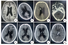
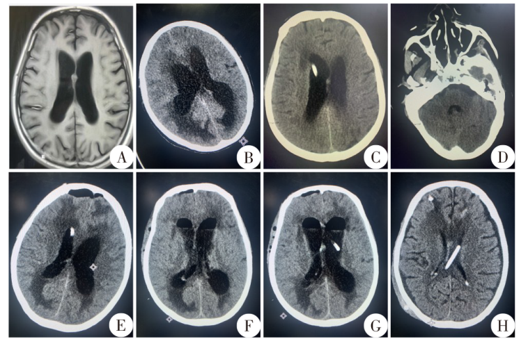
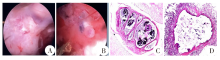
患者,男,53岁,农民,甘肃宕昌人。2021年11月19日因“头痛、头晕伴恶心、呕吐1周”就诊于甘肃省人民医院神经内科门诊。入院颅脑CT平扫示透明隔结节状稍高密度灶,头颅MRI示脑室轻度扩张。行腰椎穿刺术,颅内压180 mmH2O(1 mmH2O = 9.779 Pa);取脑脊液进行检测,总蛋白0.74 g/L。次日再行腰椎穿刺术,颅内压为300 mmH2O,遂转至神经外科作进一步治疗。患者颅压高,呈嗜睡状态,唤醒后不能正确对答,复查CT提示侧脑室扩张,考虑脑积水形成,存在脑疝风险。患者近年来有食未熟肉史,有肝棘球蚴病史。为降低患者颅压,11月26日行第1次脑室穿刺引流术,术后予重症监护。期间血清学检查提示猪囊尾蚴IgG抗体和弓形虫IgG抗体阳性,予吡奎酮(400 mg/8 h)和阿苯达唑(0.4 g/d)治疗3个疗程(7 d/疗程,疗程间隔5 d)。为改善患者脑积水症状,12月13日行第三脑室造瘘术(ETV),术后予驱虫治疗的同时行腰大池引流,但治疗效果不佳。12月28日和2022年1月11日行第2、3次脑室穿刺术以降低颅内压,术后驱虫治疗的同时予替加环素(50 mg/12 h)和舒普深(3 g/8 h)抗感染。1月25日患者颅内感染指标转阴,增强颅脑MRI未见明显脑囊尾蚴病灶。考虑堵管概率小,1月27日行脑室-腹腔分流术,术后患者意识清楚,复查头颅CT示脑室积水较前明显改善。患者于2月11日出院,出院时患者神志清楚,无明显头晕头痛、恶心呕吐和癫痫等症状。出院3个月后随访,患者病情恢复良好,生活可自理。

关键词: 脑囊尾蚴病, 脑室, 脑积水, 颅内感染

Abstract:

A 53-year-old male patient, who is a farmer, lived in Tanchang of Gansu, was treated at the Neurology Clinic of Gansu Provincial People’s Hospital due to “headache, dizziness, nausea, and vomiting for one week” on November 19, 2021. On admission, CT plain scan of the brain showed nodular and slightly high-density lesions in the transparent septum, while MRI of the head showed mild ventricular dilation. Lumbar puncture result showed that the intracranial pressure was 180 mmH2O (1 mmH2O = 9.779 Pa). The cerebrospinal fluid laboratory examination showed that the total protein in cerebrospinal fluid was 0.74 g/L. The second lumbar puncture result showed that the intracranial pressure was 300 mmH2O and the patient was transferred to neurosurgery on the following day. The patient was drowsy with high cranial pressure and unable to speak correctly upon awakening. Repeat CT scan showed lateral ventricular dilatation, which suggested high risks of hydrocephalus and cerebral hernia. The patient had eaten undercooked meat in recent years and a history of liver echinococcosis. To reduce patient’s cranial pressure, ventriculocentesis was performed on November 26 and the patient got the intensive care after surgery. The serum samples were positive for cysticercosis IgG and Toxoplasma gondii IgG. Therefore, the patient was treated with praziquantel (400 mg/8 h) and albendazole (0.4 g/d) for 3 courses (7 d/course, each treatment interval of 5 d). Endoscopic third ventriculostomy (ETV) was performed for the hydrocephalus on December 13. The patient got the antiparasitic therapy after ETV and lumbar ampullary drainage was performed at the same time, but the condition was not improved. The ventriculocentesis was performed twice on December 28 and January 11, 2022 respectively to reduce patient’s cranial pressure. The patient was treated with antiparasitic therapy and tigecycline (50 mg/12 h) and sulperazon (3 g/8 h). The patient’s intracranial infection indicators turned negative on January 25 and enhanced brain MRI showed no obvious tapeworm lesions. Laparoscopic assisted ventriculoperitoneal shunt was performed on January 27. The patient’s was consciouse and the cranial CT scan showed improved hydrocephalus after the surgery. The patient was discharged from the hospital with clear consciousness and no obvious dizziness, headache, nausea, vomiting or seizures on February 11. The patient recovered well after 3 months.

Key words: Neurocysticercosis, Intraventricular, Hydrocephalus, Intracranial infection

中图分类号: