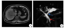
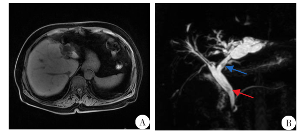
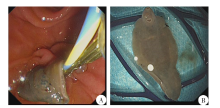
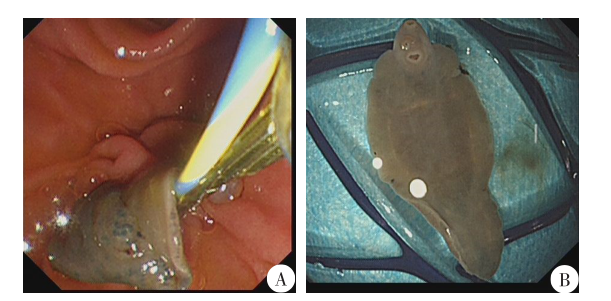
| [1] | Alejandra Caravedo M, Cabada MM. Human fascioliasis: current epidemiological status and strategies for diagnosis, treatment, and control[J]. Res Rep Trop Med, 2020, 11: 149-158. | | [2] | Agin M, Kayar Y, Dertli R. Diagnosis and treatment of Fasciola hepatica with endoscopic retrograde cholangiopancreatography in a child patient: case report[J]. Cureus, 2020, 12(9): e10486. | | [3] | Zhang YH, Zhang BY, Song ZJ. Effect of Fasciola hepatica on hepatic fibrosis and its treatment[J]. Chem Life, 2020, 40(2): 256-261. (in Chinese) | | | (张亚红, 张宝月, 宋正己. 肝片形吸虫对肝纤维化的影响及治疗[J]. 生命的化学, 2020, 40(2): 256-261.) | | [4] | Machicado C, Machicado JD, Maco V, et al. Association of Fasciola hepatica infection with liver fibrosis, cirrhosis, and cancer: a systematic review[J]. PLoS Negl Trop Dis, 2016, 10(9): e0004962. | | [5] | Fu XY, Ding JT. A case of biliary tract infection caused by Fasciola hepatica[J]. Chin J Hepatobiliary Surg, 2005, 11(9): 623. (in Chinese) | | | (付歆颖, 丁俊涛. 肝片形吸虫导致胆道感染一例[J]. 中华肝胆外科杂志, 2005, 11(9): 623.) | | [6] | Xie YL, Li L, Han DK, et al. One case of human fascioliasis hepatica[J]. J Clin Hepatol, 2014, 30(2): 166-167. (in Chinese) | | | (谢玉兰, 李丽, 韩大康, 等. 人肝片形吸虫病1例报告[J]. 临床肝胆病杂志, 2014, 30(2): 166-167.) | | [7] | Ramanan RV. Human fascioliasis: Diagnosis by typical computed tomography features and response to nitazoxanide in 16 patients from India[J]. Trop Gastroenterol, 2018, 39(3): 149-154. | | [8] | Perrodin S, Walti L, Gottstein B, et al. Fasciola hepatica in a country of low incidence: a tricky diagnosis[J]. Hepatobiliary Surg Nutr, 2019, 8(6): 597-603. | | [9] | Dolay K, Hasbah?eci M, Hatipo?lu E, et al. Endoscopic diagnosis and treatment of biliary obstruction due to acute cholangitis and acute pancreatitis secondary to Fasciola hepatica infection[J]. Ulus Travma Acil Cerrahi Derg, 2018, 24(1): 71-73. | | [10] | Sapunar J, Gallo G, Csendes A, et al. Hepatic fascioliasis diagnosed by endoscopic cholangiography[J]. Bol Chil Parasitol, 1983, 38(1/2): 17-20. | | [11] | Apt W, Tiselj R. Fascioliasis hepatica: diagnosis by endoscopic retrograde cholangiopancreatography[J]. Rev Med Chil, 1987, 115(6): 564-568. | | [12] | Tu K, Zhao LJ, Gu J. A case of Fasciola hepatica infection in common bile duct[J]. Chin J Parasitol Parasit Dis, 2021, 39(1): 133. (in Chinese) | | | (涂奎, 赵礼金, 顾进. 人胆总管肝片形吸虫病1例[J]. 中国寄生虫学与寄生虫病杂志, 2021, 39(1): 133.) | | [13] | Swain B, Otta S, Sahu MK, et al. Fasciola hepatica association with gallbladder malignancy: a rare case report[J]. Trop Parasitol, 2021, 11(1): 42-45. |
|

 ), 罗燕英, 曹立军, 周先宝, 黄应文, 贺学强*(