中国寄生虫学与寄生虫病杂志 ›› 2019, Vol. 37 ›› Issue (3): 279-285.doi: 10.12140/j.issn.1000-7423.2019.03.007

• 论著 • 上一篇    下一篇

粉尘螨过敏原Der f 32的克隆、表达、免疫学鉴定及其对树突状细胞的调节作用研究

江贝1,2(), 肖小军2, 欧阳春艳2, 罗新萍2, 孙宝清3, 李靖4, 刘志刚2,*()   

  1. 1 深圳大学生命与海洋科学学院,深圳 518055
    2 深圳大学过敏反应与免疫学研究所,深圳 518055
    3 广州医科大学呼吸疾病国家重点实验室,广州 510120
    4 广州医科大学第一附属医院,广州 510120
  • 收稿日期:2019-01-17 出版日期:2019-06-30 发布日期:2019-07-10
  • 通讯作者: 刘志刚
  • 作者简介:

    作者简介:江贝(1992-),女,硕士研究生,主要从事过敏性疾病及过敏原的研究。E-mail:578679260@qq.com

  • 基金资助:
    国家自然科学基金海外及港澳学者合作研究延续项目(No. 31729002)、广州市科学研究计划重点项目(No. 201804020043)、广东省科技计划产学研合作项目(No. 2014B090901041)、广东省医学科学技术研究基金(No. B2017081)、深圳市孔雀计划团队项目(No. KQTD20170331145453160)、深圳市科技计划国际科技合作项目(No. GJHZ2018041819053)、深圳市科技计划基础研究项目(No. JCYJ20160429171931438,No. JCYJ20160429114659119)

Cloning and expression of must mite allergen Der f 32 and its activation on mouse dendritic cells

Bei JIANG1,2(), Xiao-jun XIAO2, Chun-yan OUYANG2, Xin-ping LUO2, Bao-qing SUN3, Jing LI4, Zhi-gang LIU2,*()   

  1. 1 College of Life Sciences and Oceanography, Shenzhen University, Shenzhen 518055, China
    2 Institute of Allergy and Immunology, Shenzhen University, Shenzhen 518055, China
    3 State Key Laboratory of Respiratory Disease, Guangzhou Medical University, Guangzhou 510120, China
    4 The First Affiliated Hospital of Guangzhou Medical University, Guangzhou 510120, China
  • Received:2019-01-17 Online:2019-06-30 Published:2019-07-10
  • Contact: Zhi-gang LIU
  • Supported by:
    Supported by Joint Research Fund for Overseas Chinese Scholars and Scholars in Hong Kong and Macao Young Scholars(No. 31729002), Science and Technology Program of Guangzhou (No. 201804020043), Science and Technology Planning Project of Guangdong Province (No. 2014B090901041), Medical Scientific Research Foundation of Guangdong Province (No. B2017081), Shenzhen Peacock Team Project (No. KQTD20170331145453160), Science and Technology Program of Shenzhen (No. GJHZ2018041819053, No. JCYJ20160429171931438 and No. JCYJ20160429114659119)

摘要:

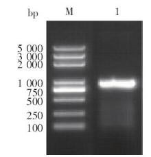
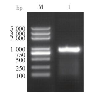
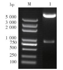
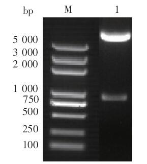
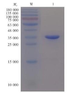
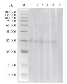
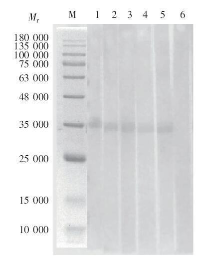
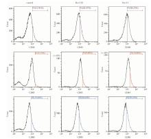
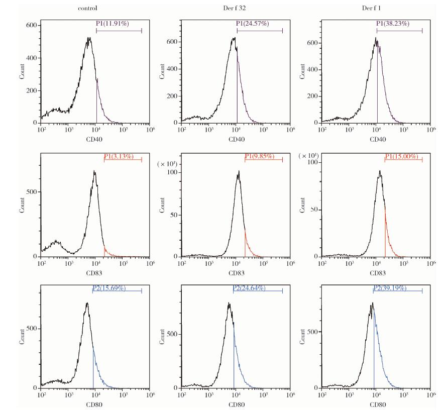
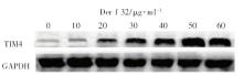
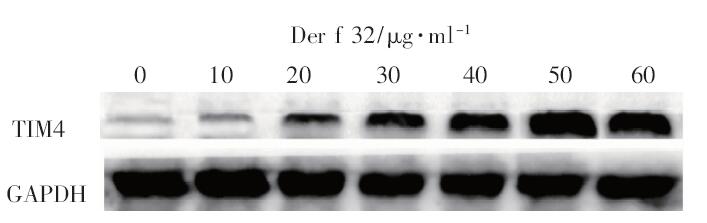
目的 克隆、表达粉尘螨Der f 32蛋白,并鉴定其免疫原性及其对树突状细胞(DC)的调节作用。方法 合成粉尘螨过敏原Der f 32基因,与pET-24a(+)载体连接,转入大肠埃希菌BL21诱导表达,经亲和层析法纯化后,十二烷基硫酸钠-聚丙烯酰氨凝胶电泳(SDS-PAGE)分析重组蛋白表达情况,蛋白质印迹(Western blotting)、临床皮肤点刺实验检测重组蛋白的免疫原性。将重组蛋白Der f 32与小鼠来源的DC2.4共培养24 h,设阳性对照组(加等量重组蛋白Der f 1)和阴性对照组(加等量PBS),流式细胞术检测细胞表面共刺激分子(CD40、CD83和CD80)的表达。将重组蛋白稀释为10、20、30、40、50、60 μg/ml等不同浓度,分别与DC2.4共培养48 h后,Western blotting分析T细胞免疫球蛋白及黏蛋白域蛋白4(TIM4)的表达情况。结果 SDS-PAGE结果显示,重组蛋白Der f 32相对分子质量(Mr)约35 000。临床皮肤点刺实验表明,42位尘螨过敏患者中有6例对重组蛋白Der f 32过敏,阳性率为14.2%。Western blotting 结果显示,重组蛋白Der f 32能与皮试阳性患者血清IgE抗体特异性结合。流式细胞术检测结果显示,重组蛋白Der f 32与DC2.4共培养后,CD40、CD83、CD80的表达水平分别为24.5%、9.6%和24.6%,高于阴性对照组(11.9%、3.1%和15.6%)(P < 0.01),低于阳性对照组(38.2%、15.0%和39.1%)(P < 0.05)。Western blotting检测结果显示,TIM4的表达量随重组蛋白Der f 32浓度的升高逐渐增加,且在Der f 32浓度为50 μg/ml时,TIM4的表达量达到最高,TIM4相对灰度值为1.112。结论 获得了具有较强免疫原性的粉尘螨新过敏原Der f 32,该蛋白能促进DC表面分子和TIM4的表达。

关键词: 粉尘螨, Der f 32, 免疫原性, 树突状细胞

Abstract:

Objective To clone and express must mite allergen Der f 32 protein and identify its antigenicity and its activation on dendritic cellline (DC2.4). Methods The DNA coding for allergen Der f 32 of dust mite was synthesized by genetic engineering technology, and cloned into expression vector pET-24a(+), and then transformed into Escherichia coli BL21 for expression. The expressed recombinant Der f 32 protein(rDer f 23)with His-tag was purified by nickel affinity chromatography and analyzed by SDS-PAGE and Western blotting with sera from patients allergic to dust mite. The rDer f 32 was used for skin prick test to detect its antigenicity in patients with allergic to dust mite. The rDer f 32 was co-cultured with mouse-derived DC2.4 for 24 h, flow cytometry was used to detect the expression of cell surface costimulatory molecules(CD40, CD83 and CD80)on the DC2.4. The DC2.4 cells were incubated with different amount of rDer f 32(10, 20, 30, 40, 50, 60 μg/ml)for 48 h and the expression of T cell immunoglobulin and mucin 4(TIM4)on DC2.4 was detected by Western blotting. Results The SDS-PAGE results showed that the rDer f 32 was successfully expressed in E. coli BL21 with a relative molecular mass of about 35 000. Clinical skin prick test showed that 6 of the 42 patients allergic to dust mite were positive with a positive rate of 14.2%. Western blotting showed that rDer f 32 was specifically recognized by IgE in sera of patients with positive in skin test. The results of flow cytometry showed that the expression levels of CD40, CD83 and CD80 were 24.5%, 9.6% and 24.6% on DC2.4 after being co-cultured with rDer f 32, which was significant higher than that of DC2.4 co-cultured with PBS(11.9%, 3.1% and 15.6%)(P < 0.01), however lower than that of the positive control group with stimulation of Der f 1(38.2%, 15.0%, and 39.1%)(P < 0.05). The results of Western blotting showed that the expression of TIM4 was increased in DC2.4 cells after being co-cultured with rDer f 32 at a dose-dependent pattern, with highest expression when Der f 32 was added at 50 μg/ml. The relative density of TIM4 expression was 1.112. Conclusion The dust mite allergen Der f 32 was successfully cloned and expressed as recombinant protein which could be recognized by the IgE in sera of patients with allergic to dust mite. rDer f 32 could stimulate DC maturation with increased expression of surface costimulatory molecules and TIM4.

Key words: Dust mites, Der f 32, Immunogenicity, Dendritic cell

中图分类号: