| [1] | World Health Organization. World Malaria Report 2021[R]. Geneva: WHO, 2021. | | [2] | Zhang L,, Feng J,, Tu H, et al. Malaria epidemiology in China in 2020[J]. Chin J Parasitol Parasit Dis, 2021, 39(2): 195-199. (in Chinese) | | [2] | ( 张丽,, 丰俊,, 涂宏, 等. 2020年全国疟疾疫情分析[J]. 中国寄生虫学与寄生虫病杂志, 2021, 39(2): 195-199.) | | [3] | Goldberg DE,, Zimmerberg J. Hardly vacuous: the parasitophorous vacuolar membrane of malaria parasites[J]. Trends Parasitol, 2020, 36(2): 138-146. | | [4] | Matz JM,, Beck JR,, Blackman MJ. The parasitophorous vacuole of the blood-stage malaria parasite[J]. Nat Rev Microbiol, 2020, 18(7): 379-391. | | [5] | Vaughan AM,, Kappe SHI. Malaria parasite liver infection and exoerythrocytic biology[J]. Cold Spring Harb Perspect Med, 2017, 7(6): a025486. | | [6] | Ingmundson A,, Nahar C,, Brinkmann V, et al. The exported Plasmodium berghei protein IBIS1 delineates membranous structures in infected red blood cells[J]. Mol Microbiol, 2012, 83(6): 1229-1243. | | [7] | Bannister LH,, Hopkins JM,, Fowler RE, et al. A brief illustrated guide to the ultrastructure of Plasmodium falciparum asexual blood stages[J]. Parasitol Today, 2000, 16(10): 427-433. | | [8] | Meis JF,, Verhave JP,, Jap PH, et al. Ultrastructural observations on the infection of rat liver by Plasmodium berghei sporozoites in vivo[J]. J Protozool, 1983, 30(2): 361-366. | | [9] | Nagao E,, Seydel KB,, Dvorak JA. Detergent-resistant erythrocyte membrane rafts are modified by a Plasmodium falciparum infection[J]. Exp Parasitol, 2002, 102(1): 57-59. | | [10] | Egea PF. Crossing the vacuolar Rubicon: structural insights into effector protein trafficking in api complexan parasites[J]. Microorganisms, 2020, 8(6): 865. | | [11] | Sá E Cunha C,, Nyboer B,, Heiss K, et al. Plasmodium berghei EXP-1 interacts with host Apolipoprotein H during Plasmodium liver-stage development[J]. Proc Natl Acad Sci USA, 2017, 114(7): E1138-E1147. | | [12] | Elmendorf HG,, Haldar K. Plasmodium falciparum exports the Golgi marker sphingomyelin synthase into a tubovesicular network in the cytoplasm of mature erythrocytes[J]. J Cell Biol, 1994, 124(4): 449-462. | | [13] | Nessel T,, Beck JM,, Rayatpisheh S, et al. EXP1 is required for organisation of EXP2 in the intraerythrocytic malaria parasite vacuole[J]. Cell Microbiol, 2020, 22(5): e13168. | | [14] | Garten M,, Beck JR,, Roth R, et al. Contacting domains segregate a lipid transporter from a solute transporter in the malarial host-parasite interface[J]. Nat Commun, 2020, 11(1): 3825. | | [15] | Spycher C,, Rug M,, Klonis N, et al. Genesis of and trafficking to the maurer’s clefts of Plasmodium falciparum-infected erythrocytes[J]. Mol Cell Biol, 2006, 26(11): 4074-4085. | | [16] | Hanssen E,, Sougrat R,, Frankland S, et al. Electron tomography of the maurer’s cleft organelles of Plasmodium falciparum-infected erythrocytes reveals novel structural features[J]. Mol Microbiol, 2008, 67(4): 703-718. | | [17] | Külzer S,, Rug M,, Brinkmann K, et al. Parasite-encoded Hsp40 proteins define novel mobile structures in the cytosol of the P. falciparum-infected erythrocyte[J]. Cell Microbiol, 2010, 12(10): 1398-1420. | | [18] | McHugh E,, Carmo OMS,, Blanch A, et al. Role of Plasmodium falciparum protein GEXP07 in maurer’s cleft morphology, knob architecture, and P. falciparum EMP1 trafficking[J]. mBio, 2020, 11(2): e03320-e03319. | | [19] | Matz JM,, Goosmann C,, Brinkmann V, et al. The Plasmodium berghei translocon of exported proteins reveals spatiotemporal dynamics of tubular extensions[J]. Sci Rep, 2015, 5: 12532. | | [20] | Spielmann T,, Fergusen DJP,, Beck HP. Etramps, a new Plasmodium falciparum gene family coding for developmentally regulated and highly charged membrane proteins located at the parasite-host cell interface[J]. Mol Biol Cell, 2003, 14(4): 1529-1544. | | [21] | MacKellar DC,, Vaughan AM,, Aly ASI, et al. A systematic analysis of the early transcribed membrane protein family throughout the life cycle of Plasmodium yoelii[J]. Cell Microbiol, 2011, 13(11): 1755-1767. | | [22] | Lee SK,, Han JH,, Park JH, et al. Evaluation of antibody responses to the early transcribed membrane protein family in Plasmodium vivax[J]. Parasit Vectors, 2019, 12(1): 594. | | [23] | Saito H,, Lund-Katz S,, Phillips MC. Contributions of domain structure and lipid interaction to the functionality of exchangeable human apolipoproteins[J]. Prog Lipid Res, 2004, 43(4): 350-380. | | [24] | Vignali M,, McKinlay A,, LaCount DJ, et al. Interaction of an atypical Plasmodium falciparum ETRAMP with human apolipoproteins[J]. Malar J, 2008, 7: 211. | | [25] | Mesén-Ramírez P,, Reinsch F,, Blancke Soares A, et al. Stable translocation intermediates jam global protein export in Plasmodium falciparum parasites and link the PTEX component EXP2 with translocation activity[J]. PLoS Pathog, 2016, 12(5): e1005618. | | [26] | Vincensini L,, Richert S,, Blisnick T, et al. Proteomic analysis identifies novel proteins of the maurer’s clefts, a secretory compartment delivering Plasmodium falciparum proteins to the surface of its host cell[J]. Mol Cell Proteomics, 2005, 4(4): 582-593. | | [27] | Currà C,, di Luca M,, Picci L, et al. The ETRAMP family member SEP2 is expressed throughout Plasmodium berghei life cycle and is released during sporozoite gliding motility[J]. PLoS One, 2013, 8(6): e67238. | | [28] | Currà C,, Pace T,, Franke-Fayard BMD, et al. Erythrocyte remodeling in Plasmodium berghei infection: the contribution of SEP family members[J]. Traffic, 2012, 13(3): 388-399. | | [29] | Andreadaki M,, Hanssen E,, Deligianni E, et al. Sequential membrane rupture and vesiculation during Plasmodium berghei gametocyte egress from the red blood cell[J]. Sci Rep, 2018, 8(1): 3543. | | [30] | Venkatesh A,, Jain A,, Davies H, et al. Hospital-derived antibody profiles of malaria patients in Southwest India[J]. Malar J, 2019, 18(1): 138. | | [31] | Milner DA Jr,, Lee JJ,, Frantzreb C, et al. Quantitative assessment of multiorgan sequestration of parasites in fatal pediatric cerebral malaria[J]. J Infect Dis, 2015, 212(8): 1317-1321. | | [32] | Heiber A,, Kruse F,, Pick C, et al. Identification of new PNEPs indicates a substantial non-PEXEL exportome and underpins common features in Plasmodium falciparum protein export[J]. PLoS Pathog, 2013, 9(8): e1003546. | | [33] | Marti M,, Good RT,, Rug M, et al. Targeting malaria virulence and remodeling proteins to the host erythrocyte[J]. Science, 2004, 306(5703): 1930-1933. | | [34] | Sun XD,, Zhao X,, Tu ZW, et al. Advances in the study of protein export by Plasmodium falciparum[J]. J Pathogen Biol, 2014, 9(9): 852-855. (in Chinese) | | [34] | ( 孙喜东,, 赵欣,, 土志伟, 等. 恶性疟原虫蛋白输出机制的研究进展[J]. 中国病原生物学杂志, 2014, 9(9): 852-855.) | | [35] | AhYoung AP,, Koehl A,, Cascio D, et al. Structural mapping of the ClpB ATPases of Plasmodium falciparum: targeting protein folding and secretion for antimalarial drug design[J]. Protein Sci, 2015, 24(9): 1508-1520. | | [36] | Florentin A,, Stephens DR,, Brooks CF, et al. Plastid biogenesis in malaria parasites requires the interactions and catalytic activity of the Clp proteolytic system[J]. Proc Natl Acad Sci USA, 2020, 117(24): 13719-13729. | | [37] | Hakamada K,, Watanabe H,, Kawano R, et al. Expression and characterization of the Plasmodium translocon of the exported proteins component EXP2[J]. Biochem Biophys Res Commun, 2017, 482(4): 700-705. | | [38] | Ho CM,, Beck JR,, Lai M, et al. Malaria parasite translocon structure and mechanism of effector export[J]. Nature, 2018, 561(7721): 70-75. | | [39] | Chisholm SA,, Kalanon M,, Nebl T, et al. The malaria PTEX component PTEX88 interacts most closely with HSP101 at the host-parasite interface[J]. FEBS J, 2018, 285(11): 2037-2055. | | [40] | Matthews K,, Kalanon M,, Chisholm SA, et al. The Plasmodium translocon of exported proteins (PTEX) component thioredoxin-2 is important for maintaining normal blood-stage growth[J]. Mol Microbiol, 2013, 89(6): 1167-1186. | | [41] | Chisholm SA,, McHugh E,, Lundie R, et al. Contrasting inducible knockdown of the auxiliary PTEX component PTEX88 in P. falciparum and P. berghei unmasks a role in parasite virulence[J]. PLoS One, 2016, 11(2): e0149296. | | [42] | Peng M,, Cascio D,, Egea PF. Crystal structure and solution characterization of the thioredoxin-2 from Plasmodium falciparum, a constituent of an essential parasitic protein export complex[J]. Biochem Biophys Res Commun, 2015, 456(1): 403-409. | | [43] | Elsworth B,, Sanders PR,, Nebl T, et al. Proteomic analysis reveals novel proteins associated with the Plasmodium protein exporter PTEX and a loss of complex stability upon truncation of the core PTEX component, PTEX150[J]. Cell Microbiol, 2016, 18(11): 1551-1569. | | [44] | Low LM,, Azasi Y,, Sherling ES, et al. Deletion of Plasmodium falciparum protein RON3 affects the functional translocation of exported proteins and glucose uptake[J]. mBio, 2019, 10(4): e01460-e01419. | | [45] | Morita M,, Nagaoka H,, Ntege EH, et al. PV1, a novel Plasmodium falciparum merozoite dense granule protein, interacts with exported protein in infected erythrocytes[J]. Sci Rep, 2018, 8(1): 3696. | | [46] | Gold DA,, Kaplan AD,, LIS A, et al. The Toxoplasma dense granule proteins GRA17 and GRA23 mediate the movement of small molecules between the host and the parasitophorous vacuole[J]. Cell Host Microbe, 2015, 17(5): 642-652. | | [47] | Garten M,, Nasamu AS,, Niles JC, et al. EXP2 is a nutrient-permeable channel in the vacuolar membrane of Plasmodium and is essential for protein export via PTEX[J]. Nat Microbiol, 2018, 3(10): 1090-1098. | | [48] | Mesén-Ramírez P,, Bergmann B,, Tran TT, et al. EXP1 is critical for nutrient uptake across the parasitophorous vacuole membrane of malaria parasites[J]. PLoS Biol, 2019, 17(9): e3000473. | | [49] | Pal C,, Kundu MK,, Bandyopadhyay U, et al. Synthesis of novel heme-interacting acridone derivatives to prevent free heme-mediated protein oxidation and degradation[J]. Bioorg Med Chem Lett, 2011, 21(12): 3563-3567. | | [50] | Saha SJ,, Siddiqui AA,, Pramanik S, et al. Hydrazonophenol, a food vacuole-targeted and ferriprotoporphyrin IX-interacting chemotype prevents drug-resistant malaria[J]. ACS Infect Dis, 2019, 5(1): 63-73. | | [51] | Iriko H,, Ishino T,, Otsuki H, et al. Plasmodium falciparum exported protein 1 is localized to dense granules in merozoites[J]. Parasitol Int, 2018, 67(5): 637-639. | | [52] | Lisewski AM,, Quiros JP,, Ng CL, et al. Supergenomic network compression and the discovery of EXP1 as a glutathione transferase inhibited by artesunate[J]. Cell, 2014, 158(4): 916-928. | | [53] | Cooper RA,, Papakrivos J,, Lane KD, et al. PfCG2, a Plasmodium falciparum protein peripherally associated with the parasitophorous vacuolar membrane, is expressed in the period of maximum hemoglobin uptake and digestion by trophozoites[J]. Mol Biochem Parasitol, 2005, 144(2): 167-176. | | [54] | Wicht KJ,, Mok S,, Fidock DA. Molecular mechanisms of drug resistance in Plasmodium falciparum malaria[J]. Annu Rev Microbiol, 2020, 74: 431-454. | | [55] | Yang B,, Sun YF,, Lei Y, et al. Research progress on the treatment of malaria with artemisinin and its derivatives[J]. Chin J Parasitol Parasit Dis, 2021, 39(3): 393-402. (in Chinese) | | [55] | ( 杨博,, 孙毅凡,, 雷瑶, 等. 青蒿素及其衍生物治疗疟疾的研究进展[J]. 中国寄生虫学与寄生虫病杂志, 2021, 39(3): 393-402.) | | [56] | Li J,, Wu LO,, Yang ZQ. Comparison of efficacy of artemisinin antimalarials and combined use the drugs[J]. Chin Trop Med, 2009, 9(1): 157-159, 195. (in Chinese) | | [56] | ( 李佳,, 吴兰鸥,, 杨照青. 青蒿素类抗疟药药效的比较及联合用药[J]. 中国热带医学, 2009, 9(1): 157-159, 195.) | | [57] | Boddey JA,, Cowman AF. Plasmodium nesting: remaking the erythrocyte from the inside out[J]. Annu Rev Microbiol, 2013, 67: 243-269. | | [58] | Hiller NL,, Akompong T,, Morrow JS, et al. Identification of a stomatin orthologue in vacuoles induced in human erythrocytes by malaria parasites: a role for microbial raft proteins in api complexan vacuole biogenesis[J]. J Biol Chem, 2003, 278(48): 48413-48421. | | [59] | Counihan NA,, Chisholm SA,, Bullen HE, et al. Plasmodium falciparum parasites deploy RhopH2 into the host erythrocyte to obtain nutrients, grow and replicate[J]. eLife, 2017, 6: e23217. | | [60] | Gupta A,, Thiruvengadam G,, Desai SA. The conserved clag multigene family of malaria parasites: essential roles in host-pathogen interaction[J]. Drug Resist Updat, 2015, 18: 47-54. | | [61] | Comeaux CA,, Coleman BI,, Bei AK, et al. Functional analysis of epigenetic regulation of tandem RhopH1/clag genes reveals a role in Plasmodium falciparum growth[J]. Mol Microbiol, 2011, 80(2): 378-390. | | [62] | Parish LA,, Mai DW,, Jones ML, et al. A member of the Plasmodium falciparum PHIST family binds to the erythrocyte cytoskeleton component band 4.1[J]. Malar J, 2013, 12: 160. | | [63] | Shakya B,, Penn WD,, Nakayasu ES, et al. The Plasmodium falciparum exported protein PF3D7_0402000 binds to erythrocyte ankyrin and band 4.1[J]. Mol Biochem Parasitol, 2017, 216: 5-13. | | [64] | Thieleke-Matos C,, Lopes da Silva M,, Cabrita-Santos L, et al. Host cell autophagy contributes to Plasmodium liver development[J]. Cell Microbiol, 2016, 18(3): 437-450. | | [65] | Wolanin K,, Fontinha D,, Sanches-Vaz M, et al. A crucial role for the C-terminal domain of exported protein 1 during the mosquito and hepatic stages of the Plasmodium berghei life cycle[J]. Cell Microbiol, 2019, 21(10): e13088. | | [66] | Mikolajczak SA,, Jacobs-Lorena V,, MacKellar DC, et al. L-FABP is a critical host factor for successful malaria liver stage development[J]. Int J Parasitol, 2007, 37(5): 483-489. | | [67] | Real E,, Rodrigues L,, Cabal GG, et al. Plasmodium UIS3 sequesters host LC3 to avoid elimination by autophagy in hepatocytes[J]. Nat Microbiol, 2018, 3(1): 17-25. | | [68] | Lu F,, Zhuo XH,, Lu SH, et al. Research progress on the interaction between host cell autophagy and apicomplexa protozoa infection[J/OL]. Chin J Parasitol Parasit Dis: 1-6[2022-01-11]. http://kns.cnki.net/kcms/detail/31.1248.R.20211213.0856.002.html. (in Chinese) | | [68] | ( 鲁飞,, 卓洵辉,, 陆绍红. 顶复门原虫感染与宿主细胞自噬相互作用的研究进展[J/OL]. 中国寄生虫学与寄生虫病杂志: 1-6[2022-01-11]. http://kns.cnki.net/kcms/detail/31.1248.R.20211213.0856.002.html. ) | | [69] | Burda PC,, Roelli MA,, Schaffner M, et al. A Plasmodium phospholipase is involved in disruption of the liver stage parasitophorous vacuole membrane[J]. PLoS Pathog, 2015, 11(3): e1004760. | | [70] | de Niz M,, Caldelari R,, Kaiser G, et al. Hijacking of the host cell Golgi by Plasmodium berghei liver stage parasites[J]. J Cell Sci, 2021, 134(10): jcs252213. | | [71] | Ross A,, Koepfli C,, Schoepflin S, et al. The incidence and differential seasonal patterns of Plasmodium vivax primary infections and relapses in a cohort of children in Papua new Guinea[J]. PLoS Negl Trop Dis, 2016, 10(5): e0004582. | | [72] | Schafer C,, Dambrauskas N,, Steel RW, et al. A recombinant antibody against Plasmodium vivax UIS4 for distinguishing replicating from dormant liver stages[J]. Malar J, 2018, 17(1): 370. | | [73] | Chawla J,, Oberstaller J,, Adams JH. Targeting gametocytes of the malaria parasite Plasmodium falciparum in a functional genomics era: next steps[J]. Pathogens, 2021, 10(3): 346. | | [74] | Baker DA,, Daramola O,, McCrossan MV, et al. Subcellular localization of Pfs16, a Plasmodium falciparum gametocyte antigen[J]. Parasitology, 1994, 108 (Pt 2): 129-137. | | [75] | Kongkasuriyachai D,, Fujioka H,, Kumar N. Functional analysis of Plasmodium falciparum parasitophorous vacuole membrane protein (Pfs16) during gametocytogenesis and gametogenesis by targeted gene disruption[J]. Mol Biochem Parasitol, 2004, 133(2): 275-285. | | [76] | Deligianni E,, Andreadaki M,, Koutsouris K, et al. Sequence and functional divergence of gametocyte-specific parasitophorous vacuole membrane proteins in Plasmodium parasites[J]. Mol Biochem Parasitol, 2018, 220: 15-18. | | [77] | Lanfrancotti A,, Bertuccini L,, Silvestrini F, et al. Plasmodium falciparum: MRNA co-expression and protein co-localisation of two gene products upregulated in early gametocytes[J]. Exp Parasitol, 2007, 116(4): 497-503. | | [78] | Furuya T,, Mu JB,, Hayton K, et al. Disruption of a Plasmodium falciparum gene linked to male sexual development causes early arrest in gametocytogenesis[J]. Proc Natl Acad Sci USA, 2005, 102(46): 16813-16818. | | [79] | Janse CJ,, Haghparast A,, Speranca MA, et al. Malaria parasites lacking eef1a have a normal S/M phase yet grow more slowly due to a longer G1 phase[J]. Mol Microbiol, 2003, 50(5): 1539-1551. | | [80] | Ponzi M,, Sidén-Kiamos I,, Bertuccini L, et al. Egress of Plasmodium berghei gametes from their host erythrocyte is mediated by the MDV-1/PEG3 protein[J]. Cell Microbiol, 2009, 11(8): 1272-1288. | | [81] | Schnider CB,, Bausch-Fluck D,, Brühlmann F, et al. BioID reveals novel proteins of the Plasmodium parasitophorous vacuole membrane[J]. mSphere, 2018, 3(1): e00522-e00517. |
|

 ), 刘蕾2, 孙毅凡1, 程洋1,*(
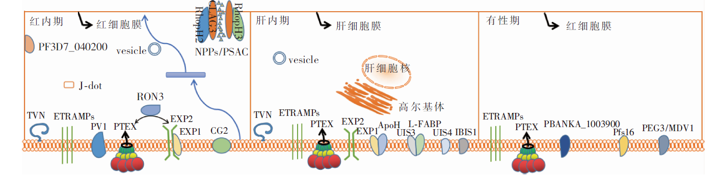