中国寄生虫学与寄生虫病杂志 ›› 2018, Vol. 36 ›› Issue (4): 361-366.

• 论著 • 上一篇    下一篇

TLR4/NF-κB信号通路在微小隐孢子虫感染中的作用机制

汲蕊, 梁瑞文, 管志玉*(), 李瑞芳, 付玉荣, 王红艳   

  1. 潍坊医学院病原生物学教研室,潍坊 261053
  • 收稿日期:2018-01-25 出版日期:2018-08-30 发布日期:2018-09-06
  • 通讯作者: 管志玉
  • 基金资助:
    山东省自然科学基金(No. ZR2015HQ030);山东省高等学校科技计划项目(No. J13LK04);山东省中医药管理局项目(No. 2015-238)

The role of TLR4/NF-κB signaling pathway in Cryptosporidim parvum infection

Rui JI, Rui-wen LIANG, Zhi-yu GUAN*(), Rui-fang LI, Yu-rong FU, Hong-yan WANG   

  1. Department of Pathogen Biology, Weifang Medical College, Weifang 261053, China
  • Received:2018-01-25 Online:2018-08-30 Published:2018-09-06
  • Contact: Zhi-yu GUAN
  • Supported by:
    Supported by Natural Science Fund of Shandong Province (No. ZR2015HQ030), Higher Education Science and Technology Plan Project of Shandong Province (No. J13LK04), Administration of Traditional Chinese Medicine Project of Shandong Province (No. 2015-238)

摘要:

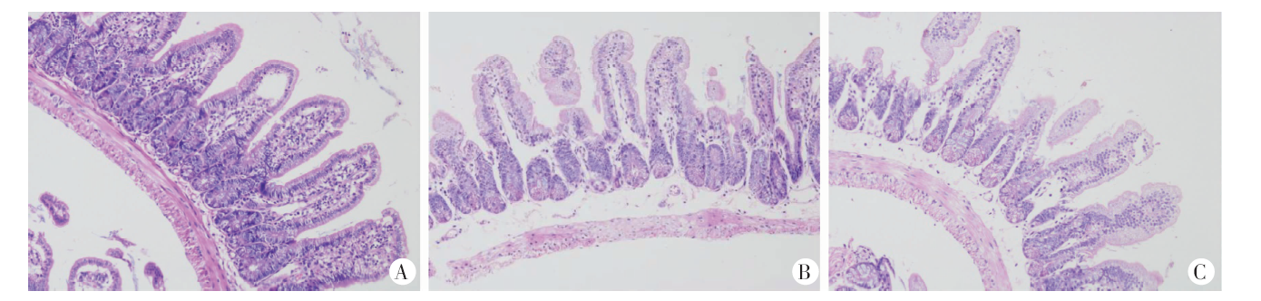
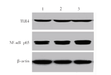
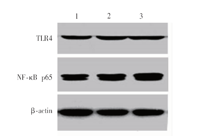
目的 探讨Toll样受体4/核因子κB(TLR4/NF-κB)信号通路在微小隐孢子虫(Cryptosporidium parvum)感染小鼠致肠黏膜损伤中作用的分子机制。方法 30只4周龄雄性BALB/c小鼠随机分成感染1周组、感染2周组和未感染组,每组10只。感染组每鼠经口灌胃微小隐孢子虫卵囊(1×105个/只),未感染组小鼠正常饮食、饮水。感染组小鼠分别于感染后第1周和第2周剖杀,未感染组小鼠于第2周剖杀。取小鼠肠组织,HE染色观察小鼠肠黏膜的病理变化。抽提肠黏膜组织总RNA,逆转录成cDNA,实时荧光定量PCR(qRT-PCR)检测TLR4和NF-κB p65的mRNA相对表达量。蛋白质印迹(Western blotting)检测肠黏膜组织中TLR4和NF-κB p65的相对表达量。ELISA检测肠黏膜组织中白细胞介素1β(IL-1β)和肿瘤坏死因子α(TNF-α)的表达水平。结果 HE染色结果显示,感染组小鼠肠绒毛明显萎缩变短、脱落,黏膜下层水肿,与肌层间形成明显的间隙;未感染组小鼠肠绒毛结构完整。qRT-PCR结果显示,感染1周组和感染2周组小鼠肠黏膜组织中TLR4的mRNA相对表达量分别为2.3 ± 0.4、3.5 ± 0.1,均高于未感染组(1.0 ± 0.0)(P < 0.05,P < 0.01);NF-κB p65的mRNA相对表达量分别为2.6 ± 0.3、3.6 ± 0.2,均高于未感染组(1.1 ± 0.1)(P < 0.05,P < 0.01)。Western blotting结果显示,感染1周组和感染2周组小鼠肠黏膜组织中TLR4的相对表达量分别为0.4 ± 0.0、0.6 ± 0.0,均高于未感染组(0.2 ± 0.0)(P < 0.05,P < 0.01);NF-κB p65的表达量分别为0.6 ± 0.0、0.8 ± 0.1,均高于未感染组(0.4 ± 0.0)(P < 0.05,P < 0.01)。ELISA检测结果显示,感染1周组和感染2周组小鼠肠黏膜组织中IL-1β的表达量分别为33.3 ± 2.2、46.1 ± 2.5,均高于未感染组(22.3 ± 5.0)(P < 0.01);TNF-α的表达量分别为45.7 ± 2.0、55.4 ± 3.6,均高于未感染组(25.7 ± 9.3)差异有统计学意义(P < 0.01)。结论 微小隐孢子虫感染小鼠后,通过激活TLR4/NF-κB信号通路,可上调TLR4和NF-κB p65的表达,促进IL-1β和TNF-α的释放,诱发肠黏膜炎症反应。

关键词: 微小隐孢子虫, Toll样受体, TLR4/NF-κB信号通路, 白细胞介素1β, 肿瘤坏死因子α

Abstract:

Objective To explore the role of the Toll-like receptor 4/ nuclear factor κB(TLR4/NF-κB)pathway in intestinal mucosal damage induced by Cryptosporidium parvum infection in mice. Methods Thirty 4-week-old BALB/c male mice were randomly divided into 1-week infection group and 2-week infection group and the non-infection group (n = 10 in each group). The mouse model of Cryptosporidium parvum infection was established intragastrically with 1×10⁵ oocysts. Uninfected mice have normal diet and drinking water. Ten infected mice were sacrificed at 7 days after infection (1-week infection group) and 14 days after infection (2-week infection group), respectively. The normal control mice were sacrificed at 14 days after infection. The intestinal tissue was collected for observing pathological alterations of intestinal mucosa. The relative expression of TLR4 mRNA and NF-κB p65 mRNA in intestinal mucosa was detected by qRT-PCR. The relative expression of TLR4 and NF-κB p65 in intestinal mucosa was detected by Western blotting. The expression of interleukin 1β (IL-1β) and tumor necrosis factor α (TNF-α) in intestinal mucosa was detected by ELISA. Result HE staining showed atrophy and shedding of intestinal villi in the infection group, with submucosal edema and a significant gap between muscle layers, while the intestinal villi were intact in the non-infection group. qRT-PCR showed that the relative expression of TLR4 mRNA in the intestinal mucosa in infection group at week 1 and week 2 was 2.3 ± 0.4 and 3.5 ± 0.1, respectively, higher than the non-infection group (1.0 ± 0.0) (P < 0.05, P < 0.01), and the relative expression of NF-κB p65 mRNA in the intestinal mucosa in infection group at week 1 and week 2 was 2.6 ± 0.3 and 3.6 ± 0.2, higher than the non-infection group (1.1 ± 0.1) (P < 0.05, P < 0.01). Western blotting showed that the protein level of TLR4 in the intestinal mucosa in infection group at week 1 and week 2 was 0.4 ± 0.0 and 0.6 ± 0.0, higher than the non-infection group (0.2 ± 0.0) (P < 0.05, P < 0.01), and that of NF-κB p65 in infection group at week 1 and week 2 was and 0.6 ± 0.0 and 0.8 ± 0.1, higher than the non-infection group (0.4 ± 0.0) (P < 0.05, P < 0.01). ELISA results showed that the expression level of IL-1β in the intestinal mucosa in infection group at week 1 and week 2 was 33.4 ± 2.2 and 46.1 ± 2.5, higher than the non-infection group (22.3 ± 5.0) (P < 0.01), and that of TNF-α in infection group at week 1 and week 2 was 45.7 ± 2.0 and 55.4 ± 3.6, higher than the non-infection group (25.7 ± 9.3) (P < 0.01). Conclusion Cryptosporidium parvum infection may activate the TLR4/NF-κB signaling pathway, up-regulate the expression of TLR4 and NF-κB p65, and promote the release of inflammatory factors TNF-α and IL-1β, thus inducing intestinal mucosal inflammation.

Key words: Cryptosiporidium parvum, Toll-like receptor, TLR4/NF-κB pathway;, Interleukin 1β, Tumor necrosis factor α

中图分类号: