中国寄生虫学与寄生虫病杂志 ›› 2022, Vol. 40 ›› Issue (4): 493-499.doi: 10.12140/j.issn.1000-7423.2022.04.012

• 论著 • 上一篇    下一篇

田鼠巴贝虫丽水分离株小鼠感染模型的建立及其病理变化

宋鹏(), 蔡玉春, 卢艳, 艾琳, 陈木新, 陈韶红, 陈家旭*()   

  1. 中国疾病预防控制中心寄生虫病预防控制所(国家热带病研究中心),国家卫生健康委员会寄生虫病原与媒介生物学重点实验室,世界卫生组织热带病合作中心,国家级热带病国际联合研究中心,上海 200025
  • 收稿日期:2022-03-04 修回日期:2022-04-28 出版日期:2022-08-30 发布日期:2022-09-07
  • 通讯作者: 陈家旭
  • 作者简介:宋鹏(1989-),男,硕士,助理研究员,从事巴贝虫病防治研究。E-mail: songpeng@nipd.chinacdc.cn
  • 基金资助:
    2019年上海市卫生健康委青年基金(20194Y0046)

Establishment of mouse infection model of Babesia microti Lishui isolate and consequent pathological changes

SONG Peng(), CAI Yu-chun, LU Yan, AI Lin, CHEN Mu-xin, CHEN Shao-hong, CHEN Jia-xu*()   

  1. National Institute of Parasitic Diseases, Chinese Center for Disease Control and Prevention (Chinese Center for Tropical Diseases Research);NHC Key Laboratory of Parasite and Vector Biology; WHO Collaborating Centre for Tropical Diseases; National Center for International Research on Tropical Diseases, Shanghai 200025, China
  • Received:2022-03-04 Revised:2022-04-28 Online:2022-08-30 Published:2022-09-07
  • Contact: CHEN Jia-xu
  • Supported by:
    Shanghai Municipal Health Commission(20194Y0046)

摘要:

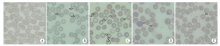
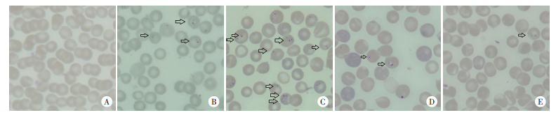
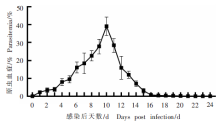
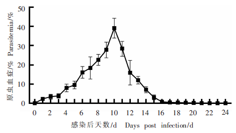
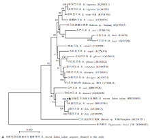
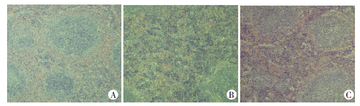
目的 建立田鼠巴贝虫丽水分离株(分离自浙江丽水患者,简称丽水分离株)BALB/c小鼠感染模型,研究小鼠感染后的原虫血症动态变化和病理特征。 方法 用浙江丽水田鼠巴贝虫病患者的全血血样腹腔接种NOD-SCID小鼠进行保种、分离田鼠巴贝虫。50只BALB/c小鼠随机分为感染组和对照组(每组25只),感染组每鼠腹腔接种1.0 × 107个NOD-SCID小鼠田鼠巴贝虫感染红细胞,对照组小鼠接种等量生理盐水。每日采小鼠尾静脉血制备薄血膜片,吉氏染色后显微镜下观察原虫形态,分析原虫血症。用DNA提取试剂盒提取感染小鼠血样DNA,巢式PCR扩增巴贝虫18S rRNA基因,测序后进行基因型鉴定和系统进化树分析。感染后0、5、10、15和20 d,每组分别取5只小鼠,测量体质量、脾长度和质量,制备脾组织切片,HE染色显微镜下观察脾组织病理特征;采用动物全自动血液分析仪检测感染小鼠血常规。组间比较采用t检验。 结果 感染后5 d,感染组小鼠血涂片中可见单个环状体;感染后10 d,同一红细胞中可见2个或4个虫体,呈双梨形或马耳他十字形,并有细胞溶血现象;感染后15和20 d,红细胞内虫体仍以环状体为主。感染后10 d,感染组小鼠的原虫血症达到峰值,为(39.1 ± 4.6)%;感染后20 d,原虫血症降至1%以下。田鼠巴贝虫丽水分离株18S rRNA基因序列与田鼠巴贝虫(GenBank登录号MT423326)的一致性为98%,在系统进化树上聚在同一分支上。感染后10、15和20 d,感染组小鼠体质量分别为(20.60 ± 1.02)、(22.04 ± 0.77)和(22.78 ± 0.64)g,均低于对照组[(23.94 ± 0.84)、(24.50 ± 0.26)和(24.64 ± 0.54)g](t = 5.64、6.78和4.99,P < 0.01)。感染后5、10、15和20 d,感染组小鼠脾质量分别为(0.33 ± 0.02)、(0.98 ± 0.11)、(0.93 ± 0.04) 和(0.67 ± 0.05)g,均高于对照组[(0.11 ± 0.01)、(0.12 ± 0.01)、(0.10 ± 0.02)和(0.11 ± 0.01)g](t = 21.82、22.25、35.62和10.47,P < 0.01);脾长度分别为(2.40 ± 0.12)、(3.16 ± 0.06)、(3.22 ± 0.05)和(2.98 ± 0.08)cm,均高于对照组小鼠[(1.76 ± 0.09)、(1.74 ± 0.09)、(1.74 ± 0.15)和(1.80 ± 0.07)cm](t = 9.44、30.27、20.93和24.09,P < 0.01)。感染后10 d,感染组小鼠脾明显肿大,组织结构紊乱,白髓和红髓交界的边缘区模糊,脾窦充血,淋巴细胞大量浸润;感染后20 d,脾组织实质结构恢复,红髓、白髓分布清晰。血常规结果显示,感染后10 d,感染组小鼠红细胞计数、红细胞压积、血红蛋白浓度、平均血细胞体积、红细胞分布宽度标准差、红细胞分布宽度变异系数、平均血红蛋白含量和平均血红蛋白浓度分别为(4.45 ± 0.32)× 1012/L、(27.72 ± 2.03)%、(86.2 ± 6.0)g/L、(60.7 ± 1.4)fL、(84.1 ± 4.0)fL、(31.9 ± 1.3)%、(19.4 ± 0.4)pg和(320.4 ± 3.8)g/L,与对照组[(9.55 ± 0.16)× 1012/L、(47.94 ± 1.64)%、(163.0 ± 4.8)g/L、(48.2 ± 1.1)fL、(27.7 ± 1.3)fL、(13.5 ± 0.5)%、(16.7 ± 0.7)pg和(339.0 ± 3.9)g/L]相比,差异有统计学意义(t = 32.24、17.34、22.23、15.71、30.33、28.41、7.43和7.61,P < 0.01);感染后10 d,感染组小鼠白细胞计数、单核细胞计数和百分比、中性粒细胞计数和百分比分别为(6.76 ± 0.87)× 109/L、(0.78 ± 0.20)× 109/L、(9.90 ± 0.87)%、(1.92 ± 0.42)× 109/L和(27.74 ± 2.67)%,与对照组[(3.85 ± 0.26)× 109/L、(0.17 ± 0.05)× 109/L、(3.28 ± 0.40)%、(0.78 ± 0.15)× 109/L和(21.20 ± 1.18)%]相比,差异有统计学意义(t = 7.12、6.54、15.54、5.71和5.00,P < 0.01)。 结论 建立了从人体分离的田鼠巴贝虫丽水分离株小鼠感染模型,小鼠感染后体质量显著减轻、脾肿大、脾组织结构紊乱、贫血严重。

关键词: 田鼠巴贝虫, 小鼠动物模型, 脾脏, 病理特征

Abstract:

Objective To establish the mouse infection model of Babesia microti Lishui isolate (isolated from one patient in Lishui, Zhejiang Province) and evaluate the change of parasitemia and pathological features during infection in the mice. Methods NOD-SCID mice were intraperitoneally inoculated with whole blood sample from a patient infected B. microti Lishui for species preservation and isolation. Fifty BALB/c mice were randomly assigned to infection group and control group, 25 mice in each group. The BALB/c mice in the infection group were intraperitoneally inoculated with 1.0 × 10⁷ infected erythrocytes from NOD-SCID mice and the BALB/c mice in the control group were inoculated with an equal volume of normal saline. The tail vein blood was collected daily to prepare the thin blood smear. The morphology of B. microti was observed under microscope after Giemsa staining, and the parasitemia was calculated. DNA was extracted from the infected mice samples. The 18S rRNA gene was amplified by Nest-PCR for sequencing, geneotyping, and phylogenetic tree analysis. At 0, 5, 10, 15, and 20 days post-infection, 5 mice in each group were selected to measure their body weight, spleen weight, and spleen length. The spleen tissue sections were prepared, and the pathological characteristics were observed under a microscope after HE staining. The routine blood test for samples from the infected mice was detected by an animal automatic blood analyzer. T-test was used for comparison between the two groups. Results At 5 days post-infection, the ring forms of the B. microti Lishui isolate were observed in the blood smear in the infection group. At 10 days post-infection, two or four parasites, in the shape of a double pear-shaped or Maltese cross, could be identified in the same erythrocyte, and hemolysis occurred. The parasitemia in mice in the infection group peaked (39.1 ± 4.6)% at 10 days post-infection and decreased to less than 1% after 20 days post-infection. The 18S rRNAs of the B. microti Lishui isolate and B. microti (GenBank accession number MT423326) share 98% sequence identity at the nucleotide level, and they also clustered in the same branch on the phylogenetic tree. At 10, 15 and 20 days post-infection, the body weight of mice in the infection group was (20.60 ± 1.02), (22.04 ± 0.77) and (22.78 ± 0.64) g, which was lower than that of the control group [(23.94 ± 0.84), (24.50 ± 0.26) and (24.64 ± 0.54) g] (t = 5.64, 6.78 and 4.99, P < 0.01). At 5, 10, 15 and 20 days post-infection, the spleen weight of mice in the infection group was (0.33 ± 0.02), (0.98 ± 0.11), (0.93 ± 0.04) and (0.67 ± 0.05) g, which was higher than that of the control group [(0.11 ± 0.01), (0.12 ± 0.01), (0.10 ± 0.02) and (0.11 ± 0.01) g] (t = 21.82, 22.25, 35.62 and 10.47, P < 0.01); the spleen length of the infected mice was (2.40 ± 0.12), (3.16 ± 0.06), (3.22 ± 0.05) and (2.98 ± 0.08) cm, which was higher than that of the control group [(1.76 ± 0.09), (1.74 ± 0.09), (1.74 ± 0.15) and (1.80 ± 0.07) cm] (t = 9.44, 30.27, 20.93 and 24.09, P < 0.01). At 10 days post-infection, splenomegaly, architectural distortion, blurring of the white pulp/red pulp border, massive lymphoproliferation, and congestion of splenic sinus were recognized in the spleen of mice in the infection group. The microanatomical structure of the spleen and the border region between the red and white pulp were recovered at 20 days post-infection. The rountin blood test results showed, at 10 days post-infection, the erythrocyte count, hematocrit, hemoglobin concentration, mean corpuscular volume, erythrocyte distribution width-standard deviation, erythrocyte distribution width-coefficient of variation, mean corpuscular hemoglobin, and mean corpuscular hemoglobin concentration of mice in the infection group were (4.45 ± 0.32) × 1012/L, (27.72 ± 2.03)%, (86.2 ± 6.0) g/L, (60.7 ± 1.4) fL, (80.1 ± 4.0) fL, (31.9 ± 1.3)%, (19.4 ± 0.4) pg and (320.4 ± 3.8) g/L, respectively, which was higher than that of the control group [(9.55 ± 0.16) × 1012/L, (47.94 ± 1.64)%, (163.0 ± 4.8) g/L, (48.2 ± 1.1) fL, (27.7 ± 1.3) fL, (13.5 ± 0.5)%, (16.7 ± 0.7) pg and (339.0 ± 3.9) g/L] (t = 32.24, 17.34, 22.23, 15.71, 30.33, 28.41, 7.43 and 7.61, P < 0.01). At 10 days post-infection, the white blood cell count, monocyte count, monocyte percent, neutrophil count, and neutrophil percent of the mice in the infection group were (6.76 ± 0.87) × 109/L, (0.78 ± 0.20) × 109/L, (9.90 ± 0.87)%, (1.92 ± 0.42) × 109/L and (27.74 ± 2.67)%, which was higher than that of the control group [(3.85 ± 0.26) × 109/L, (0.17 ± 0.05) × 109/L, (3.28 ± 0.40)%, (0.78 ± 0.15) × 109/L and (21.20 ± 1.18)%] (t = 7.12, 6.54, 15.54, 5.71 and 5.00, P < 0.01). Conclusion A mouse infection model with B. microti isolated from patient in Zhejiang Lishui was established. After infection, the mice body weight was significantly reduced, accompanying with splenomegaly, spleen structural disorder and anemia.

Key words: Babesia microti, Mice model, Spleen, Pathological characteristics

中图分类号: