中国寄生虫学与寄生虫病杂志 ›› 2017, Vol. 35 ›› Issue (3): 254-258.

• 论著 • 上一篇    下一篇

感染大片形吸虫水牛肝脏的病理变化

魏志勇1, 盛兆安1, 梁艺颖1, 张凯1, 黄维义1,*()   

  1. 广西大学动物科学技术学院,南宁 530004
  • 收稿日期:2016-11-07 出版日期:2017-03-30 发布日期:2017-09-07
  • 通讯作者: 黄维义
  • 基金资助:
    国家自然科学基金(No. 31260605);国家重点基础研究发展计划“973计划”(No. 2015CB150300)

Hepatic pathological changes in buffaloes infected with Fasciola gigantica

Zhi-yong WEI, Zhao-an SHENG, Yi-ying LIANG, Kai ZHANG, Wei-yi HUANG*()   

  1. College of Animal Science and Technology, Guangxi University, Nanning, 530004, China
  • Received:2016-11-07 Online:2017-03-30 Published:2017-09-07
  • Contact: Wei-yi HUANG
  • Supported by:
    Supported by the National Natural Science Foundation of China(No. 31260605) and National Key Basic Research Program(973 Program) of China (No. 2015CB150300)

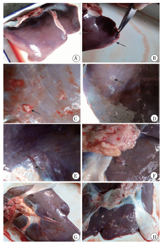
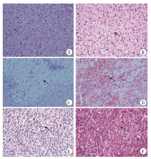
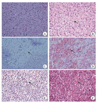
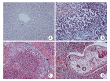
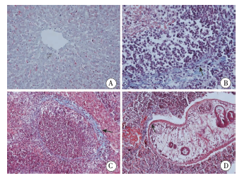
摘要: 目的 观察沼泽型水牛感染大片形吸虫(Fasciola gigantica)囊蚴后其肝脏在不同感染阶段的病理变化。 方法 29头健康沼泽型水牛分为感染组(24)和对照组(5头),感染组每头一次性经口感染500个大片形吸虫囊蚴,对照组不作任何处理。感染后第3、10、28、42和70天解剖水牛(感染组第42天4头,其余时间点5头;对照组各1头),肉眼观察肝脏的病理变化;采集肝脏组织按常规方法制作组织切片,苏木素-伊红(HE)染色,光学显微镜下观察组织病理结构;Masson三色染色,光学显微镜下观察肝组织中胶原蛋白。 结果 感染组水牛在感染囊蚴后第3天,肝表面可见散在的灰白色片状病变区域,第10天灰白色片状病变更加明显并出现瘢痕,第28天肝脏表面开始出现化脓性结节,第42和70天化脓性病灶逐渐加重;对照组水牛肝表面光滑,无其他病变。肝组织切片HE染色后结果显示,感染后第3天,肝索结构轻微紊乱和肝细胞发生颗粒变性,第10天局部肝细胞核溶解、破碎,第28天肝小叶内开始出现嗜酸粒细胞浸润和肝小叶内局部出血,第42天肝小叶内大面积出血,第70天肝窦间隙内出现纤维化,肝细胞开始修复;对照组肝组织切片的肝索结构清晰,肝细胞结构正常。肝组织切片Masson三色染色结果显示,感染组水牛第42天在汇管区和肝细胞窦状隙间开始出现少量胶原纤维,第70天胶原纤维逐渐增多;对照组肝窦间隙内有少量的胶原纤维。 结论 水牛感染大片形吸虫囊蚴后,肝组织出现动态病理损伤,在感染囊蚴后第3天出现早期肝损伤,随后损伤逐步加重并呈现明显的炎性反应,至感染第42天开始出现肝纤维化的典型病理变化.

关键词: 大片形吸虫, 水牛, 肝脏, 病理变化

Abstract: Objective To observe the hepatic pathological changes in buffaloes at different times after infection with Fasciola gigantica metacercariae. Methods Twenty-nine healthy marshy water buffaloes were divided into infection group (n = 24) and control group (n = 5). Buffaloes in the infection group were orally given 500 metacercariae and sacrificed at day 3, 10, 28, 42, 70 (n = 5, 5, 5, 4, 5) post infection. The control group received no treatment. The hepatic pathological changes were examined by visual observation and hematoxylin-eosin (HE) staining on liver tissue sections. Masson’s trichrome stain was performed to visualize collagen protein in liver tissue. Results A gray-white tablet-shaped lesion was seen scattering on liver surface at day 3 post infection, which became more significant at day 10, accompanied by the formulation of scar. Purulent nodules began to appear on the liver surface from day 28 and increased at day 42 and day 70. The control group showed a smooth liver surface with no pathological signs. HE staining showed structural disorder of hepatic cord and granular degeneration of hepatic cells at day 3, karyolysis and karyorrhexis at day 10, eosinophil infiltration and localized hemorrhage in hepatic lobules at day 28, massive hemorrhage in hepatic lobules at day 42, as well as fibrosis between sinusoids and hepatic cell repair at day 70. The control group showed a clear structure of hepatic cord and intact hepatic cells. The Masson’s trichrome stain showed formation of a small amount of collagen fibers in the portal areas and sinusoids at day 42, which increased at day 70, while the control group had only a slight sign of collagen fibers. Conclusion There occur dynamic hepatic pathological changes in buffaloes at different times after F. gigantica infection. The hepatic lesions begin to appear at as early as at day 3, then become more serious with time, accompanied with inflammatory reactions, until fibrosis, a typical sign of hepatic pathology, at day 42.

Key words: Fasciola gigantica, Bufflao, Liver, Pathological change

中图分类号: