
CHINESE JOURNAL OF PARASITOLOGY AND PARASITIC DISEASES ›› 2022, Vol. 40 ›› Issue (5): 579-586.doi: 10.12140/j.issn.1000-7423.2022.05.003
• ORIGINAL ARTICLES • Previous Articles Next Articles
LI Jia-ming1( ), WANG Yi-xuan1, YANG Ning-ai2, MA Hui-hui1, LAN Min1, LIU Chun-lan1, ZHAO Zhi-jun2,3,*()
Received:2022-03-22
Revised:2022-05-04
Online:2022-10-30
Published:2022-09-14
Contact:
ZHAO Zhi-jun
E-mail:1151143915@qq.com;z15815z@163.com
CLC Number:
LI Jia-ming, WANG Yi-xuan, YANG Ning-ai, MA Hui-hui, LAN Min, LIU Chun-lan, ZHAO Zhi-jun. Effects of ROP16 protein of Toxoplasma gondii on polarization and apoptosis of MH-S cells and their related mechanisms[J]. CHINESE JOURNAL OF PARASITOLOGY AND PARASITIC DISEASES, 2022, 40(5): 579-586.
Add to citation manager EndNote|Ris|BibTeX
URL: https://www.jsczz.cn/EN/10.12140/j.issn.1000-7423.2022.05.003
Table 1
Primer sequences of genes in ROP16-MH-S cells for RT-qPCR analysis
| 基因名称 Gene name | 引物序列(5′→3′) Primer sequence(5′→3′) |
|---|---|
| GAPDH | F: GGTTGTCTCCTGCGACTTCA R: TGGTCCAGGGTTTCTTACTCC |
| Bax | F: TTGCCCTCTTCTACTTTGCTAG R: CCATGATGGTTCTGATCAGCTC |
| Bcl-2 | F: GATGACTTCTCTCGTCGCTAC R: GAACTCAAAGAAGGCCACAATC |
| Caspase 3 | F: AACAAAATGATTCTGTGAGCCC R: TTGACAAATGCTTTTCCCTGAG |
| Caspase 9 | F: TGTGAATATCTTCAACGGGAGC R: GAGTAGGACACAAGGATGTCAC |
| IL-1β | F: TGGACCTTCCAGGATGAGGACA R: GTTCATCTCGGAGCCTGTAGTG |
| IL-6 | F: CTTGCAAGACTTCCATCCAC R: AGTGGTAGACAGGTCTGTTGG |
| IL-10 | F: ACCTGGTAGAAGTGATGCCCCAGGCA R: CTATGCAGTTGATGAAGATGTCAAA |
| IL-12 | F: TTGAACTGGCGTTGGAAGCACG R: CCACCTGTGAGTTCTTCAAAGGC |
| IL-18 | F: AGACCTGGAATCAGACAACTTT R: TCAGTCATATCCTCGAACACAG |
| TNF-α | F: CCTGTAGCCCACGTCGTAC R: GGGAGTAGACAAGGTACAACCC |
| TGF-β | F: ACGTCACTGGAGTTGTACGG R: GGGGCTGATCCCGTTGATT |
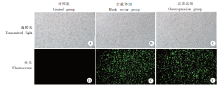
Fig. 1
Fluorescence protein expression of MH-S cells transfected with T. gondii ROP16 overexpressing lentivirus (× 200)
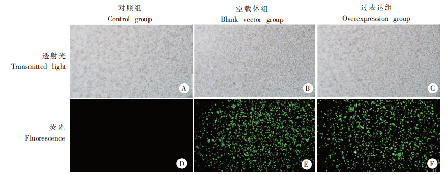
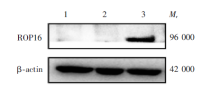
Fig. 2
Relative expression of T. gondii ROP16 protein in ROP16-MH-S cells by Western blotting1: Control group; 2: Blank vector group; 3: Overexpression group.
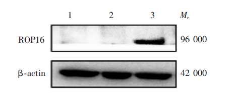
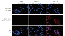
Fig. 3
Location of T. gondii ROP16 in ROP16-MH-S cells by immunofluorescence assay (× 600)
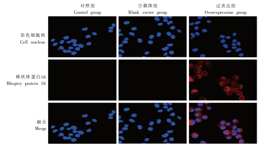
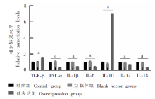
Fig. 4
mRNA relative transcription level of polarization-related genes in ROP16-MH-S cells by RT-qPCR
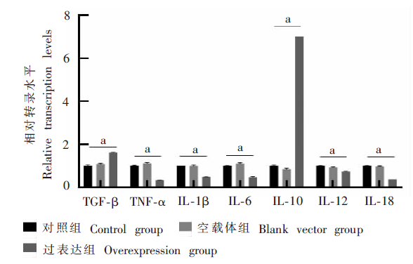
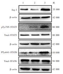
Fig. 5
Relative expression of polarized protein and STAT signalling pathway protein in ROP16-MH-S cells by Western blotting1: Control group; 2: Blank vector group; 3: Overexpression group.

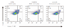
Fig. 6
Apoptosis of ROP16-MH-S cells determined by flow cytometry

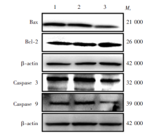
Fig. 7
Relative expression of apoptotic protein in ROP16-MH-S cells by Western blotting1: Control group; 2: Blank vector group; 3: Overexpression group.

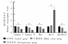
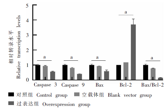
Fig. 8
mRNA relative transcription level of apoptosis-related genes in ROP16-MH-S cells by RT-qPCR
| [1] |
Kim K,, Weiss LM. Toxoplasma gondii: the model apicomplexan[J]. Int J Parasitol, 2004, 34(3): 423-432.
pmid: 15003501 |
| [2] |
Zhang Y,, Lai BS,, Juhas M, et al. Toxoplasma gondii secretory proteins and their role in invasion and pathogenesis[J]. Microbiol Res, 2019, 227: 126293.
doi: 10.1016/j.micres.2019.06.003 |
| [3] | Zheng B,, Lu SH. Research progress on immune evasion related molecules of Toxoplasma gondii[J]. Chin J Parasitol Parasit Dis, 2012, 30(5): 396-400. (in Chinese) |
| ( 郑斌,, 陆绍红. 刚地弓形虫免疫逃避相关分子的研究进展[J]. 中国寄生虫学与寄生虫病杂志, 2012, 30(5): 396-400.) | |
| [4] |
Lima TS,, Lodoen MB. Mechanisms of human innate immune evasion by Toxoplasma gondii[J]. Front Cell Infect Microbiol, 2019, 9: 103.
doi: 10.3389/fcimb.2019.00103 |
| [5] | Liu GZ,, Wang B,, Wang HF. Advances in research of Toxoplasma gondii rhoptry protein ROP16[J]. Chin J Schisto Control, 2015, 27(2): 217-220. (in Chinese) |
| ( 刘功振,, 王彬,, 王洪法. 弓形虫棒状体蛋白ROP16的研究进展[J]. 中国血吸虫病防治杂志, 2015, 27(2): 217-220.) | |
| [6] |
Jensen KDC,, Wang Y,, Wojno EDT, et al. Toxoplasma polymorphic effectors determine macrophage polarization and intestinal inflammation[J]. Cell Host Microbe, 2011, 9(6): 472-483.
doi: 10.1016/j.chom.2011.04.015 pmid: 21669396 |
| [7] |
Xu YW,, Xing RX,, Zhang WH, et al. Toxoplasma ROP16 Ⅰ/Ⅲ ameliorated inflammatory bowel diseases via inducing M2 phenotype of macrophages[J]. World J Gastroenterol, 2019, 25(45): 6634-6652.
doi: 10.3748/wjg.v25.i45.6634 |
| [8] |
Chtanova T,, Schaeffer M,, Han SJ, et al. Dynamics of neutrophil migration in lymph nodes during infection[J]. Immunity, 2008, 29(3): 487-496.
doi: 10.1016/j.immuni.2008.07.012 pmid: 18718768 |
| [9] |
Boothroyd JC. Have it your way: how polymorphic, injected kinases and pseudokinases enable Toxoplasma to subvert host defenses[J]. PLoS Pathog, 2013, 9(4): e1003296.
doi: 10.1371/journal.ppat.1003296 |
| [10] | Su YJ,, Dong H,, Qiao X, et al. Difference of expression profiling of A549 cells induced by Toxoplasma effector ROP16Ⅱ[J]. Chin J Zoonoses, 2018, 34(4): 323-329. (in Chinese) |
| ( 苏雅静,, 董辉,, 乔霞, 等. 弓形虫ROP16 Ⅱ效应分子对宿主A549细胞基因表达谱的影响[J]. 中国人兽共患病学报, 2018, 34(4): 323-329.) | |
| [11] |
Ong YC,, Reese ML,, Boothroyd JC. Toxoplasma rhoptry protein 16 (ROP16) subverts host function by direct tyrosine phosphorylation of STAT6[J]. J Biol Chem, 2010, 285(37): 28731-28740.
doi: 10.1074/jbc.M110.112359 |
| [12] |
Butcher BA,, Fox BA,, Rommereim LM, et al. Toxoplasma gondii rhoptry kinase ROP16 activates STAT3 and STAT6 resulting in cytokine inhibition and arginase-1-dependent growth control[J]. PLoS Pathog, 2011, 7(9): e1002236.
doi: 10.1371/journal.ppat.1002236 |
| [13] |
Saeij JPJ,, Coller S,, Boyle JP, et al. Toxoplasma co-opts host gene expression by injection of a polymorphic kinase homologue[J]. Nature, 2007, 445(7125): 324-327.
doi: 10.1038/nature05395 |
| [14] |
Yamamoto M,, Standley DM,, Takashima S, et al. A single polymorphic amino acid on Toxoplasma gondii kinase ROP16 determines the direct and strain-specific activation of Stat3[J]. J Exp Med, 2009, 206(12): 2747-2760.
doi: 10.1084/jem.20091703 |
| [15] |
Rosowski EE,, Lu D,, Julien L, et al. Strain-specific activation of the NF-κB pathway by GRA15, a novel Toxoplasma gondii dense granule protein[J]. J Exp Med, 2011, 208(1): 195-212.
doi: 10.1084/jem.20100717 |
| [16] |
Martinez FO,, Gordon S,, Locati M, et al. Transcriptional profiling of the human monocyte-to-macrophage differentiation and polarization: new molecules and patterns of gene expression[J]. J Immunol, 2006, 177(10): 7303-7311.
doi: 10.4049/jimmunol.177.10.7303 pmid: 17082649 |
| [17] | Rutschman R,, Lang R,, Hesse M, et al. Cutting edge: Stat6-dependent substrate depletion regulates nitric oxide production[J]. J Immuno, 2001, 166(4): 2173-2177. |
| [18] | Kochanowsky JA,, Thomas KK,, Koshy AA. ROP16-mediated activation of STAT6 suppresses host cell reactive oxygen species production, facilitating type Ⅲ Toxoplasma gondii growth and survival[J]. mBio, 2021, 12(2): e03305-e03320. |
| [19] |
Rutschman R,, Lang R,, Hesse M, et al. Cutting edge: STAT6-dependent substrate depletion regulates nitric oxide production[J]. J Immunol, 2001, 166(4): 2173-2177.
pmid: 11160269 |
| [20] |
Robben P M,, Mordue D G,, Truscott S M, et al. Production of IL-12 by macrophages infected with Toxoplasma gondii depends on the parasite genotype[J]. J Immunol, 2004, 172(6): 3686-3694.
pmid: 15004172 |
| [21] |
Modolell M,, Corraliza IM,, Link F, et al. Reciprocal regulation of the nitric oxide synthase/arginase balance in mouse bone marrow-derived macrophages by Th1 and Th2 cytokines[J]. Eur J Immunol, 1995, 25(4): 1101-1104.
pmid: 7537672 |
| [22] |
Alvarez C,, De-La-Torre A,, Vargas M, et al. Striking divergence in Toxoplasma ROP16 nucleotide sequences from human and meat samples[J]. J Infect Dis, 2014, 211(12): 2006-2013.
doi: 10.1093/infdis/jiu833 |
| [23] | Han M,, Wu H. Research progress of toxoplasmosis in China[J]. Med Inf, 2018, 31(2): 33-36. (in Chinese) |
| ( 韩梅,, 吴寒. 我国弓形虫病研究进展[J]. 医学信息, 2018, 31(2): 33-36.) | |
| [24] | Zhu Y,, Yang QL. Interaction between Toxoplasma gondii and host cell[J]. Prog Microbiol Immunol, 2014, 42(5): 77-80. (in Chinese) |
| ( 朱勇,, 杨秋林. 弓形虫与宿主细胞相互作用研究进展[J]. 微生物学免疫学进展, 2014, 42(5): 77-80.) | |
| [25] |
Carmen JC,, Hardi L,, Sinai AP. Toxoplasma gondii inhibits ultraviolet light-induced apoptosis through multiple interactions with the mitochondrion-dependent programmed cell death pathway[J]. Cell Microbiol, 2006, 8(2): 301-315.
pmid: 16441440 |
| [26] |
Jia ZH,, Jia Y,, Guo FJ, et al. Phosphorylation of STAT3 at Tyr705 regulates MMP-9 production in epithelial ovarian cancer[J]. PLoS One, 2017, 12(8): e0183622.
doi: 10.1371/journal.pone.0183622 |
| [27] |
Hirano T,, Ishihara K,, Hibi M. Roles of STAT3 in mediating the cell growth, differentiation and survival signals relayed through the IL-6 family of cytokine receptors[J]. Oncogene, 2000, 19(21): 2548-2556.
pmid: 10851053 |
| [28] |
Nash PB,, Purner MB,, Leon RP, et al. Toxoplasma gondii-infected cells are resistant to multiple inducers of apoptosis[J]. J Immunol, 1998, 160(4): 1824-1830.
pmid: 9469443 |
| [29] |
Earnshaw WC,, Martins LM,, Kaufmann SH. Mammalian caspases: structure, activation, substrates, and functions during apoptosis[J]. Annu Rev Biochem, 1999, 68(1): 383-424.
doi: 10.1146/annurev.biochem.68.1.383 |
| [1] | XUE Yushan, LIN Ping, CHENG Xunjia, FENG Meng. Damage caused by chronic infection of Toxoplasma gondii on the host central nervous system and its mechanism [J]. CHINESE JOURNAL OF PARASITOLOGY AND PARASITIC DISEASES, 2023, 41(5): 527-531. |
| [2] | JIANG Wenjing, MENG Yali, ZHAO Lina, WANG Chunmiao, ZHANG Xiaolei. Immunoprotection of nuclei acid vaccine dual-targeting rhoptry protein 18 and surface antigen 30 of Toxoplasma gondii in mice [J]. CHINESE JOURNAL OF PARASITOLOGY AND PARASITIC DISEASES, 2023, 41(5): 532-538. |
| [3] | ZHAO Ziqi, LV Fangli. Study on the inhibitory effect of artemether liposome on Toxoplasma gondii proliferation in vitro [J]. CHINESE JOURNAL OF PARASITOLOGY AND PARASITIC DISEASES, 2023, 41(4): 446-451. |
| [4] | ZHANG Chi, CHEN Jiating, XIN Zixuan, YANG Lili, YANG Zihan, PENG Hongjuan. Transcriptome analysis of mice brain chronically infected with Toxoplasma gondii and validation of the kynurenine pathway associated with depression [J]. CHINESE JOURNAL OF PARASITOLOGY AND PARASITIC DISEASES, 2023, 41(3): 270-278. |
| [5] | OU Yangran, LIU Xingzhuo, HUANG Shiguang, LYU Fangli. Effect of locking galectin-receptor interaction on the immunopathology of small intestine of Toxoplasma gondii-infected mice [J]. CHINESE JOURNAL OF PARASITOLOGY AND PARASITIC DISEASES, 2023, 41(3): 279-285. |
| [6] | DU Juan, LI Jia, WU Di, YU Qi, ZHANG Wei, BAI Runian, GUO Junlin, LIU Qingbin, LEI Qili, GU Chuanhui, WANG Meng, ZHAO Haojun. Seroepidemiological survey of Toxoplasma gondii infection in dogs and cats in Beijing 2022 [J]. CHINESE JOURNAL OF PARASITOLOGY AND PARASITIC DISEASES, 2023, 41(3): 389-392. |
| [7] | ZOU Wei-hao, WU Wei-ling, LIAO Yuan-peng, CHEN Min, PENG Hong-juan. Preparation and application of monoclonal antibody against Toxoplasma gondii bradyzoite antigen 1 [J]. CHINESE JOURNAL OF PARASITOLOGY AND PARASITIC DISEASES, 2022, 40(5): 587-593. |
| [8] | DAI Li-sha, ZHANG Li-xin, YIN Kun. Research advances in Toxoplasma gondii induced host mental-behavioural disorders [J]. CHINESE JOURNAL OF PARASITOLOGY AND PARASITIC DISEASES, 2022, 40(5): 642-646. |
| [9] | WANG Jie, WEN Hong-yang, CHEN Ying, AN Ran, LUO Qing-li, SHEN Ji-long, DU Jian. Construction and identification of macrophage migration inhibitory factor gene knockout strain of Toxoplasma gondii [J]. CHINESE JOURNAL OF PARASITOLOGY AND PARASITIC DISEASES, 2022, 40(3): 349-354. |
| [10] | WANG Zhen-xun, XIONG Si-si, SUN Xia-hui, WANG Yong-liang, PAN Ge, HE Shen-yi, CONG Hua. Differential expression and action mechanism of lncRNA102796 in the brain of mice with chronic infection of Toxoplasma gondii [J]. CHINESE JOURNAL OF PARASITOLOGY AND PARASITIC DISEASES, 2022, 40(2): 187-193. |
| [11] | JIANG Feng, CHEN Run, DU Ning-ning, ZHU Meng-yi, ZHONG Hao, CHEN Hui, XI Xu-xia, ZHAN Xiao-dong, LI Chao-pin. Investigation of Toxoplasma gondii infection in pet dogs and cats in Wuhu City [J]. CHINESE JOURNAL OF PARASITOLOGY AND PARASITIC DISEASES, 2022, 40(1): 124-126. |
| [12] | SHAO Han, LI Si-yuan, LI Jun. The affect of metformin on autophagy and apoptosis of Echinococcus multilocularis cysts and protoscoleces [J]. CHINESE JOURNAL OF PARASITOLOGY AND PARASITIC DISEASES, 2022, 40(1): 43-49. |
| [13] | LU Fei, ZHUO Xun-hui, LU Shao-hong. Research progress on the interaction between host cell autophagy and apicomplexa protozoa infection [J]. CHINESE JOURNAL OF PARASITOLOGY AND PARASITIC DISEASES, 2021, 39(6): 826-831. |
| [14] | ZHANG Xiao-han, FENG Ying, CHEN Ran, SANG Xiao-yu, YANG Na. Advances in research of structure, function and regulatory mechanism of Toxoplasma gondii conoid [J]. CHINESE JOURNAL OF PARASITOLOGY AND PARASITIC DISEASES, 2021, 39(6): 832-835. |
| [15] | ZHOU Wen-zheng, SUN Jun-gang, ZHAO Xi-bin, CAO Li. Therapeutic effect of intensity modulated radiation therapy on secondary femur infection with Echinococcus granulosus in rats [J]. CHINESE JOURNAL OF PARASITOLOGY AND PARASITIC DISEASES, 2021, 39(4): 443-448. |
| Viewed | ||||||
|
Full text |
|
|||||
|
Abstract |
|
|||||